
UrOops3D, danger-zone atlas
Operative animation, Transplant ureteric reconstruction

Transplant ureter: leak, stricture and reconstruction

The transplant ureter has a single blood supply from the renal artery and no nerves, so it leaks in the first weeks when its distal end dies and strictures months later at the same place, and the graft does not hurt when it obstructs.

This page is how the leak and the stricture present, the ultrasound and the nephrostomy that define them, the stent and the endoscopic options that buy time, and the redo reimplant, the native ureter and the Boari flap that fix them for good.

Advanced tier FRCS transplant and specialist urology, transplant ureteric complications Interactive 3D

Operative visualisation

Schematic scene preview: Transplant ureteric reconstruction
Preparing the 3D anatomy…

UrOops3D · UroRef · © Nity G

Click me next Section 2 of 8 Anatomy The structures this operation works between, and the one it must not cross.

The structures in the scene

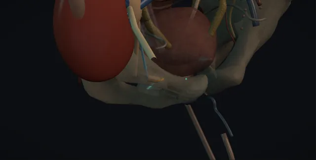
Transplant ureteric reconstruction: how the structures relateRight iliac fossa under the scar, graft to bladder. Transplant ureter, ischaemic distal end: fed from the renal artery alone Graft renal artery and vein. Transplant ureter, ischaemic distal end: high anterolateral; retrograde access is hard Transplant orifice. Transplant ureter, ischaemic distal end: substitute, mobilised with its fat and adventitia Native ureter on the psoas (danger). Transplant ureter, ischaemic distal end: psoas hitch or Boari flap, if capacity allows Bladder. Bladder: beside the hitch; the iliac vein tears in scar Recipient iliac vessels (danger). Graft renal artery and vein: nephrostomy planned on ultrasound away from them Nephrostomy and stent (danger).Transplant ureter,ischaemic distal end!Graft renal arteryand veinTransplant orifice!Native ureter on thepsoasBladder!Recipient iliacvesselsNephrostomy andstentfed from the renalartery alonehigh anterolateral;retrograde accessis hardsubstitute,mobilised with itsfat and adventitiapsoas hitch orBoari flap, ifcapacity allowsbeside the hitch; theiliac vein tears in scarnephrostomy planned onultrasound away fromthemTargetDangerLandmarkContext
Right iliac fossa under the scar, graft to bladder. Schematic, not to scale; relationships only.
  • The graft sits in the right iliac fossa under the scar with its artery and vein on the anterior surface joining the external iliac vessels, and its ureter curving from the lower pole medially and down to the anterolateral wall of the bladder, where the distal centimetre is the ischaemic segment.
  • The native ureter runs on the psoas medial to the graft toward the same bladder, and the bladder itself can be drawn up to the psoas or cut into a flap that reaches the transplant pelvis.
  • The nephrostomy enters the graft from the front, away from the vessels.
  • ›The transplant ureter and its blood supply - fed from the renal artery alone through the hilar and periureteric fat; the distal end is the furthest from it and the first to die.
  • ›The graft renal artery and vein - anterior and superficial in the iliac fossa, just under the scar; found and protected before any dissection of the ureter.
  • ›The transplant orifice - high on the anterolateral bladder wall, pointing the wrong way for a cystoscope; retrograde access is difficult.
  • ›The native ureter - medial to the graft on the psoas with its own blood supply; the first substitute when the transplant ureter cannot reach.
  • ›The bladder - the source of a psoas hitch or a Boari flap, if it has the capacity, which the long-dialysed bladder may not.
  • ›The recipient iliac vessels - beneath the graft anastomoses and beside the bladder hitch; the external iliac vein and the genitofemoral nerve at the psoas.
  • ›The nephrostomy and the stent - into a graft just under the skin, planned on the ultrasound away from the vessels; the stent across the defect for six weeks with a date.
Click me next Section 3 of 8 Steps The operation in order, with the landmark that guards each move.

Operative steps

  1. Who: the leak early, the stricture late Urological complications follow about one transplant in twenty, and nearly all of them are the distal ureter. The leak comes in the first two weeks, from a distal end that has lost its supply, a ureter cut too long, a hilar dissection that took the golden triangle, or an anastomosis under tension; the necrotic segment separates and urine collects beside the graft. The stricture comes months to years later at the same distal segment or the tunnel, from ischaemia, scar, a tunnel that was too tight, a kink, a lymphocele pressing on it, BK virus or rejection. Both threaten the graft rather than the patient, and the graft cannot report either: it is denervated and does not hurt, so the creatinine is the symptom.
  2. Presentation and the creatinine A rising creatinine, a falling urine output or urine from the drain or the wound in the first weeks is a leak until proven otherwise; a rising creatinine with a hydronephrosis on a routine scan a year on is a stricture until proven otherwise. Fever, tenderness over the graft and a swollen leg point to an infected collection or a lymphocele. The drain fluid creatinine is sent: urine has a creatinine many times the serum, lymph the same as serum. Rejection, tacrolimus toxicity, dehydration and infection cause the same creatinine rise, so the nephrologist is in the conversation from the first hour and the ultrasound is done the same day, because the graft is losing nephrons while the cause is debated.
  3. Imaging: ultrasound, nephrostomy and the antegrade study Ultrasound with Doppler first: the pelvicalyceal system compared with the baseline scan, because mild dilatation is common in a denervated, refluxing transplant and means little on its own; a collection beside the graft; the bladder volume, since a full bladder dilates a transplant pelvis; and the perfusion and resistive index of the graft. A MAG3 renogram shows obstruction as a curve that does not fall with the bladder emptied. Where obstruction or a leak is likely, a percutaneous nephrostomy under ultrasound into the graft, which lies just under the skin, drains the kidney and gives an antegrade nephrostogram that shows the level, the length and the site of the leak or stricture. CT is used for a collection, with contrast only if the graft can take it.
    where it bites: mild dilatation of a transplant pelvis is common and means little; a rising creatinine with dilatation is obstruction until the renogram or the antegrade study says otherwise.
  4. First control: nephrostomy and stent Every leak and every obstruction is first decompressed. A urethral catheter keeps the bladder empty so that nothing refluxes to the leak. The nephrostomy drains the kidney, the creatinine is watched for the fall that proves the problem was the drainage, and an antegrade guidewire is passed across the leak or the stricture under screening and a JJ stent placed over it, with the nephrostomy left on free drainage for a day and then clamped and removed if the antegrade study is clean. A small leak with a stent and a catheter often closes in a few weeks; a short stricture stays open on a stent while the plan is made. Immunosuppression is reviewed with the nephrologist and antibiotic cover given for every intervention.
  5. Endoscopic options A stricture under about a centimetre and less than a few months old can be treated from above: balloon dilatation through the nephrostomy tract under screening, or an antegrade incision with the holmium laser or a cutting balloon through the full thickness of the stricture into the periureteric fat, with a stent for six weeks after. About half of endoscopic treatments hold in the long term; the rest recur and are then reconstructed. A long stricture, a stricture that recurs after one endoscopic treatment, or a leak that does not close on a stent is an operation, and a long-term stent changed every few months is a choice for the patient too frail for one.
    where it bites: the transplant vessels lie on the anterior surface of the graft just under the skin; a nephrostomy or an incision that is not planned on the ultrasound can enter them.
  6. Redo ureteroneocystostomy Through the old Gibson incision, with the graft vessels found and protected first because they lie superficial and anterior in a scarred field, the transplant ureter is traced from the lower pole, keeping its fat, to the stricture or the necrotic end. The diseased segment is excised back to a bleeding, pink end; if enough healthy ureter remains to reach the bladder without tension it is spatulated and reimplanted into the filled bladder over a stent, extravesically with a short tunnel or as a simple refluxing anastomosis, the bladder mobilised and hitched to the psoas if the reach is short. In the first weeks the planes are still open; a year on they are not.
    where it bites: in the reoperative fossa the graft artery and vein are the first structures found and protected; a ureteric dissection that meets them unprepared costs the kidney.
  7. Native ureter to the transplant When the transplant ureter is too short or too ischaemic to reach, the recipient's own ureter on that side is the best substitute: it has its own blood supply, it is usually intact and dilated enough to handle, and it lies nearby on the psoas medial to the graft. It is divided as high as needed. The renal end is ligated, or the native kidney is removed if it is infected or refluxing, and the bladder end, still attached to the bladder with its own blood supply, is brought to the graft and joined end to side to the transplant renal pelvis, a ureteropyelostomy, or end to end to the stump of transplant ureter, spatulated, over a stent, mucosa to mucosa with fine absorbable suture. The native kidney is left in place unless it is infected or refluxing badly; a native ureter that drains a kidney with little function is not missed.
  8. Boari flap and psoas hitch Where there is no native ureter, or it has been used or is diseased, the bladder is the source of length. A psoas hitch alone, the bladder mobilised on its contralateral pedicle and fixed to the psoas tendon above the iliac vessels with the genitofemoral nerve protected, gains a few centimetres. A Boari flap, a tongue of bladder with a broad base cut from the anterolateral wall, tubularised and brought up to the transplant pelvis or the short ureteric stump, gains ten centimetres or more, provided the bladder is of good capacity, which in a long-dialysed patient with a defunctioned bladder it may not be. An ileal interposition is the last reconstruction, and graft nephrectomy is the honest end when the kidney is not worth it.
  9. After A catheter for a week and a stent for six weeks with its removal date written down, a drain until it is dry, and an antegrade study through the nephrostomy before it is removed if one was left. The creatinine is followed daily and then at each clinic, because the graft, not the wound, is the outcome. Ultrasound at three months after the stent is out, and a MAG3 if there is any dilatation, then at six and twelve months, because recurrence is silent. Every intervention is covered with antibiotics and immunosuppression is reviewed with the nephrologist, particularly for BK virus where that was the cause. The patient is told that the ureter is the weak point of the graft and that the same problem can return.
Click me next Section 4 of 8 Danger zones Where this operation bites, and why it bites there.
Critical relationship

Danger zones

The spatial relationships the animation is built to make automatic. Before you read each one, ask yourself: at this point in the operation, what is about to go wrong, and what is my first move? The save for each sits in the Complications tab.

  • RiskThe graft hilum and vessels - superficial, anterior and scarred in; the first structures found and protected in any reoperation and the reason a nephrostomy is planned on ultrasound.
  • RiskThe recipient iliac vessels - under the anastomoses and beside the hitch; the external iliac vein tears in a scarred field.
  • RiskThe native ureteric blood supply - the substitute ureter is mobilised with its adventitia and fat, medial above the brim, or it fails as the first one did.
  • RiskBowel and bladder in the reoperative pelvis - peritoneum adherent to the graft with small bowel behind it, and a thin defunctioned bladder that tears when it is mobilised.
Practise it Take me into the scene Opens the animation at a danger moment and asks you to predict the mechanism and your first move before it reveals.
Click me next Section 5 of 8 Complications The save for each one: the first move, the escalation trigger, the op-note line.

Complications and immediate saves

Trigger each of these live in the Animation tab, then read the save: the first move, the escalation trigger, and the line that goes in the op note.

Graft vascular injury and haemorrhage

  • the graft artery or vein entered during a nephrostomy, an antegrade incision or a reoperative dissection: haematuria through the nephrostomy, a haematoma compressing the graft, or bleeding in the field.
The save
  1. Pressure on the field or the nephrostomy clamped, the patient resuscitated and cross-matched, and the transplant surgeon called. first move
  2. Duplex ultrasound for the perfusion of the graft and the size of the haematoma; a graft with no flow is theatre now.
  3. Bleeding from the nephrostomy tract that does not settle with a larger tube: angiography and selective embolisation.
  4. A torn graft vessel in the field, or a graft losing perfusion: vascular help, repair under control, and graft nephrectomy if the iliac artery is threatened. escalation trigger
  5. Op note: the vessel, the control, the transfusion and the perfusion of the graft at the end. document

Persistent leak or fistula

  • urine in the drain or a collection that does not resolve on a stent, or a fistula to the skin or the vagina: the necrotic segment is longer than the study suggested, or the reconstruction has failed.
The save
  1. Catheter draining, stent position on a plain film, and drain fluid creatinine; a full bladder refluxes urine through the leak. first move
  2. An antegrade study through the nephrostomy to see the length of the leaking segment and whether the stent crosses it.
  3. A collection is drained percutaneously and the nephrostomy kept on free drainage so the kidney is diverted.
  4. A leak that does not close in a few weeks with full diversion, or a fistula: reconstruction with the native ureter or the bladder, in the early window while the planes are open. escalation trigger
  5. Note: the level and length of the leak, the drains and stents with their dates, and the operation planned. document

Recurrent stricture and graft obstruction

  • hydronephrosis and a rising creatinine after an endoscopic treatment or a reconstruction: continued ischaemia, scar, tension, BK virus or a lymphocele pressing on the new join.
The save
  1. Ultrasound compared with the baseline, and a MAG3 with the bladder emptied; decompress with a nephrostomy if the creatinine is rising. first move
  2. Antegrade study to measure the recurrence; BK virus PCR and a review of the immunosuppression with the nephrologist.
  3. A first recurrence after endoscopic treatment is reconstructed, not dilated again; a long-term stent is the option for the frail.
  4. A recurrence after reconstruction: the next operation up the ladder, native ureter, Boari flap or ileal interposition, at a transplant centre. escalation trigger
  5. Note: the length and level, the graft function, the cause found and the surveillance interval. document

Infection and sepsis

  • an obstructed or leaking transplant with a nephrostomy, a stent and immunosuppression: pyelonephritis of the graft, an infected collection, and sepsis that comes fast and quietly.
The save
  1. Sepsis six with cultures from blood, urine and the nephrostomy, broad antibiotics by the transplant protocol, and drainage of the obstructed system if it is not already drained. first move
  2. Ultrasound or CT for a collection and drain it; a stent in an infected system is changed, not left.
  3. Immunosuppression reduced with the nephrologist for the duration, and the antiproliferative held.
  4. Sepsis not settling with drainage and antibiotics: critical care, and graft nephrectomy if the graft is the unresolvable source. escalation trigger
  5. Note: the organisms, the drains, the immunosuppression changes and the date the antibiotics stop. document

Loss of graft function

  • the graft that was obstructed for too long, leaked into an infected collection, or lost its vessels at the reconstruction: a creatinine that does not fall and a patient back on dialysis.
The save
  1. Every rising creatinine with dilatation is decompressed within a day; the graft has no reserve and does not complain. first move
  2. A biopsy with the nephrologist when the creatinine does not recover after decompression, for rejection, BK virus or infarction.
  3. The reconstruction chosen for reliability rather than elegance; the native ureter over a second attempt at a short transplant ureter.
  4. A graft with no recoverable function that is infected, bleeding or painful: graft nephrectomy and a return to the waiting list once sensitisation is assessed. escalation trigger
  5. Note: the timeline from the first creatinine rise to decompression, the interventions, and what the patient was told about the graft. document
Click me next Section 6 of 8 Troubleshooting When the case turns: the problem, then the next move.

Intra-operative troubleshooting

When the case turns: the problem, then the next move.

  • ActionRising creatinine, mild dilatation, and the nephrologist says rejection - both can be true; a MAG3 with the bladder empty, or a nephrostomy with the creatinine watched for 48 hours, settles the obstruction question while the biopsy answers the other.
  • ActionThe nephrostomy needle keeps hitting the hilum - stop; replan on the ultrasound from a lateral, more posterior calyx with the vessels on the screen; a radiologist who does transplants if the first two passes fail.
  • ActionThe antegrade wire will not cross the stricture - a hydrophilic wire and an angled catheter under screening, with a retrograde cystoscope in the bladder to meet it; if it will not pass, the stricture is complete and is an operation.
  • ActionThe transplant ureter is too short after excision - do not stretch it; find the native ureter on the psoas and bring it to the transplant pelvis, or mobilise and hitch the bladder.
  • ActionThe bladder is tiny and will not mobilise for a flap - the long-dialysed bladder; the native ureter is the answer, and an ileal interposition if there is none.
  • ActionUrine leak at the new anastomosis on the drain - catheter and stent checked, drain fluid creatinine, antegrade study; a stented leak with the bladder empty is given weeks before anyone reoperates.
Click me next Section 7 of 8 FRCS viva Say it out loud, then mark yourself against the model answer.

FRCS viva questions

The examiner stems this scene prepares you for. Try to answer out loud first, then reveal, and learn the discriminator, the detail that turns a pass into a strong pass.

QWhy does the transplant ureter leak early and stricture late?reveal answer
  • Both come from the same anatomy.
  • The native ureter has a segmental supply from the renal artery, the aorta, the gonadal and the iliac vessels, but the transplant ureter is cut from all of those except the renal artery, so its entire supply runs through the hilar and periureteric fat, the golden triangle, and the distal end is the furthest from it.
  • If the fat was stripped on the bench, the ureter was cut long, or the anastomosis was under tension, the distal segment necroses in the first two weeks and separates, and urine collects beside the graft: that is the early leak.
  • A distal segment that survives with a marginal supply heals with fibrosis and contracts over the following months, or the detrusor tunnel scars, or a lymphocele, a kink, BK virus infection or rejection narrows it: that is the late stricture, at the same distal centimetre.
  • Because the graft is denervated neither produces pain, so the presentation is a rising creatinine, a falling output or urine in the drain early, and hydronephrosis on a routine scan late.
  • Prevention is on the bench and at the anastomosis, and a stent at the time of transplantation roughly halves the early complications.
Strong pass

Say both

  • The single supply through the golden triangle given as the reason for both
  • The denervated graft as the reason the creatinine is the symptom
Examiner note
The single supply through the golden triangle given as the reason for both, and the denervated graft as the reason the creatinine is the symptom.
QA transplant recipient has a rising creatinine and a dilated transplant pelvis on ultrasound at nine months. How do you manage her?reveal answer
  • I would treat this as obstruction of a graft that cannot report itself, and I would act the same day while the nephrologist considers rejection, tacrolimus toxicity and infection, because the two enquiries run in parallel.
  • I would compare the ultrasound with her baseline, since mild dilatation is common in a refluxing denervated transplant, and look at the Doppler and for a collection or lymphocele beside the graft.
  • A MAG3 renogram with the bladder drained shows whether the curve falls;
  • if it does not, I would ask for a percutaneous nephrostomy under ultrasound, planned on the screen away from the hilar vessels, which decompresses the graft and gives an antegrade study to show the level and length of the stricture.
  • A fall in creatinine after decompression proves the obstruction was the cause.
  • I would send BK virus PCR and review the immunosuppression with the nephrologist.
  • With the stricture defined I would pass an antegrade wire and stent it, and then plan: a stricture under a centimetre may be balloon dilated or laser incised from above with about a fifty per cent long-term success, and a longer, older or recurrent stricture is reconstructed, with a redo reimplant, the native ureter to the transplant pelvis, or a Boari flap according to what reaches.
Strong pass

Say both

  • The nephrostomy used as both treatment and diagnosis with the creatinine fall as the proof
  • The parallel nephrology enquiry named
Examiner note
The nephrostomy used as both treatment and diagnosis with the creatinine fall as the proof, and the parallel nephrology enquiry named.
QWhat are the reconstructive options when a transplant ureter cannot be reimplanted directly, and how do you choose?reveal answer
  • The order is by reliability and by what remains.
  • If enough healthy transplant ureter remains after excision to reach the bladder without tension, I would reimplant it, mobilising the bladder and hitching it to the psoas if the reach is short.
  • If the transplant ureter is too short or too ischaemic, I would use the ipsilateral native ureter, which has its own blood supply and lies close on the psoas, dividing it high and joining it end to side to the transplant renal pelvis as a ureteropyelostomy, or end to end to a healthy stump of transplant ureter, spatulated over a stent;
  • the native kidney is left unless it is infected or refluxing badly.
  • If there is no native ureter, the bladder is the source of length: a psoas hitch gains a few centimetres and a Boari flap ten or more, provided the bladder has the capacity, which a long-dialysed defunctioned bladder may not.
  • An ileal interposition is the last reconstruction, and graft nephrectomy is the honest choice for a kidney with poor function or an unresolvable infection.
  • In every reconstruction the graft vessels are found and protected first, the ureter is mobilised with its fat, the anastomosis is spatulated and tension-free over a stent, and the creatinine is the measure of success.
Strong pass

Say both

  • The native ureter placed above the bladder flap in the order with its blood supply as the reason
  • The graft vessels found first in every reoperation
Examiner note
The native ureter placed above the bladder flap in the order with its blood supply as the reason, and the graft vessels found first in every reoperation.
QWhat is the role of nephrostomy and stenting in transplant ureteric complications, and what are their risks?reveal answer
  • Decompression is the first step for every leak and every obstruction, because the graft has no reserve and loses function while the cause is being debated.
  • A percutaneous nephrostomy into the graft is planned on ultrasound from a lateral or posterior calyx away from the hilar vessels, which lie anteriorly and can be entered by an unplanned puncture with haemorrhage or an arteriovenous fistula.
  • It drains the kidney, allows an antegrade nephrostogram that defines the level and length of the problem better than any other study, and gives the route for an antegrade wire, a JJ stent and, for a short stricture, balloon dilatation or laser incision.
  • A stent across a leak with the bladder kept empty by a catheter allows many small leaks to close;
  • a stent across a stricture holds the graft open while the reconstruction is planned, and a long-term stent changed every few months is a choice for the patient too frail for surgery.
  • The risks are bleeding, infection in an immunosuppressed patient, for which every intervention is covered with antibiotics, stent symptoms, encrustation and migration, and the forgotten stent, which is why the removal date is recorded in a register.
  • Endoscopic treatment of a stricture holds in about half of cases; a recurrence is reconstructed rather than dilated again.
Strong pass

Say both

  • The nephrostomy planned away from the anterior hilar vessels with the reason
  • The stent register named as part of the treatment
Examiner note
The nephrostomy planned away from the anterior hilar vessels with the reason, and the stent register named as part of the treatment.
Click me next Section 8 of 8 Evidence The guidance each of these answers is built on.

Evidence and guidance

Two minutes

Close the lap: recall it before you leave.

Say it aloud, then reveal. Reading is not remembering.

  1. 01 The relationship Say the critical spatial relationship out loud, then reveal. reveal
    Memory frame

    The graft sits in the right iliac fossa under the scar with its artery and vein on the anterior surface joining the external iliac vessels, and its ureter curving from the lower pole medially and down to the anterolateral wall of the bladder, where the distal centimetre is the ischaemic segment.

    Why?
    The native ureter runs on the psoas medial to the graft toward the same bladder, and the bladder itself can be drawn up to the psoas or cut into a flap that reaches the transplant pelvis. The nephrostomy enters the graft from the front, away from the vessels.
  2. 02 The first move Graft vascular injury and haemorrhage. Your first move? reveal
    First move

    Pressure on the field or the nephrostomy clamped, the patient resuscitated and cross-matched, and the transplant surgeon called.

  3. 03 The escalation trigger Graft vascular injury and haemorrhage. What makes you call for help? reveal
    Escalate when

    A torn graft vessel in the field, or a graft losing perfusion: vascular help, repair under control, and graft nephrectomy if the iliac artery is threatened.

  4. 04 The discriminator What turns a pass into a strong pass? reveal
    Strong pass

    Say both

    • The single supply through the golden triangle given as the reason for both
    • The denervated graft as the reason the creatinine is the symptom
    Examiner note
    The single supply through the golden triangle given as the reason for both, and the denervated graft as the reason the creatinine is the symptom.
  5. 05 The pitfall Where does this operation bite first? reveal
    Pitfall

    The graft hilum and vessels

    Why?
    superficial, anterior and scarred in; the first structures found and protected in any reoperation and the reason a nephrostomy is planned on ultrasound.

Revision practice with self-marked answers, not an assessment of clinical competence. Kept in this browser only; it does not sync between devices.