The bladder that stores at high pressure or holds nothing, opened like a clam and patched with a length of ileum that has been cut open so it cannot contract.
The operation trades a bladder that empties for one that is safe and large, and the patient pays with a catheter, mucus and a lifetime of follow-up. This page is the selection, the bivalve, the patch and the ward after.
Advanced tierFRCS functional and reconstructive urologyInteractive 3D
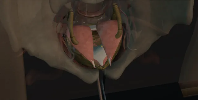
Lower midline, the filled bladder from the assistant's side. Schematic, not to scale; relationships only.
From the assistant's side the bladder stands filled in the middle of a lower midline wound with the bowel packed up under the Deaver.
The bivalve runs in the sagittal plane and opens the bladder into two shells joined at the base, with the trigone and orifices lying at the bottom of the posterior shell.
The ileal cup comes down from the right on its mesentery, is sewn into the clam edge to edge, and the suprapubic catheter leaves through the native bladder in front of it.
›The bladder dome and the sagittal line - the bivalve runs from two centimetres above the neck anteriorly, over the dome, to two centimetres above the interureteric ridge posteriorly.
›Trigone and ureteric orifices - the posterior limit; the cut stops above the ridge and the orifices are catheterised if they lie near it.
›Terminal ileum and the ileocaecal valve - the segment is taken at least fifteen centimetres proximal to the valve, sparing the last ileum for B12 and bile salts.
›The mesentery of the patch - transilluminated to choose the arcade; enters the back of the plate along one side and must reach the pelvis flat and untwisted.
›The antimesenteric border - the line of the detubularisation, exactly opposite the mesentery, from staple line to staple line.
›The suprapubic catheter - through native bladder and out through a separate stab; never through the patch.
›Retractors - Deaver to hold the packed bowel up, a Balfour or ring retractor for the wound, a Morris on the bladder.
Who is offered itThe small, poorly compliant or overactive bladder that has failed everything else: anticholinergics and beta-3 agonists, intermittent catheterisation, botulinum toxin more than once, neuromodulation where it was appropriate. Most are neuropathic, spina bifida and spinal injury, with a detrusor pressure that threatens the kidneys; a few are idiopathic overactivity or a contracted bladder after tuberculosis or radiation. The second condition is as important as the first: the patient who can and will catheterise for the rest of their life, with the hands, the eyesight, the urethra or the channel and the will, because a large floppy augmented bladder does not empty itself. Renal function, the upper tracts and the bowel are assessed before the decision.
Preparation and consentThe consent is long and written down. Mucus in the urine and the need to irrigate; bladder stones in about one in ten to one in five over the years; infection; a hyperchloraemic metabolic acidosis from urine sitting on ileum, and B12 deficiency if the terminal ileum is used; spontaneous perforation, rare but life-threatening, presenting as an acute abdomen; intermittent catheterisation in most neuropathic patients and about a third of the idiopathic; bowel complications; and a small long-term risk of malignancy in the patch or at the anastomosis. Bowel preparation is not routine. Antibiotics at induction, calf compression, a urethral catheter on the field, and the stoma nurse involved if a channel is planned.
Lower midline, packs, the bladder filledA lower midline incision from symphysis to above the umbilicus, the layers as for any lower midline, the peritoneum opened high. The small bowel is packed into the upper abdomen under a Deaver, the table tipped head-down and a self-retaining retractor set. The bladder is mobilised: the urachus divided, the peritoneum swept off the dome and the lateral walls freed from the pelvic side wall enough to bring the dome up into the wound. It is then filled through the catheter with 200 to 300 millilitres of saline so that it stands up as a globe and the line of the bivalve can be marked on it with the diathermy. Stay sutures of 2-0 Vicryl go on either side of the marked line. where it bites: a scarred neuropathic pelvis may have the peritoneum stuck to the dome and small bowel adherent to it; the peritoneum is opened at the top of the wound and every adhesion is taken sharply.
The clamThe bladder is bivalved in the sagittal plane with the diathermy, beginning on the dome and carried anteriorly down to about two centimetres above the bladder neck, then posteriorly down to a point short of the trigone, stopping about two centimetres above the interureteric ridge. The result is two half-shells joined at the base, opened like a clam, with a long ellipse to be filled. The orifices are looked at from inside and, if they lie close to the posterior limit of the cut, a ureteric catheter is passed up each before it is extended. The mucosal edges bleed; they are picked with the diathermy and the shells are left resting in the wound on their stay sutures. where it bites: the posterior limb of the bivalve stops above the interureteric ridge; a cut carried into the trigone takes the orifices and the closing mechanism of the neck with it.
The ileal segmentThe terminal ileum is delivered and a segment of twenty to twenty-five centimetres is chosen at least fifteen centimetres proximal to the ileocaecal valve, on a mesentery that reaches the pelvis without tension when the loop is dropped onto the bladder. The mesentery is held to the light so the arcades show and the segment is taken on a good arcade; windows are made between the arcades at each end and the mesentery divided between them with the LigaSure or between ties. The bowel is divided at each end with a linear stapler and continuity restored above the segment with a stapled side-to-side anastomosis, the mesenteric window closed with interrupted 3-0 Vicryl. The isolated loop is irrigated with saline through its open end until clear. where it bites: the loop has to reach the bladder base without pulling on its mesentery; a loop that lies tight at this stage is an ischaemic patch at the end.
Detubularising the segmentThe loop is laid on a swab and opened along its antimesenteric border, exactly opposite the mesentery, with the cutting diathermy or scissors, from one stapled end to the other, the staple lines excised as it goes. The purpose is to destroy the tube: a tube of ileum contracts as a ring and generates pressure, which is what the operation is trying to abolish. Opened flat it becomes a rectangle of mucosa about twenty centimetres long and eight to ten wide, its mesentery entering the back of the plate along one side. Bleeding from the cut edge is taken with the bipolar, the mucosa is wiped of mucus and the plate is measured against the open bladder.
Reconfiguring the patchThe plate is folded on itself into a U, the two limbs lying side by side, and the adjacent inner edges are sewn together with a running 3-0 Vicryl, mucosa to mucosa, from the fold to the open ends. Folded again and sewn, if the length allows, the plate becomes a cup, and either shape has the same property: no line of ileum runs the full length of the patch as a tube, so no coordinated contraction can raise the pressure in it. The cup is oriented so that the mesentery enters from its convex side without twisting, and the free edge of the cup is now an ellipse of about the same length as the clam.
Sewing the patch to the clamThe posterior edge first, because it is the deepest and hardest to see. Beginning at the posterior apex of the bivalve, one running 2-0 or 3-0 Vicryl is carried along each side, edge to edge, full thickness of ileum to full thickness of bladder, the bites close and the suture pulled just enough to appose, the assistant following. The two sutures meet and are tied at the anterior apex. At the corners the bites are placed carefully so the patch is not gathered. It is a watertight closure that is wanted, not a tight one, and any gap found on the fill is closed with a single interrupted suture rather than the whole line being redone. where it bites: the mesentery of the patch must lie flat with no twist under the bladder; a patch that turns blue before the wound is closed is taken down now.
Catheters, drain, the test fillA suprapubic catheter, 16 to 20 French, is brought through the native bladder, not through the patch, and out through a separate stab in the abdominal wall; the urethral catheter is left in as well, because mucus blocks one and the other will save the night. A tube drain is placed beside the suture line. The bladder is filled with 150 to 200 millilitres of saline and the suture line inspected all round: a leak is closed with an interrupted suture and the fill repeated. Omentum is brought down over the suture line if it reaches. The bowel is checked, the count confirmed, and the abdomen closed as a mass closure with looped 1 PDS.
After: mucus, catheters, the lifetime planThe catheters are flushed with 30 to 50 millilitres of saline three times a day from the first day, because mucus blocks them and a blocked augmented bladder leaks or bursts its suture line. The drain comes out when its output is low and clear. The catheters stay for two to three weeks and a cystogram before they come out is usual. Intermittent catheterisation is taught before discharge and the patient is told that they empty by the clock, not by sensation. Lifelong follow-up: irrigation for mucus, renal function and electrolytes, a bicarbonate for acidosis, B12 yearly after five years if terminal ileum was used, upper tract ultrasound, and a low threshold for cystoscopy for haematuria or infection, with the perforation warning repeated at every visit.
The spatial relationships the animation is built to make automatic. Before you read each one, ask yourself: at this point in the operation, what is about to go wrong, and what is my first move? The save for each sits in the Complications tab.
RiskThe trigone - the posterior limb of the bivalve stops two centimetres above the interureteric ridge; beyond it are the orifices and the neck.
RiskThe mesentery of the patch - short, tight or twisted under the bladder it strangles the ileum; the patch must lie without pull before it is sewn.
RiskThe bowel anastomosis and its window - a leak on day three to five, or an internal hernia through an unclosed mesenteric window.
RiskThe outlet and the channel - an augmented bladder does not empty; a urethra or channel that cannot be catheterised means retention, leakage and eventually perforation.
Trigger each of these live in the Animation tab, then read the save: the first move, the escalation trigger, and the line that goes in the op note.
Urine leak from the suture line
urine in the drain in the first week, or a fluid collection with ileus, from a patch-to-bladder line that was not watertight or a blocked catheter that pressurised it.
The save
Check both catheters are draining and flush them; a blocked catheter is the commonest cause of a leak.first move
Send the drain fluid for creatinine to confirm urine.
Keep the drain and both catheters, and image with a cystogram or CT to size the leak and exclude an obstructed system.
Generalised peritonitis, sepsis or a large intraperitoneal collection: return to theatre for washout and repair.escalation trigger
Op note and discharge summary: the catheters, the flush regime, the cystogram date and the leak if there was one.document
Bowel anastomotic leak or obstruction
pain, fever, distension and a rising white cell count on day three to five, or a bowel that will not work at all.
The save
A patient who is not progressing after a bowel anastomosis has a leak until proven otherwise; examine, bloods, nil by mouth.first move
CT with oral contrast to separate a contained leak from free peritonitis and to look for an internal hernia through the mesenteric window.
A contained collection drains radiologically with antibiotics and nutrition.
Free leak, peritonitis or an obstructed loop: laparotomy, washout, repair or resection with senior help.escalation trigger
Op note: the anastomotic technique, the window closure, and the plan for feeding and review.document
Spontaneous perforation of the augmented bladder
months or years later, an over-full bladder, a missed catheterisation or blunt trauma; abdominal pain, distension and sepsis in a patient whose sensation may be absent.
The save
Suspect it in any augmented patient with abdominal pain, sepsis or a rising creatinine, however vague; catheterise and drain the bladder first.first move
CT cystogram: the intraperitoneal leak is usually from the patch or the suture line.
A small contained leak in a well patient may be managed with catheter drainage, antibiotics and observation.
Peritonitis, free fluid or a patient who is unwell: laparotomy, washout and repair of the defect the same day.escalation trigger
Note: the cause found, the repair, and the catheterisation schedule reinforced with the patient and their carers.document
Mucus, infection and stones
a bladder lined partly by bowel makes mucus that blocks catheters, harbours bacteria and nucleates stones over the years.
The save
Daily irrigation with saline, taught before discharge and continued for life; the frequency goes up when the mucus does.first move
Asymptomatic bacteriuria is not treated; symptomatic infection is cultured and treated and its cause, usually incomplete emptying, sought.
Recurrent infection, haematuria or a catheter that catches: cystoscopy for stones, which are removed endoscopically or through a small open cystotomy.
Sepsis with an obstructed upper tract, or a stone burden that endoscopy cannot clear: admit, drain and plan the clearance.escalation trigger
Note: the irrigation regime, the infection history, and the imaging interval for stones.document
Metabolic acidosis and B12 deficiency
ileum in contact with urine reabsorbs chloride and ammonium: a hyperchloraemic acidosis, worse with a poorly emptying bladder or renal impairment; B12 falls years later if terminal ileum was taken.
The save
Bicarbonate, chloride and renal function at every review; a low bicarbonate is treated with oral sodium bicarbonate before symptoms appear.first move
Improve emptying: more frequent catheterisation reduces the time urine sits on the patch.
B12 measured yearly from five years and replaced when low.
A falling creatinine clearance, bone pain or growth failure in a child: renal and metabolic review and reconsider the reconstruction.escalation trigger
Note: the bowel segment used and its distance from the valve, so that the follow-up bloods are the right ones.document
When the case turns: the problem, then the next move.
ActionThe ileal loop will not reach the pelvis - divide the mesentery deeper between the arcades or choose a more mobile loop; never sew a patch under tension.
ActionThe patch is longer than the clam - extend the bivalve a little, both limbs, within the limits; or gather the patch by taking wider bites on the ileum, not by cutting the mesentery side.
ActionThe mucosal edge of the bladder bleeds everywhere - the bipolar on points, not a run of monopolar; the suture line itself is haemostatic once done.
ActionLeak on the test fill - find the gap and close it with an interrupted full-thickness suture; refill and check again; do not redo the whole line.
ActionCatheter blocked with mucus on the ward - flush 50 millilitres of saline and aspirate; if it will not clear, change the urethral catheter and leave the suprapubic; a blocked bladder is a leaking bladder.
ActionThe patient cannot catheterise the urethra after the operation - the outlet was the wrong route; a suprapubic catheter for now, and a catheterisable channel planned as a second procedure.
The examiner stems this scene prepares you for. Try to answer out loud first, then reveal, and learn the discriminator, the detail that turns a pass into a strong pass.
QWho would you offer an augmentation cystoplasty to, and what would you consent them for?reveal answer
I would offer it to a patient with a small, poorly compliant or overactive bladder that has failed conservative and less invasive treatment: anticholinergics or beta-3 agonists, intermittent catheterisation, botulinum toxin and, where appropriate, neuromodulation.
Most are neuropathic, with a detrusor pressure that threatens the upper tracts; some have refractory idiopathic overactivity or a contracted bladder after tuberculosis or radiation.
The essential second criterion is the ability and the will to catheterise for life, because an augmented bladder may not empty.
I would assess renal function, the upper tracts, the bowel and, in a woman of childbearing age, the plans for pregnancy.
I would consent for mucus and lifelong irrigation, bladder stones in about one in ten to one in five, urinary infection, hyperchloraemic metabolic acidosis and B12 deficiency, spontaneous perforation as a rare but lethal risk, intermittent catheterisation in most neuropathic and about a third of idiopathic patients, bowel complications including leak and obstruction, and a small long-term risk of malignancy, with lifelong follow-up.
I would name the alternatives: continued botulinum toxin, a catheter, or diversion.
Strong pass
Say both
The ability to catheterise for life stated as a selection criterion equal to the bladder
The consent including perforation and malignancy rather than stopping at stones and mucus
Examiner note
The ability to catheterise for life stated as a selection criterion equal to the bladder, and the consent including perforation and malignancy rather than stopping at stones and mucus.
QDescribe the steps of a clam ileocystoplasty.reveal answer
Through a lower midline incision I would pack the bowel away, mobilise the bladder and fill it so that it stands up.
I would bivalve it in the sagittal plane with diathermy from about two centimetres above the bladder neck anteriorly, over the dome, to about two centimetres above the interureteric ridge posteriorly, so that the trigone and orifices are preserved, catheterising the ureters if the orifices lie close to the cut.
I would isolate twenty to twenty-five centimetres of ileum at least fifteen centimetres from the ileocaecal valve on a mesentery that reaches the pelvis without tension, restore bowel continuity with a stapled side-to-side anastomosis and close the mesenteric window.
I would open the segment along its antimesenteric border to detubularise it, fold it into a U or a cup with a running absorbable suture, and sew the free edge of the patch to the clam edge to edge with running 2-0 or 3-0 Vicryl, posterior edge first, watertight.
I would leave a suprapubic catheter through native bladder, a urethral catheter and a drain, test the closure with 150 to 200 millilitres, bring omentum down, and close the abdomen with a mass closure.
Strong pass
Say both
The limits of the bivalve stated in centimetres from the neck and the ridge
The antimesenteric detubularisation given as the reason the patch cannot generate pressure
Examiner note
The limits of the bivalve stated in centimetres from the neck and the ridge, and the antimesenteric detubularisation given as the reason the patch cannot generate pressure.
QWhy is the bowel detubularised, and what happens if it is not?reveal answer
A segment of intact bowel is a tube of circular and longitudinal muscle with intact myenteric coordination, so it contracts as a ring in peristaltic waves and generates pressure;
a tube of ileum can produce pressures well above the 40 centimetres of water that threatens the upper tracts.
Opening it along the antimesenteric border and reconfiguring it as a U or a cup does two things.
It breaks the circular continuity so that no coordinated ring contraction can occur, and any contraction that does happen is local and of low amplitude.
And it increases the radius of the resulting reservoir, which by Laplace's law means a larger volume is stored at a lower pressure for the same wall tension.
The clam itself contributes: bivalving the bladder interrupts the detrusor as a ring as well, so the native bladder can no longer contract effectively against the patch.
If the bowel were left as a tube and simply sewn on, the patient would have a larger bladder that still generated high-pressure waves, with persistent incontinence and the same risk to the kidneys, which is the historical lesson from the tubular cystoplasties that preceded the clam.
Strong pass
Both mechanisms, the broken ring and the larger radius through Laplace, and noting that the bivalve interrupts the native detrusor as well as the bowel.
QWhat is the long-term follow-up after an augmentation, and why?reveal answer
The follow-up is lifelong and structured around the four things that go wrong.
Emptying: the patient catheterises by the clock, and residual volume and the technique are reviewed, because retention causes infection, stones, acidosis and perforation.
Metabolism: serum bicarbonate, chloride and creatinine at each visit with oral bicarbonate for acidosis, and B12 annually from about five years if terminal ileum was used, because deficiency is slow and neurological damage is not reversible.
Structure: upper tract ultrasound and renal function to confirm the pressures are safe and no stricture or reflux has developed, with urodynamics if there is any doubt, and imaging or cystoscopy for stones when there is recurrent infection, haematuria or catheter difficulty.
Malignancy: adenocarcinoma at the anastomosis and urothelial tumours in the augmented bladder are reported at a low rate a decade or more later;
routine surveillance cystoscopy is debated, but haematuria, new pain or recurrent infection after the first decade earns a cystoscopy.
Every visit repeats the perforation warning: abdominal pain in an augmented patient is a surgical emergency until proven otherwise.
Strong pass
Organising the follow-up around emptying, metabolism, structure and malignancy, and stating the perforation warning as a standing instruction to the patient.
Say it aloud, then reveal. Reading is not remembering.
01The relationshipSay the critical spatial relationship out loud, then reveal.reveal
Memory frame
From the assistant's side the bladder stands filled in the middle of a lower midline wound with the bowel packed up under the Deaver.
Why?
The bivalve runs in the sagittal plane and opens the bladder into two shells joined at the base, with the trigone and orifices lying at the bottom of the posterior shell. The ileal cup comes down from the right on its mesentery, is sewn into the clam edge to edge, and the suprapubic catheter leaves through the native bladder in front of it.
02The first moveUrine leak from the suture line. Your first move?reveal
First move
Check both catheters are draining and flush them; a blocked catheter is the commonest cause of a leak.
03The escalation triggerUrine leak from the suture line. What makes you call for help?reveal
Escalate when
Generalised peritonitis, sepsis or a large intraperitoneal collection: return to theatre for washout and repair.
04The discriminatorWhat turns a pass into a strong pass?reveal
Strong pass
Say both
The ability to catheterise for life stated as a selection criterion equal to the bladder
The consent including perforation and malignancy rather than stopping at stones and mucus
Examiner note
The ability to catheterise for life stated as a selection criterion equal to the bladder, and the consent including perforation and malignancy rather than stopping at stones and mucus.
05The pitfallWhere does this operation bite first?reveal
Pitfall
The trigone
Why?
the posterior limb of the bivalve stops two centimetres above the interureteric ridge; beyond it are the orifices and the neck.
Revision practice with self-marked answers, not an assessment of clinical competence. Kept in
this browser only; it does not sync between devices.