
UrOops3D, danger-zone atlas
Operative animation, Bladder neck closure

Bladder neck closure for the devastated outlet

The urethra that has been eroded by decades of catheters, burnt by radiation, opened by fistula or scarred by failed repairs, leaking urine that no operation on the sphincter will hold. The neck is closed and the bladder drains another way, decided before the first suture.

This page is the selection, the plan for drainage, the retropubic dissection, the closure in two layers with omentum between, and the leak that follows if any of it was skimped.

Advanced tier FRCS reconstructive urology, the devastated outlet Interactive 3D

Operative visualisation

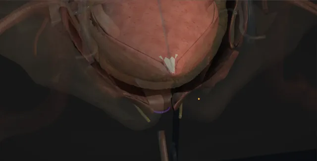
Schematic scene preview: Bladder neck closure
Preparing the 3D anatomy…

UrOops3D · UroRef · © Nity G

Click me next Section 2 of 8 Anatomy The structures this operation works between, and the one it must not cross.

The structures in the scene

Bladder neck closure: how the structures relateRetropubic, from the assistant's side, the neck at the bottom of the space. Bladder neck, trimmed and closed: just behind; a suture through one obstructs Ureteric orifices and trigone (danger). Bladder neck, trimmed and closed: stuck after radiation; a finger guides the plane Vagina, or prostate and rectum, behind (danger). Bladder neck, trimmed and closed: anterior; divided before the neck is dissected Dorsal venous complex. Bladder neck, trimmed and closed: lies at the bottom of the space, behind the pubis Retropubic space of Retzius. Bladder neck, trimmed and closed: laid between the closure and the urethral stump Greater omentum. Bladder neck, trimmed and closed: the only route out; blocked, the closure leaks Suprapubic catheter through the dome (danger).Bladder neck, trimmedand closed!Ureteric orificesand trigone!Vagina, or prostateand rectum, behindDorsal venouscomplexRetropubic space ofRetziusGreater omentum!Suprapubic catheterthrough the domejust behind; asuture through oneobstructsstuck afterradiation; a fingerguides the planeanterior; dividedbefore the neck isdissectedlies at the bottomof the space,behind the pubislaid between theclosure and theurethral stumpthe only route out;blocked, theclosure leaksTargetDangerLandmarkContext
Retropubic, from the assistant's side, the neck at the bottom of the space. Schematic, not to scale; relationships only.
  • From the assistant's side the field is the retropubic space with the bladder held down by a Morris and the symphysis in front, the bladder neck at the bottom of the space with the catheter balloon sitting on it and the dorsal vein above.
  • Behind the neck lies the vagina or the prostate and rectum, and the orifices are just inside the neck on the trigone.
  • When the urethra is divided the neck is closed as a two-layer line facing the pelvis, the urethral stump is closed below it, and omentum comes down from above to lie between them, with the suprapubic catheter leaving the dome.
  • ›Retropubic space of Retzius - between the bladder and the back of the symphysis; obliterated in the irradiated or operated pelvis and taken sharply.
  • ›The bladder neck - found by the catheter balloon pulled down onto it; dissected circumferentially and trimmed back to healthy bladder before closure.
  • ›Ureteric orifices and trigone - a centimetre or two behind the neck, closer in the scarred bladder; identified and catheterised before the posterior wall is closed.
  • ›Dorsal venous complex - anterior to the neck and urethra; divided between sutures or sealed before the neck is dissected.
  • ›The plane behind the neck - vagina in a woman, prostate and rectum in a man; developed on the bladder side with a finger in the vagina or rectum as a guide.
  • ›The greater omentum - brought down on the right gastroepiploic pedicle to lie between the neck closure and the urethral stump.
  • ›The suprapubic catheter - 16 to 20 French silicone through the dome, away from the closure; the only route out for three weeks and, for most, for life.
Click me next Section 3 of 8 Steps The operation in order, with the landmark that guards each move.

Operative steps

  1. Who: the outlet that cannot be saved The devastated outlet is a urethra and bladder neck that have been destroyed rather than weakened: the woman with a neuropathic bladder and a long-term urethral catheter whose urethra has eroded to a patulous gutter, the pelvis after radiotherapy with a fistula or a fixed open neck, the man after multiple failed sphincters, slings and urethroplasties whose urethra is a scarred tube that leaks continuously. Sphincters, slings and bulking have failed or cannot be offered. The alternatives are laid out honestly: a long-term suprapubic catheter alone, a catheterisable channel, an ileal conduit, or cystectomy with diversion. Bladder capacity and compliance are measured beforehand, because a small high-pressure bladder closed at the neck needs augmentation as well or it will leak or harm the kidneys.
  2. The plan: how the urine will leave The bladder neck is never closed until the route out has been decided and, where possible, built in the same operation. For most patients it is a suprapubic catheter for life, changed every twelve weeks, and the patient has lived with one for a trial period beforehand to see whether they can. For the patient with the hands and the will it is a continent catheterisable channel, a Mitrofanoff from the appendix or a Monti from ileum, with augmentation if the bladder is small. The stoma site is marked by the stoma nurse. The consent names recurrent leakage through the closure in about one in five, fistula, stones, infection, the catheter changes, and the possibility of a conduit later if the closure fails twice.
  3. Incision and the retropubic space Lower midline or Pfannenstiel, the choice made by the previous scars and by whether bowel is needed. The rectus sheath is opened and the muscles separated in the midline; the transversalis fascia is divided and the retropubic space of Retzius entered with a finger sweeping the bladder off the back of the symphysis, staying extraperitoneal unless omentum or bowel is wanted. In the irradiated or many-times operated pelvis the space is obliterated and is taken sharply with scissors on the bone, the bladder held down with a Morris retractor. The endopelvic fascia is opened on each side of the neck so the lateral surfaces of the bladder base are seen. A urethral catheter with its balloon inflated identifies the bladder neck as the catheter is pulled and pushed.
    where it bites: in a scarred, irradiated pelvis the bladder is stuck to the bone and the plane is found sharply, not with a finger; a finger tears the bladder and enters the wrong plane.
  4. Dissecting the neck and dividing the urethra With the balloon pulled down onto the neck the junction of bladder and urethra is dissected circumferentially: anteriorly the dorsal venous complex is divided between sutures or with the LigaSure, laterally the pillars of the bladder are taken with the bipolar, and posteriorly, the dangerous side, the plane between the bladder base and the vagina in a woman, or the prostate and rectum in a man, is developed with the diathermy on the bladder side. In a woman a finger in the vagina and in a man a finger in the rectum guide the posterior dissection. The catheter balloon is deflated and the urethra transected just below the neck. The ureteric orifices are looked for from inside and catheterised if they lie within a centimetre of the cut.
    where it bites: the ureteric orifices sit a centimetre or two behind the neck in a scarred trigone; they are identified and catheterised before the posterior wall of the neck is closed.
  5. Closing the urethral stump The distal urethra is closed so it does not become a blind pocket that collects secretions or, in a woman, a sinus to the vagina. In a man the stump is oversewn with two or three interrupted 2-0 Vicryl sutures through its full thickness, taking care not to include the rectum behind it. In a woman the urethra is short and the stump is closed with interrupted sutures and buried with the periurethral tissue over it; some units excise the urethra to the meatus instead and close the vaginal side, which is the choice when the urethra has eroded to the vagina already. The closed stump is checked with a swab for bleeding and left alone.
  6. Closing the bladder neck in two layers The neck is trimmed back to healthy bladder. The first layer is a running or interrupted 2-0 or 3-0 Vicryl or PDS through the full thickness of the neck, inverting the mucosa into the bladder so no mucosa faces the pelvis, tied without tension. The second layer is interrupted 2-0 Vicryl in the muscle and adventitia, imbricating the first so that it lies buried. The closure is then tested by filling the bladder with 100 to 150 millilitres through the suprapubic route and looking for a leak at the neck; any leak is closed with an interrupted suture and the fill repeated. The suture lines of the neck and the urethral stump are deliberately offset so they do not lie against each other.
    where it bites: tension and the wrong tissue are what open a neck closure; the edge is trimmed to healthy bladder and the two layers sit without pull, or the closure fails.
  7. Omentum between the closure and the urethra The peritoneum is opened and the greater omentum brought down on its right gastroepiploic pedicle, mobilised off the transverse colon and lengthened if needed by dividing the left gastroepiploic and the short branches, until it reaches the pelvis without tension. It is laid between the closed neck and the closed urethral stump and around the neck closure, and sutured in place with a few 3-0 Vicryl sutures, so that the two suture lines never touch, the closure has a blood supply beside it, and any leak is absorbed rather than becoming a fistula. In a woman with a vaginal repair the omentum lies between bladder and vagina. Where omentum is absent a Martius labial fat flap or a rectus flap does the same work.
  8. Suprapubic catheter, drain, the test fill A 16 to 20 French silicone suprapubic catheter is brought through the dome of the bladder, well away from the closed neck, and out through a separate stab in the abdominal wall, fixed with a purse-string in the bladder and a suture at the skin. If a catheterisable channel has been made it is catheterised and its own catheter left in. A tube drain is placed in the retropubic space. The bladder is filled once more through the suprapubic catheter and the neck watched; the drain and the wound must stay dry. The rectus sheath is closed with looped 1 PDS and the skin with staples or a subcuticular suture. The suprapubic catheter is the only route out for the next three weeks.
  9. After: leakage, stones, the channel The suprapubic catheter stays for three weeks and a cystogram before it is changed shows the neck holding. Then the pattern for life: the suprapubic catheter changed every twelve weeks, or the channel catheterised by the clock. Leakage per urethra in the first weeks is a small fistula through the closure and is managed by keeping the bladder empty; leakage that persists after three months is a failed closure and is re-closed with a fresh interposition, or diverted. Stones form on the catheter balloon and in the bladder and are looked for with ultrasound; the kidneys are imaged for pressure and the bladder pressure remeasured if leakage or infection returns. The patient is told plainly that this is the last operation on the urethra.
Click me next Section 4 of 8 Danger zones Where this operation bites, and why it bites there.
Critical relationship

Danger zones

The spatial relationships the animation is built to make automatic. Before you read each one, ask yourself: at this point in the operation, what is about to go wrong, and what is my first move? The save for each sits in the Complications tab.

  • RiskThe ureteric orifices - a centimetre or two behind the neck, closer in the scarred trigone; a suture through an orifice obstructs a kidney.
  • RiskThe rectum or vagina behind the neck - stuck to the bladder base after radiation or repeated surgery; a finger inside guides the plane and an injury is repaired at once.
  • RiskThe tissue at the edge - irradiated, eroded or scarred neck does not heal; the edge is trimmed to healthy bladder or the closure opens.
  • RiskThe route out - a neck closed with no dependable drainage is a bladder that leaks through the closure, and a high-pressure bladder that harms the kidneys.
Practise it Take me into the scene Opens the animation at a danger moment and asks you to predict the mechanism and your first move before it reveals.
Click me next Section 5 of 8 Complications The save for each one: the first move, the escalation trigger, the op-note line.

Complications and immediate saves

Trigger each of these live in the Animation tab, then read the save: the first move, the escalation trigger, and the line that goes in the op note.

Closure breakdown and urethral leakage

  • urine per urethra after the operation: a small fistula through the closure in the first weeks, or a closure that has opened, in about one in five over time, more after radiation.
The save
  1. Keep the bladder empty: check the suprapubic catheter is draining and flush it; a full bladder pushes urine through the weakest point. first move
  2. A cystogram to see the leak; a small track with good drainage is given three months to close.
  3. Bladder pressures rechecked; a high-pressure bladder needs anticholinergics, botulinum toxin or augmentation before any re-closure.
  4. Leakage persisting after three months, or a large defect: re-closure with a fresh interposition, a vaginal approach in a woman, or conversion to a conduit. escalation trigger
  5. Op note: the tissue quality at the edge, the layers, the interposition used, and the cystogram date; the discharge summary names the leak and its plan. document

Ureteric obstruction or injury

  • loin pain, a rising creatinine or a leak from the pelvis after the neck closure: an orifice caught in the posterior suture line of a scarred trigone.
The save
  1. Identify and catheterise the orifices before the posterior wall is closed; look at the drain and the creatinine on the first day. first move
  2. Ultrasound both kidneys; a new hydronephrosis after neck closure is a suture until proven otherwise.
  3. Nephrostomy and an antegrade study; a stent passed antegrade if the ureter opens.
  4. A ureter that is ligated or divided: reimplantation into the bladder, with a psoas hitch if needed, with senior help. escalation trigger
  5. Op note: whether the orifices were seen and catheterised, the distance from the closure, and the imaging plan. document

Rectal or vaginal injury

  • the plane behind the neck stuck by radiation or previous surgery: a hole in the vagina or rectum found at the time, or a fistula found later.
The save
  1. Recognised at the time: repair in two layers with absorbable suture, with omentum laid over the repair. first move
  2. A rectal injury in an irradiated or contaminated field: a defunctioning colostomy with colorectal help, decided in theatre.
  3. A vaginal injury is closed from the vaginal side as well if it can be reached, and the omentum kept between vagina and bladder.
  4. Faeces or gas per urethra or per stoma, or a discharge later: image, divert and plan a staged repair. escalation trigger
  5. Op note: the injury, the layers of the repair, the interposition, and whether the bowel was diverted. document

High-pressure bladder and the upper tracts

  • a small, poorly compliant bladder that now has no outlet at all: pressure that leaks through the closure or goes up the ureters to the kidneys.
The save
  1. Measure capacity and compliance before the operation; a low-compliance bladder is augmented at the same time or the closure is not done. first move
  2. After the operation, drain continuously through the suprapubic catheter until the pressures have been rechecked.
  3. Anticholinergics or botulinum toxin for overactivity; more frequent emptying of a channel.
  4. Hydronephrosis, a rising creatinine or recurrent leakage with a high-pressure trace: augmentation, or a conduit. escalation trigger
  5. Note: the pre-operative urodynamic findings and the pressure plan, so that a later leak is read against them. document

Stones, infection and the failing route out

  • a lifetime with a suprapubic catheter or a channel: encrustation on the balloon, bladder stones, recurrent infection, and a catheter or channel that blocks or will not pass.
The save
  1. Catheter changes every twelve weeks on a written schedule, irrigation if the bladder was augmented, and the patient taught what a blocked catheter feels like. first move
  2. Symptomatic infection is cultured and treated; bacteriuria in a catheterised bladder is not.
  3. Ultrasound of the bladder and kidneys yearly; stones are cleared through the suprapubic tract or the channel with a cystoscope.
  4. A tract or channel that has closed, or a blocked bladder that cannot be reached: drain percutaneously under ultrasound and revise the route. escalation trigger
  5. Note: the catheter size and change interval, the channel and its catheter, and the imaging schedule. document
Click me next Section 6 of 8 Troubleshooting When the case turns: the problem, then the next move.

Intra-operative troubleshooting

When the case turns: the problem, then the next move.

  • ActionThe retropubic space will not open - sharp dissection on the back of the symphysis with scissors, not a finger; open the endopelvic fascia laterally and come at the neck from the sides.
  • ActionThe bladder neck cannot be found - pull and push the catheter with the balloon up; open the bladder on the dome and put a finger through the neck from inside.
  • ActionThe orifices lie right at the cut edge - catheterise both with 5 French ureteric catheters from inside and close the neck with the catheters in view; stent them if a bite comes close.
  • ActionThe neck edge is white, thin and irradiated - trim it back until it bleeds; a closure in dead tissue is a fistula in three weeks.
  • ActionLeak at the neck on the test fill - an interrupted full-thickness suture at the point, refill, and bring the omentum down over it.
  • ActionThe omentum will not reach - mobilise it off the transverse colon and divide the left gastroepiploic to lengthen it on the right pedicle; failing that, a rectus flap or a Martius flap.
Click me next Section 7 of 8 FRCS viva Say it out loud, then mark yourself against the model answer.

FRCS viva questions

The examiner stems this scene prepares you for. Try to answer out loud first, then reveal, and learn the discriminator, the detail that turns a pass into a strong pass.

QWhat is the devastated outlet, and when would you close the bladder neck?reveal answer
  • The devastated outlet describes a urethra and bladder neck destroyed beyond reconstruction: the eroded patulous urethra of long-term catheterisation in the neuropathic woman, the irradiated pelvis with a fixed open neck or a fistula, the man after multiple failed artificial sphincters, slings or urethroplasties.
  • The leakage is continuous and no procedure on the sphincter will hold it.
  • I would consider bladder neck closure when those options have failed or cannot be offered, after a multidisciplinary discussion, and only with a dependable alternative route for drainage decided beforehand: a suprapubic catheter for life, which most patients will have trialled, or a continent catheterisable channel for the patient who can catheterise.
  • I would measure bladder capacity and compliance first, because a small high-pressure bladder closed at the neck will leak through the closure or damage the kidneys, and I would augment it in the same operation if needed.
  • I would put the alternatives to the patient plainly: a suprapubic catheter with the urethra left as it is, an ileal conduit, or cystectomy with diversion, and I would consent for breakdown and recurrent leakage in about one in five, fistula, ureteric injury, stones and infection.
Strong pass

Say both

  • The drainage route and the bladder pressure settled before the neck is closed
  • Rather than closure presented as an operation on its own
Examiner note
The drainage route and the bladder pressure settled before the neck is closed, rather than closure presented as an operation on its own.
QDescribe a transabdominal bladder neck closure.reveal answer
  • Through a lower midline or Pfannenstiel incision I would enter the retropubic space, sharply in a scarred pelvis, and open the endopelvic fascia on each side to see the bladder base.
  • With a catheter balloon pulled onto the neck I would dissect the vesicourethral junction circumferentially, dividing the dorsal venous complex anteriorly, the bladder pillars laterally and developing the posterior plane on the bladder side with a finger in the vagina or rectum as a guide.
  • I would transect the urethra just below the neck, identify the ureteric orifices from inside and catheterise them if they lie within a centimetre.
  • I would close the urethral stump with interrupted absorbable sutures.
  • I would trim the neck back to healthy bladder and close it in two layers, a full-thickness inverting layer of 2-0 or 3-0 absorbable suture and a second imbricating seromuscular layer, without tension, and test it by filling the bladder.
  • I would bring the greater omentum down on its right gastroepiploic pedicle and fix it between the neck closure and the urethral stump.
  • I would place a 16 to 20 French silicone suprapubic catheter through the dome away from the closure, or catheterise the channel if one has been made, leave a retropubic drain, and keep the bladder empty for three weeks before a cystogram.
Strong pass

Say all three

  • The orifices identified from inside before the posterior closure
  • The edge trimmed to healthy tissue
  • The omentum interposed with its pedicle named
Examiner note
The orifices identified from inside before the posterior closure, the edge trimmed to healthy tissue, and the omentum interposed with its pedicle named.
QWhy does a bladder neck closure fail, and what do you do when it does?reveal answer
  • A closure fails for four reasons, and each is a decision at the operation.
  • Tissue: the neck closed in irradiated, eroded or scarred bladder that cannot heal, avoided by trimming the edge back until it bleeds.
  • Tension: a closure that is pulled together rather than laid together, avoided by mobilising the bladder base and by two layers that sit without pull.
  • Contact: the closure lying against the closed urethral stump so that any leak has a track to follow, avoided by offsetting the suture lines and interposing well-vascularised omentum or a Martius flap between them.
  • Pressure: a small, poorly compliant or overactive bladder with no outlet at all, avoided by measuring the pressures before the operation and augmenting if they are high, and by continuous drainage while the closure heals.
  • When leakage per urethra follows, I would first confirm the suprapubic catheter or channel is draining and keep the bladder empty, image the leak with a cystogram, and recheck the bladder pressures.
  • A small early leak with good drainage is given three months.
  • A persistent or large defect is re-closed with a fresh interposition, transvaginally in a woman where the anatomy allows, after the pressure problem has been dealt with;
  • a second failure, or a patient who has had enough, is offered an ileal conduit.
Strong pass

The four causes, tissue, tension, contact and pressure, each paired with the operative decision that prevents it, and the pressure corrected before any re-closure.

QWhat are the long-term consequences of living with a closed bladder neck?reveal answer
  • The patient now has a bladder with no natural outlet, and everything follows from that.
  • Drainage: a suprapubic catheter changed every twelve weeks by a trained team, with a plan for the blocked catheter and a tract that must never be allowed to close, or a catheterisable channel used by the clock with the same discipline.
  • Stones: encrustation on the balloon and stones in the bladder, particularly if the bladder was augmented and makes mucus, so ultrasound yearly and a low threshold for cystoscopy through the tract or channel.
  • Infection: bacteriuria is universal and not treated, symptomatic infection is treated and its cause sought in a blocked catheter or a residual.
  • Pressure: the bladder pressure is measured if leakage or infection returns and the kidneys are imaged for hydronephrosis, because a closed high-pressure bladder damages them silently.
  • Leakage: recurrent leakage per urethra is a failed closure until proven otherwise.
  • And the psychological consequence, which is the end of urethral function for good: the patient should have understood that this was the last operation on the urethra, and that the alternative if the closure fails twice is diversion.
  • Follow-up is lifelong, shared between urology, the district nursing team and the stoma nurse.
Strong pass

Say both

  • Treating bacteriuria as expected rather than as infection
  • Framing the bladder pressure check as the reason the kidneys stay safe after a closed outlet
Examiner note
Treating bacteriuria as expected rather than as infection, and framing the bladder pressure check as the reason the kidneys stay safe after a closed outlet.
Click me next Section 8 of 8 Evidence The guidance each of these answers is built on.

Evidence and guidance

Two minutes

Close the lap: recall it before you leave.

Say it aloud, then reveal. Reading is not remembering.

  1. 01 The relationship Say the critical spatial relationship out loud, then reveal. reveal
    Memory frame

    From the assistant's side the field is the retropubic space with the bladder held down by a Morris and the symphysis in front, the bladder neck at the bottom of the space with the catheter balloon sitting on it and the dorsal vein above.

    Why?
    Behind the neck lies the vagina or the prostate and rectum, and the orifices are just inside the neck on the trigone. When the urethra is divided the neck is closed as a two-layer line facing the pelvis, the urethral stump is closed below it, and omentum comes down from above to lie between them, with the suprapubic catheter leaving the dome.
  2. 02 The first move Closure breakdown and urethral leakage. Your first move? reveal
    First move

    Keep the bladder empty: check the suprapubic catheter is draining and flush it; a full bladder pushes urine through the weakest point.

  3. 03 The escalation trigger Closure breakdown and urethral leakage. What makes you call for help? reveal
    Escalate when

    Leakage persisting after three months, or a large defect: re-closure with a fresh interposition, a vaginal approach in a woman, or conversion to a conduit.

  4. 04 The discriminator What turns a pass into a strong pass? reveal
    Strong pass

    Say both

    • The drainage route and the bladder pressure settled before the neck is closed
    • Rather than closure presented as an operation on its own
    Examiner note
    The drainage route and the bladder pressure settled before the neck is closed, rather than closure presented as an operation on its own.
  5. 05 The pitfall Where does this operation bite first? reveal
    Pitfall

    The ureteric orifices

    Why?
    a centimetre or two behind the neck, closer in the scarred trigone; a suture through an orifice obstructs a kidney.

Revision practice with self-marked answers, not an assessment of clinical competence. Kept in this browser only; it does not sync between devices.