
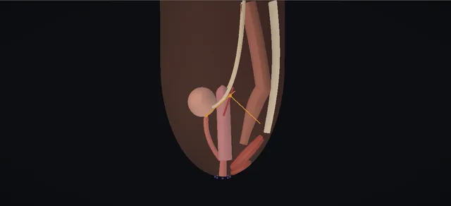
UrOops3D, danger-zone atlas

Schematic model.

Operative animation, Cloacal reconstruction

Urogenital sinus and cloacal reconstruction: posterior sagittal repair with total urogenital mobilisation

Indications

  • A persistent cloaca: urethra, vagina and rectum join in one common channel with a single perineal opening.
  • A urogenital sinus: urethra and vagina share a channel and the anus opens separately.
  • Hydrocolpos or a bladder that does not empty, needing drainage in the newborn.

Alternatives

  • In the newborn: colostomy and drainage only, with the definitive repair deferred.
  • Partial urogenital mobilisation for a suitable urogenital sinus.
  • Laparotomy with separation of the vagina and a pull-through or replacement for a long channel.

Risks

  • Injury to the urethra, bladder, ureters or rectum.
  • Urethrovaginal, vaginal or rectal fistula.
  • Stenosis of the urethra, vagina or anus.
  • Urinary or faecal incontinence, often needing catheterisation or bowel management.
  • Infection, hydrocolpos or upper tract deterioration.
  • Complications of a bowel or skin graft used for the vagina.
  • Sexual, reproductive and psychological harm.

A girl born with one perineal opening into which the urethra, the vagina and the rectum all drain. The newborn is decompressed, not repaired: a divided colostomy, and drainage of the hydrocolpos and, if needed, the bladder.

Endoscopy measures the common channel, and its length sets the plan. Months later, prone, the posterior sagittal repair separates the rectum from the vagina, brings the urethra and vagina down together as one block, and places the rectum inside the sphincter complex.

The lesson is the plane between rectum and vagina, the blood supply of the block, and a channel too long for this to be enough.

Advanced tier FRCS paediatric urology, disorders of the urogenital sinus and anorectal malformations Interactive 3D

Operative visualisation

Schematic scene preview: Cloacal reconstruction
Preparing the 3D anatomy…

UrOops3D · UroRef · © Nity G

How to use this module. Play through the eight steps, or use Back and Next. The profile views ghost the skin to show the three tracts joining; the prone views look into the wound from behind. The screen in step three holds the channel decision. Triggers show a long channel, a rectal injury, a fistula and a stenosis.

Click me next Section 2 of 8 Anatomy The structures this operation works between, and the one it must not cross.

The structures in the scene

Cloacal reconstruction: how the structures relateA newborn girl with a cloaca, the pelvis in profile from the left. Common channel to the single opening: mobilised with the vagina as one block Urethra and bladder neck, in front (danger). Common channel to the single opening: joins at the confluence; length sets the plan Vagina, behind the urethra. Common channel to the single opening: separated along a common wall, no plane Rectum, on the back of the vagina (danger). Common channel to the single opening: rectum placed inside it, closed behind Sphincter complex, in the midline (danger). Common channel to the single opening: obstructed by hydrocolpos; near a high dissection Ureters, above and behind (danger). Common channel to the single opening: coccyx marks the top of the incision Sacrum and coccyx, behind.Common channel to thesingle opening!Urethra and bladderneck, in frontVagina, behind theurethra!Rectum, on the backof the vagina!Sphincter complex,in the midline!Ureters, above andbehindSacrum and coccyx,behindmobilised with thevagina as one blockjoins at theconfluence; lengthsets the planseparated along acommon wall, noplanerectum placedinside it, closedbehindobstructed byhydrocolpos; near ahigh dissectioncoccyx marks thetop of the incisionTargetDangerLandmarkContext
A newborn girl with a cloaca, the pelvis in profile from the left. Schematic, not to scale; relationships only.
  • In profile, the urethra lies in front, the vagina behind it and the rectum behind both, all running down to one confluence and one common channel that opens on the perineum.
  • The bladder lies above the urethra and the sacrum behind the rectum.
  • Prone, the incision runs up the midline from the opening to the coccyx; the rectum is met first, then the common wall it shares with the vagina, then the urogenital block and, at its upper limit, the bladder neck.
  • ›Common channel - the single passage from the confluence to the perineal opening; its length is the main decision.
  • ›Confluence - where the urethra, the vagina and, in a cloaca, the rectum join the channel.
  • ›Urethra and bladder neck - in front; the bladder neck and the urethral sphincter lie close to the upper limit of the mobilisation.
  • ›Vagina - behind the urethra; distended by a hydrocolpos at birth; its blood supply comes from above and laterally.
  • ›Rectum - behind the vagina, sharing a common wall with it, opening into the back of the channel.
  • ›Sphincter complex - the parasagittal fibres, the muscle complex and the external sphincter, mapped by the stimulator and divided in the midline.
  • ›Perineal body - rebuilt between the new introitus and the anus.
  • ›Ureters - can be obstructed by a hydrocolpos and may insert ectopically; at risk in a high dissection.
  • ›Sacrum and coccyx - behind the rectum; the coccyx marks the upper end of the incision.
Click me next Section 3 of 8 Steps The operation in order, with the landmark that guards each move.

Operative steps

  1. One perineal opening A newborn girl with a single perineal opening and a distended lower abdomen. The urethra, the vagina and the rectum join in one common channel. In a urogenital sinus only the urethra and vagina share the channel and the anus opens separately; in a cloaca the rectum opens into it as well. The vagina is often distended by retained fluid, a hydrocolpos.
    where it bites: a hydrocolpos can obstruct the bladder and the ureters; the kidneys are at risk from the first days.
  2. Decompress first Nothing is reconstructed in the newborn. After resuscitation and assessment of the associated anomalies, a divided colostomy diverts the stool and leaves enough distal colon for the later pull-through. The hydrocolpos is drained, here by a vaginostomy, so that it no longer obstructs the urinary tract. A vesicostomy is added only if the bladder still does not empty.
    where it bites: an undrained hydrocolpos leads to infection, pyocolpos and obstruction of the ureters; a colostomy placed too distally interferes with the pull-through.
  3. Measure the channel Endoscopy through the single opening passes up the common channel to the confluence and shows the urethra, the vagina and the rectal opening. The length of the common channel decides the repair: a short channel can be repaired from below, a long one usually needs the abdomen opened as well. The findings and the plan are agreed by the specialist team with the parents.
    where it bites: a channel misjudged as short leads to a repair from below that cannot be completed without tension.
  4. Posterior sagittal approach Months later, prone with the pelvis raised, the vaginostomy closed. The muscle stimulator maps the external sphincter on the skin; its centre marks the future anus. The incision runs in the midline from the coccyx down to the single opening, and the parasagittal fibres and the muscle complex are divided exactly in the midline, so that the two halves can be brought back together.
    where it bites: a cut off the midline leaves unequal halves of the sphincter mechanism.
  5. Separate the rectum The rectum opens into the back of the common channel and shares a common wall with the vagina, with no natural plane between them. With stays on its wall, it is separated from the vagina from below upward until it is free and reaches the perineum without tension. The opening it leaves in the back of the vagina is closed.
    where it bites: the rectal wall and the posterior vaginal wall are the same tissue at first; a dissection that strays opens one or the other.
  6. Total urogenital mobilisation Stays are placed round the edge of the common channel. The urethra and the vagina are dissected all round as one block, without separating them from each other, and the block is brought down to the perineum. The redundant channel is opened in the midline to form a vestibule, on which the urethra and the vagina open separately.
    where it bites: the dissection runs close to the bladder neck and the urethral sphincter, and the vaginal blood supply comes from above and laterally.
  7. Rectum into the sphincter The stimulator finds the limits of the sphincter complex again inside the wound. The perineal body is rebuilt between the vagina and the anal site. The rectum is brought down and placed within the limits of the sphincter complex, the muscle complex is closed behind it, and the anoplasty is sewn to the skin.
    where it bites: a rectum placed outside the sphincter complex, or under tension, gives poor control or dehiscence and stricture.
  8. Three openings The wound is closed from the coccyx down. At the end there are three separate openings on the perineum: the urethra, the vagina and the anus. The colostomy is closed later, after the anus has been calibrated. Follow-up is lifelong: the kidneys, bladder emptying and continence, bowel control, menstruation, and later sexual, reproductive and psychological health.
    where it bites: renal deterioration and incomplete bladder emptying can be silent; they are looked for, not waited for.
Click me next Section 4 of 8 Danger zones Where this operation bites, and why it bites there.
Critical relationship

Danger zones

The spatial relationships the animation is built to make automatic. Before you read each one, ask yourself: at this point in the operation, what is about to go wrong, and what is my first move? The save for each sits in the Complications tab.

  • RiskUrethral sphincter and bladder neck - at the upper limit of the urogenital mobilisation; a dissection carried too high risks continence.
  • RiskRectum and anal sphincter - the common wall with the vagina has no plane; the sphincter complex is divided and closed exactly in the midline.
  • RiskUreters and ectopic insertions - obstructed by a hydrocolpos in the newborn, and close to a high dissection for a long channel.
  • RiskVaginal and genital neurovascular supply - comes from above and laterally; mobilising urethra and vagina together keeps it.
Practise it Take me into the scene Opens the animation at a danger moment and asks you to predict the mechanism and your first move before it reveals.
Click me next Section 5 of 8 Complications The save for each one: the first move, the escalation trigger, the op-note line.

Complications and immediate saves

Trigger each of these live in the Animation tab, then read the save: the first move, the escalation trigger, and the line that goes in the op note.

Long channel: mobilisation not enough

  • with a long common channel the urogenital block mobilised from below does not bring the vagina to the perineum without tension; the repair needs the abdomen opened and the vagina separated, pulled through or replaced.
The save
  1. Stop pulling. Do not sew the vagina to the perineum under tension. first move
  2. Turn the child and open the abdomen, as planned when the channel was long, or with the senior surgeon and the parents told if it was not. escalation trigger
  3. Separate the vagina from the urinary tract from above, keeping its blood supply, and close the urinary side.
  4. Bring the vagina down if it will reach; if not, replace it with a segment of bowel.
  5. Op note: the channel length as measured, why the plan changed and what was used for the vagina. document

Rectal injury in the separation

  • the dissection leaves the common wall and opens the rectum; the same management applies to an anoplasty that comes apart.
The save
  1. Stop the dissection and define the edges of the hole. first move
  2. Close the rectal wall in layers, with the colostomy still protecting it.
  3. Check that the closures of the rectum and the vagina do not lie against each other; interpose tissue if they do.
  4. An anoplasty that has come apart or retracted is looked at by the senior surgeon; the colostomy stays until it has healed. escalation trigger
  5. Op note: the site and size of the injury, how it was closed and what was placed between the suture lines. document

Urethrovaginal fistula

  • a track between the urethra and the vagina; urine leaks from the vagina.
  • Separating the two with a poor blood supply was the classic cause, and total urogenital mobilisation was introduced to avoid it.
The save
  1. Keep the bladder drained with a catheter and confirm the track by examination and endoscopy under anaesthesia. first move
  2. Treat infection, and look at the kidneys and bladder emptying.
  3. Plan the repair in the specialist centre, with tissue placed between the urethral and vaginal closures. escalation trigger
  4. Record the site of the track and the bladder function before repair. document

Vaginal stenosis

  • the new introitus or the lower vagina narrows, usually after a vagina brought down under tension or short of blood supply; it can obstruct menstruation later.
The save
  1. Examine under anaesthesia and define the length and site of the narrowing. first move
  2. A short ring may respond to dilatation; a longer stricture needs revision.
  3. Revision before puberty is planned by the specialist team, timed against menstruation. escalation trigger
  4. Record the calibre found, the treatment and the plan for review at puberty. document
Click me next Section 6 of 8 Troubleshooting When the case turns: the problem, then the next move.

Intra-operative troubleshooting

When the case turns: the problem, then the next move.

  • ActionA girl with a single opening and a hydrocolpos - drain the hydrocolpos in the newborn period, by catheterisation through the channel or a vaginostomy; an undrained hydrocolpos obstructs the ureters and becomes infected.
  • ActionThe colostomy is low on the sigmoid - a colostomy placed too distally leaves too little colon for the pull-through; it may need revision before the main repair.
  • ActionA urogenital sinus with a separate anus - no colostomy is needed; the sinus can be repaired from below, and partial mobilisation, limited to the pubourethral ligament, may be enough.
  • ActionWhether to operate early or wait for the child - the separation of urinary, genital and anal tracts is functional surgery; surgery that only alters appearance is deferred until the child can take part in the decision.
  • ActionPoor bladder emptying after repair - clean intermittent catheterisation through the urethra, or through a continent channel; the upper tracts are followed for life.
  • ActionFaecal incontinence after repair - a bowel management programme keeps most children clean; the sacrum and spinal cord are checked, since their anomalies predict poorer control.
Click me next Section 7 of 8 FRCS viva Say it out loud, then mark yourself against the model answer.

FRCS viva questions

The examiner stems this scene prepares you for. Try to answer out loud first, then reveal, and learn the discriminator, the detail that turns a pass into a strong pass.

QWhat is the difference between a urogenital sinus and a cloaca?reveal answer
  • Both are failures of separation of the tracts that open on the perineum.
  • In a urogenital sinus the urethra and the vagina join in a common channel and the anus opens separately in the normal place.
  • In a persistent cloaca the rectum also opens into that channel, so a girl has a single perineal opening.
  • They form a spectrum, and the length of the common channel matters in both.
  • I would look for the associated anomalies: renal anomalies and vesicoureteric reflux, sacral and spinal anomalies, and a hydrocolpos.
  • In the series of Rink and colleagues, 14 of 23 girls with a cloaca had a renal anomaly and 13 had vesicoureteric reflux.
Strong pass

Say all three

  • Knowing that a single perineal opening in a girl is a cloaca until proved otherwise
  • Asking about the kidneys
  • The sacrum and the spine
Examiner note
Knowing that a single perineal opening in a girl is a cloaca until proved otherwise, and asking about the kidneys, the sacrum and the spine.
QA newborn girl has a single perineal opening and a distended abdomen. What do you do?reveal answer
  • I would resuscitate her and assess her with the neonatal team, with an ultrasound of the kidneys, the bladder and the pelvis, and of the spine.
  • I would not attempt any reconstruction.
  • She needs a divided colostomy, placed so that enough distal colon is left for the later pull-through, and fixed so that it does not prolapse.
  • If there is a hydrocolpos I would drain it, because it obstructs the ureters and becomes infected; and if the bladder does not empty I would add a vesicostomy.
  • In the series of Pena and colleagues, 48 of the girls referred from other centres had a hydrocolpos that had not been drained, with urinary infection, pyocolpos or vaginal perforation as a result, 24 had a colostomy placed too distally, and 23 had a prolapsed colostomy.
  • I would then plan endoscopy and imaging before the definitive repair.
Strong pass

Say both

  • Draining the hydrocolpos
  • Placing the colostomy correctly: the common errors of the newborn period
Examiner note
Draining the hydrocolpos and placing the colostomy correctly: the common errors of the newborn period.
QWhy does the length of the common channel matter?reveal answer
  • It predicts both the operation and the result.
  • In the review of 339 cloacas by Pena, Levitt, Hong and Midulla, the mean channel length was 2.3 centimetres in those repaired from below alone and 4.7 centimetres in those who needed a laparotomy as well.
  • They divided the series at 3 centimetres: 62 per cent had a channel shorter than that, and could be repaired posterior sagittally with a reproducible, relatively short operation, with a good prognosis.
  • Those with a longer channel usually needed a laparotomy, had more urological problems and worse function, and their surgeons need special training.
  • For urinary control, 78 per cent of those with a channel longer than 3 centimetres needed intermittent catheterisation, against 28 per cent of those with a shorter one.
  • So I would measure the channel at endoscopy before deciding where and how to repair it.
Strong pass

Quoting the threshold of about 3 centimetres and its effect on catheterisation, with the source.

QWhat is total urogenital mobilisation, and when is it not enough?reveal answer
  • After the rectum has been separated from the vagina, the urethra and the vagina are mobilised together as a single unit and brought down to the perineum, instead of being separated from each other.
  • Pena described it in 1997 in eleven girls: the operating time for that part fell by about 70 per cent, the blood supply of the vagina and urethra stayed excellent, and none developed a urethrovaginal fistula, vaginal stricture or acquired vaginal atresia in a short follow-up.
  • In the larger review, the 19 urethrovaginal fistulas, 17 vaginal strictures or atresias and 6 urethral strictures had all occurred before the technique was introduced.
  • Rink described a partial mobilisation for the urogenital sinus, with the dissection limited to the pubourethral ligament, to reduce the risk to continence.
  • It is not enough when the channel is long: then I would open the abdomen, separate the vagina from the urinary tract and pull it through, or replace it with rectum, ileum or colon if it will not reach.
Strong pass

Say both

  • Knowing that the block keeps its own blood supply
  • That a long channel needs separation and often vaginal replacement
Examiner note
Knowing that the block keeps its own blood supply, and that a long channel needs separation and often vaginal replacement.
QWhat do you tell the parents about the long-term outcome?reveal answer
  • I would tell them that the operation separates the three tracts but that function is the lifelong question.
  • In the series of Pena and colleagues, 54 per cent of those evaluated were continent of urine; 24 per cent were dry with intermittent catheterisation through the urethra and 22 per cent through a continent channel.
  • Sixty per cent had voluntary bowel movements, and the 40 per cent who were faecally incontinent were kept clean by a bowel management programme.
  • In the three-decade series of Rink and colleagues, age-adjusted creatinine was abnormal in four of 22 and borderline in six more, and 15 of the 18 children older than about four years were continent, several after further reconstruction.
  • I would explain that menstruation, sexual function and fertility have to be followed into adult life, with psychological support, and that the EAU guideline asks for a multidisciplinary team and shared decisions throughout.
Strong pass

Separating anatomical success from function, and planning follow-up of the kidneys, the bladder, the bowel and the reproductive tract into adulthood.

Click me next Section 8 of 8 Evidence The guidance each of these answers is built on.

Evidence and guidance

Operative Dictations in Urologic Surgery
Armenakas NA, Fracchia JA, Golan R. Wiley, 2019
The book has no chapter on urogenital sinus or cloacal repair and none is followed here. The step order follows the hub sequence and the abstracts below; the instruments, the stays, the stimulator use in the wound, the vaginostomy as the drainage shown and the colostomy closure after calibration are general practice.
Open guideline
European Association of Urology
EAU Guidelines on Paediatric Urology, 2026 edition: chapter on disorders and differences of sex development, sections 20.1, 20.4.2, 20.5 and 20.8, read 19 October 2026
Read through a fetch tool that returns a summary with quotations. A multidisciplinary approach and shared decisions (strong); referral to experienced centres (strong); the opening of the urogenital sinus to be evaluated; surgery that alters appearance is not urgent and is delayed until the patient can consent. The chapter does not describe the cloacal operation.
Open guideline
Journal of Pediatric Surgery
Pena A. Total urogenital mobilization: an easier way to repair cloacas. J Pediatr Surg 1997;32:263-268
Abstract read on PubMed 19 October 2026. Eleven girls; after the rectum is separated from the vagina, urethra and vagina mobilised together as a single unit; operating time for the reconstruction reduced by about 70%; blood supply excellent; no urethrovaginal fistula, vaginal stricture or acquired atresia at 1 to 14 months.
Open guideline
Journal of Pediatric Surgery
Pena A, Levitt MA, Hong A, Midulla P. Surgical management of cloacal malformations: a review of 339 patients. J Pediatr Surg 2004;39:470-479
Abstract read on PubMed 19 October 2026. All approached posterior sagittally, 111 also by laparotomy; mean channel 4.7 cm with laparotomy, 2.3 cm without; 62% under 3 cm. Urine: 54% continent, 24% dry with urethral and 22% with conduit catheterisation; catheterisation 78% over 3 cm against 28% under. Bowel: 60% voluntary. Neonatal errors listed.
Open guideline
Journal of Pediatric Urology
Rink RC, Metcalfe PD, Kaefer MA, Casale AJ, Meldrum KK, Cain MP. Partial urogenital mobilization: a limited proximal dissection. J Pediatr Urol 2006;2:351-356
Abstract read on PubMed 19 October 2026. Based on total urogenital mobilisation, a dissection limited to the pubourethral ligament, in 15 patients with urogenital sinus; no intraoperative complications and no voiding dysfunction in the short term.
Open guideline
BJU International
Rink RC, Herndon CDA, Cain MP, et al. Upper and lower urinary tract outcome after surgical repair of cloacal malformations: a three-decade experience. BJU Int 2005;96:131-134
Abstract read on PubMed 19 October 2026. 23 patients, mean follow-up 9.3 years: renal anomalies in 14, reflux in 13; creatinine abnormal in 4 and borderline in 6; 10 void and 11 catheterise; 15 of 18 over 47 months continent, after reconstruction in several.
Open guideline

Curriculum. Supports learning relevant to the ISCP Urology Curriculum syllabus topic Paediatric urology: urogenital sinus and cloaca. Not a workplace-based assessment and not evidence of competence.

Two minutes

Close the lap: recall it before you leave.

Say it aloud, then reveal. Reading is not remembering.

  1. 01 The relationship Say the critical spatial relationship out loud, then reveal. reveal
    Memory frame

    In profile, the urethra lies in front, the vagina behind it and the rectum behind both, all running down to one confluence and one common channel that opens on the perineum.

    Why?
    The bladder lies above the urethra and the sacrum behind the rectum. Prone, the incision runs up the midline from the opening to the coccyx; the rectum is met first, then the common wall it shares with the vagina, then the urogenital block and, at its upper limit, the bladder neck.
  2. 02 The first move Long channel: mobilisation not enough. Your first move? reveal
    First move

    Stop pulling.

    More detail
    Do not sew the vagina to the perineum under tension.
  3. 03 The escalation trigger Long channel: mobilisation not enough. What makes you call for help? reveal
    Escalate when

    Turn the child and open the abdomen, as planned when the channel was long, or with the senior surgeon and the parents told if it was not.

  4. 04 The discriminator What turns a pass into a strong pass? reveal
    Strong pass

    Say all three

    • Knowing that a single perineal opening in a girl is a cloaca until proved otherwise
    • Asking about the kidneys
    • The sacrum and the spine
    Examiner note
    Knowing that a single perineal opening in a girl is a cloaca until proved otherwise, and asking about the kidneys, the sacrum and the spine.
  5. 05 The pitfall Where does this operation bite first? reveal
    Pitfall

    Urethral sphincter and bladder neck

    Why?
    at the upper limit of the urogenital mobilisation; a dissection carried too high risks continence.

Revision practice with self-marked answers, not an assessment of clinical competence. Kept in this browser only; it does not sync between devices.