
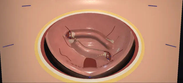
UrOops3D, danger-zone atlas

Schematic child bladder: every dimension is an authoring estimate.

Operative animation, Cohen ureteric reimplantation

Bilateral ureteric reimplantation: the Cohen cross-trigonal tunnels

Indications

  • Breakthrough febrile urinary infection despite prophylaxis.
  • High-grade reflux that has not settled with growth.
  • New renal scarring or falling function on follow-up.
  • Failed endoscopic injection, or the family’s choice after counselling.

Alternatives

  • Continued prophylaxis and surveillance, with bladder and bowel dysfunction treated.
  • Endoscopic subureteric injection: less invasive, less reliable in the higher grades.
  • Extravesical (Lich-Gregoir) repair: on both sides at once it risks temporary retention.
  • Politano-Leadbetter repair; laparoscopic or robot-assisted repair in some centres.

Risks

  • Haematuria and bladder spasm in the first days.
  • Ureteric obstruction: swelling early, stricture late.
  • Persistent reflux; urine leak; infection.
  • Harder endoscopic access to these ureters for life: a stone in an adult is awkward to reach from below.

Open antireflux surgery for the child whose vesicoureteric reflux has not settled and keeps causing febrile infection, shown here on both sides.

Through the opened bladder each refluxing ureter is freed on a cuff of mucosa, drawn across the trigone under the mucosa, and sewn to a new opening on the far side: the left ureter above the interureteric bar, the right below it.

The cross-trigonal route is chosen because it gives a long tunnel with good muscle behind it from a ureter that keeps its own hiatus. The price is paid later: each orifice faces across the bladder, so a stone in that ureter in adult life is hard to reach from below.

The lesson is the layers on the way in, the blood supply in the adventitia, the plane the tunnel lives in, and three checks before the first bite: no twist, no kink, no tension.

Advanced tier FRCS paediatric urology, vesicoureteric reflux, reconstruction Interactive 3D

Operative visualisation

Schematic scene preview: Cohen ureteric reimplantation
Preparing the 3D anatomy…

UrOops3D · UroRef · © Nity G

How to use this module. Play the ten steps, or use Back and Next. Watch the wall open layer by layer, the mucosa lift as each tunnel opens, the ridges advance as the ureters cross, and the look from behind. The triggers show a twist, a short tunnel, a pale ureter and a buttonhole.

Click me next Section 2 of 8 Anatomy The structures this operation works between, and the one it must not cross.

The structures in the scene

Cohen ureteric reimplantation: how the structures relateInto the opened bladder from in front: dome above, neck below. Submucosal tunnel across the trigone: starts where it is freed on a cuff Refluxing orifice, lateral and wide. Submucosal tunnel across the trigone: the ureter still enters here; a kink sits here Its own hiatus through the detrusor (danger). Submucosal tunnel across the trigone: kept on the ureter drawn through it Adventitia: the ureter’s vessels (danger). Submucosal tunnel across the trigone: thin roof buttonholes; needs muscle behind Mucosal roof, detrusor floor (danger). Submucosal tunnel across the trigone: ends above and lateral to it Opposite orifice: new opening above it. Submucosal tunnel across the trigone: swept off the hiatus before delivery Peritoneum, bowel and vas behind (danger).Submucosal tunnelacross the trigoneRefluxing orifice,lateral and wideIts own hiatusthrough the detrusor!Adventitia: theureter’s vessels!Mucosal roof,detrusor floorOpposite orifice:new opening above it!Peritoneum, boweland vas behindstarts where it isfreed on a cuffthe ureter stillenters here; a kinksits herekept on the ureterdrawn through itthin roofbuttonholes; needsmuscle behindends above andlateral to itswept off thehiatus beforedeliveryTargetDangerLandmarkContext
Into the opened bladder from in front: dome above, neck below. Schematic, not to scale; relationships only.
  • Seen through the cystotomy, the dome is at the top, the bladder neck at the bottom and the patient’s right on the surgeon’s left.
  • The two orifices sit at the upper corners of the trigone with the interureteric bar between them.
  • The left ureter’s tunnel runs across the trigone above that bar to a window above and lateral to the right orifice.
  • The right ureter’s tunnel runs below the bar to a window at the lower edge of the left orifice site.
  • From behind the bladder each ureter is seen to enter by its own unchanged hiatus and then turn across in the wall, the left above the right.
  • ›Trigone and interureteric bar - the floor the tunnel crosses; the bar between the two orifices is the landmark the tunnel runs above.
  • ›Refluxing orifice - sits lateral and gapes, with a short oblique course through the wall: too little ureter under mucosa to act as a valve.
  • ›Intramural ureter and hiatus - the ureter keeps its own hiatus through the detrusor in this operation; only its submucosal course and its orifice move.
  • ›Adventitial sheath - carries the longitudinal vessels that keep the distal ureter alive once it is detached; it stays on the ureter.
  • ›Mucosa and detrusor - two layers with a plane between them: the tunnel is that plane, mucosa as roof and muscle as floor.
  • ›The new orifice in later life - on the far side of the trigone, facing across the bladder, with the last part of the ureter running transversely: the reason a stone in this ureter in an adult is awkward to reach from below.
  • ›Abdominal wall layers - skin, fat and Scarpa’s fascia, anterior rectus sheath, recti parted in the midline, transversalis fascia, then the prevesical space in front of the bladder. Peritoneum lies above, swept up off the dome.
  • ›Bladder wall layers - detrusor outside, then submucosa, then mucosa. The cystotomy goes through all three; the tunnels live between the last two.
  • ›The two windows - the left ureter’s above and lateral to the right orifice; the right ureter’s at the inferior edge of the left orifice site. Each ureter ends on the far side from where it entered.
  • ›Peritoneum, vas and bowel - directly behind the hiatus; swept away, not grasped.
Click me next Section 3 of 8 Steps The operation in order, with the landmark that guards each move.

Operative steps

  1. Expose the bladder The child is supine with a catheter in the bladder, which is filled so that the dome rises and carries the peritoneum up with it. A small curved transverse incision is made in the skin crease above the pubis. The layers are taken in order: skin, fat and Scarpa’s fascia, the anterior rectus sheath opened across, the recti parted in the midline, then the transversalis fascia and the prevesical space. The peritoneal reflection is swept up off the dome with a pledget until the anterior wall is bare; the peritoneum is not opened.
    where it bites: The inferior epigastric vessels run under the recti toward the ends of the wound.
  2. Open the bladder Two Allis forceps lift the anterior wall and it is opened vertically in the midline, through detrusor and then mucosa, from the dome toward the neck. Stay stitches draw the edges apart and a stitch at the lower apex stops the cut extending. A ring retractor with padded blades holds the bladder open so that the trigone and both orifices can be seen without the blades resting on them.
    where it bites: A cystotomy that runs into the bladder neck.
  3. Intubate both ureters A fine feeding tube is passed into each orifice in turn and up the ureter. A traction stitch is placed through the lip of each orifice and tied round its tube. From this point each ureter is moved by its stitch and by its cuff of mucosa; forceps do not go on its wall.
    where it bites: A tube forced into a false passage rather than the lumen.
  4. Circumscribe the orifices With gentle traction on the tube the mucosa is scored in a full circle round the orifice, so that a cuff of mucosa stays on the end of the ureter. The cut is deepened first at six o’clock, through mucosa and submucosa, to the plane between the ureter and the detrusor. The second side is done in the same way.
  5. Mobilise both ureters Fine scissors spread in the plane between the adventitial sheath and the detrusor, and the muscle strands tethering the ureter in its hiatus are divided one at a time, staying on the muscle. Traction delivers first the intramural ureter and then a short extravesical length, with its sheath and longitudinal vessels intact. A pledget through the hiatus sweeps the peritoneum off the back of the bladder. Both ureters are delivered until each would cross the trigone with length to spare.
    where it bites: Stripping the adventitia takes the blood supply with it. The peritoneum and bowel lie behind each hiatus, and in a boy the vas crosses there.
  6. Make two tunnels The more laterally placed ureter, here the left, is given the upper tunnel: its new site is above and lateral to the opposite orifice, and a small window is opened in the mucosa there. The other ureter is given a lower tunnel to a window at the inferior edge of the first ureter’s original orifice. Each tunnel is made by passing scissors under the mucosa from the original hiatus and spreading across the trigone to the window. The roof is mucosa and the floor is detrusor. The two tunnels cross the midline one above the other, with a bridge of mucosa between them.
    where it bites: Thin trigonal mucosa buttonholes easily. Two tunnels that run into each other leave neither ureter with a roof.
  7. Draw the ureters through A fine right-angled clamp is passed back through each tunnel from its window to the original hiatus and takes the traction stitch. The feeding tube is removed and the ureter is drawn across under the mucosa. Seen from behind the bladder, each ureter still enters by its own hiatus and only then turns across. Before any stitch is placed the lie of both is checked: the vessels on the surface run straight, each turns gently into its tunnel, and neither is stretched tight.
    where it bites: A twist, a kink at the hiatus, or tension: each obstructs the ureter.
  8. Anastomose Each ureter is trimmed back to a cut edge that bleeds and its end is spatulated ventrally. Two dorsal bites take the full thickness of the ureter to the trigone muscle and mucosa at the lower edge of the window, anchoring it. Interrupted absorbable bites at nine, twelve and three o’clock then bring mucosa to ureter all round. The second ureter is sewn in the same way.
    where it bites: A pale cut edge is trimmed further. Bites that are too deep or too many narrow the lumen.
  9. Close the old sites and check The mucosa is closed over each original orifice with shallow interrupted bites, the needle staying in mucosa because a ureter now runs just beneath. The closure of the left site stops short of the right ureter’s new orifice beside it. Urine is seen to come from both new orifices, and the feeding tube is passed up each and removed to show that it runs freely.
    where it bites: A deep bite over an old site catches the ureter under it, and a closure carried too far covers the neighbouring new orifice.
  10. Close the bladder A urethral catheter is placed. The cystotomy is closed in two layers, mucosa then muscle, and tested by filling the bladder through the catheter. A drain is left in the prevesical space, brought out through its own stab away from the suture line. The rectus sheath, fat and skin are closed in turn.
Click me next Section 4 of 8 Danger zones Where this operation bites, and why it bites there.
Critical relationship

Danger zones

The spatial relationships the animation is built to make automatic. Before you read each one, ask yourself: at this point in the operation, what is about to go wrong, and what is my first move? The save for each sits in the Complications tab.

  • RiskThe adventitia of the ureter - its vessels are the distal ureter’s only supply after mobilisation; dissect on the detrusor, hold the cuff or the stitch.
  • RiskBehind the hiatus - peritoneum and bowel, and the vas in a boy, lie against the back of the bladder where the ureter enters.
  • RiskThe tunnel roof - trigonal mucosa is thin; a buttonhole leaves the ureter uncovered at that point.
  • RiskThe turn into the tunnel - where the ureter leaves its hiatus and bends across the trigone; a sharp bend or a twist here obstructs it.
  • RiskThe opposite orifice - the window sits just above it; the tunnel passes over its intramural ureter, not through it.
  • RiskThe bladder neck - the lower end of the cystotomy; an apex stitch keeps the cut out of it.
Practise it Take me into the scene Opens the animation at a danger moment and asks you to predict the mechanism and your first move before it reveals.
Click me next Section 5 of 8 Complications The save for each one: the first move, the escalation trigger, the op-note line.

Complications and immediate saves

Trigger each of these live in the Animation tab, then read the save: the first move, the escalation trigger, and the line that goes in the op note.

Obstruction of the reimplanted ureter

  • a twist, a kink at the hiatus, tension, or oedema at the new orifice; early it may be transient swelling, later it is a stricture.
The save
  1. On the table: if the ureter looks twisted or kinked, take it back through the tunnel and pass it again before any stitch. first move
  2. Prove drainage before closing: efflux at the new orifice and a fine tube that passes freely.
  3. After surgery, loin pain, vomiting, fever or a falling urine output prompts an ultrasound for new or worsening hydronephrosis.
  4. Worsening dilatation with pain, infection or a rising creatinine needs drainage and senior paediatric urology review. escalation trigger
  5. Op note: lie of the ureter, efflux seen, tube passed freely, whether a stent was left. document

Persistent or new reflux

  • reflux continues on the operated side, or appears on the other side afterwards; a short tunnel or a tunnel without muscle behind it is the technical cause.
The save
  1. On the table: if the tunnel is short, lengthen it across the trigone before the ureter is drawn through. first move
  2. Follow up clinically and with ultrasound; routine cystography after an uncomplicated open reimplant is a unit decision.
  3. Look for and treat bladder and bowel dysfunction, which undermines any antireflux operation.
  4. Recurrent febrile infection with reflux on imaging goes back to the paediatric urology team for a planned decision. escalation trigger
  5. Op note: the route and the judged length of the tunnel. document

Ischaemic distal ureter

  • a pale end after mobilisation, from stripped adventitia or instruments on the ureteric wall; it leaks early or strictures late.
The save
  1. Trim back until the cut edge bleeds before starting the anastomosis. first move
  2. Re-check the lie: a trimmed ureter that only reaches under tension has exchanged one problem for another.
  3. If it will not reach without tension, stop and get senior help before improvising. escalation trigger
  4. Op note: perfusion of the cut edge and how much was trimmed. document

Difficult later access to the ureter

  • after a cross-trigonal reimplant the orifice faces across the bladder, so retrograde catheterisation or ureteroscopy in later life is harder.
The save
  1. Counsel the family before surgery: this is a known consequence of the technique, not a failure. first move
  2. Record the operation clearly so that a future endoscopist knows where the orifice is and which way it faces.
  3. A later stone or stricture on that side is planned with someone experienced in reimplanted ureters. escalation trigger
  4. Op note and discharge letter: technique named, side, position of the new orifice. document
Click me next Section 6 of 8 Troubleshooting When the case turns: the problem, then the next move.

Intra-operative troubleshooting

When the case turns: the problem, then the next move.

  • ActionThe mucosa buttonholes while the tunnel is being made - a small hole is closed with a fine absorbable stitch; a large breach means moving the window or re-siting the tunnel so the ureter has an intact roof.
  • ActionThe ureter will not come down far enough - more length comes from freeing it further outside the bladder through the hiatus, on the muscle and with the sheath intact; it does not come from pulling.
  • ActionThe ureter looks twisted once it is through - take it back and pass it again, following the vessels on its surface; do not accept a spiral.
  • ActionThe cut end is pale - trim to bleeding ureter, then check reach and tension again before the first bite.
  • ActionThe feeding tube will not pass up the new orifice - something is kinked or a bite has crossed the lumen; find it and correct it before the bladder is closed.
  • ActionYears later, an adult with this reimplant has a stone in that ureter - expect the orifice on the opposite side of the trigone, facing across the bladder, with a transverse terminal ureter: a flexible cystoscope, an angled catheter and a hydrophilic wire, or access from above through the kidney. Plan it; do not discover it with a rigid scope.
  • ActionOnly one side refluxes - that ureter alone is crossed, through the upper route to a window above and lateral to the opposite orifice; the other is left untouched.
  • ActionWhich ureter goes above? - the more laterally placed orifice takes the upper tunnel; the other goes below the bar to the inferior edge of the first orifice’s old site. The tunnels must not meet.
Click me next Section 7 of 8 FRCS viva Say it out loud, then mark yourself against the model answer.

FRCS viva questions

The examiner stems this scene prepares you for. Try to answer out loud first, then reveal, and learn the discriminator, the detail that turns a pass into a strong pass.

QDescribe a bilateral Cohen cross-trigonal ureteric reimplantation.reveal answer
  • It is an open intravesical antireflux operation.
  • Through a low transverse incision, taken through fat and Scarpa’s fascia, the rectus sheath and between the recti into the prevesical space, I sweep the peritoneum off the dome and open the bladder vertically in the midline.
  • I pass a fine tube up each ureter and place a traction stitch through each orifice.
  • I circumscribe each orifice leaving a cuff of mucosa on the ureter and mobilise each ureter in the plane between its adventitia and the detrusor, keeping the sheath and its vessels on the ureter, until both will cross the trigone without tension.
  • The more lateral ureter takes an upper submucosal tunnel to a window above and lateral to the opposite orifice;
  • the other takes a lower tunnel to a window at the inferior edge of the first orifice’s original site, so the two cross one above the other.
  • I draw each ureter through by its stitch, check that neither is twisted, kinked or tight, trim and spatulate them, anchor each dorsally to trigone muscle and mucosa and complete each anastomosis with interrupted absorbable bites.
  • I close the mucosa over the original orifices, confirm efflux and free passage of a tube on both sides, close the bladder in two layers and leave a catheter and a drain.
Strong pass

Stating that each ureter keeps its own hiatus, saying which goes above and where each window lies, and naming the three checks before the first bite: no twist, no kink, no tension.

QWhy does lengthening the submucosal ureter stop reflux?reveal answer
  • The normal antireflux mechanism is a flap valve.
  • The ureter runs obliquely through the bladder wall and then under the mucosa, with detrusor behind it, so that as the bladder fills and contracts the pressure compresses that segment against its muscular backing and closes it.
  • A refluxing ureter typically has a lateral orifice and a short intramural and submucosal course, so there is too little ureter to compress.
  • Every open reimplantation, whatever its name, works by lengthening the submucosal segment and giving it firm muscle behind.
  • The Cohen operation does that by taking the ureter across the trigone, which gives a long tunnel with good muscle beneath it and a gentle course from the original hiatus.
  • The length the tunnel needs relative to the calibre of the ureter is a traditional teaching figure that I would quote as a rule of thumb, not as evidence.
Strong pass

Say both

  • Explaining the flap valve as length of submucosal ureter with muscle behind it
  • Treating the traditional tunnel ratio as a rule of thumb rather than a proven threshold
Examiner note
Explaining the flap valve as length of submucosal ureter with muscle behind it, and treating the traditional tunnel ratio as a rule of thumb rather than a proven threshold.
QWhat are the alternatives to this operation, and what is its main drawback?reveal answer
  • The alternatives start with not operating: continued antibiotic prophylaxis and treatment of bladder and bowel dysfunction, since much reflux resolves with growth.
  • Endoscopic subureteric injection of a bulking agent is less invasive and is widely offered, with lower success in the higher grades.
  • Among open operations the extravesical Lich-Gregoir repair avoids opening the bladder, and the Politano-Leadbetter operation makes a new hiatus and brings the ureter down a vertical tunnel to its original site.
  • Laparoscopic and robot-assisted reimplantation are done in some centres.
  • The European guideline describes the cross-trigonal operation as the most popular and reliable of the open techniques.
  • Its main drawback is that the orifice ends up on the opposite side facing across the bladder, which makes retrograde access to that ureter harder in later life, and I would say so when counselling the family.
Strong pass

Say both

  • Naming difficulty with later retrograde ureteric access as the specific drawback of the cross-trigonal route
  • Saying it is part of consent
Examiner note
Naming difficulty with later retrograde ureteric access as the specific drawback of the cross-trigonal route and saying it is part of consent.
QA 30-year-old who had a Cohen reimplant as a child presents with a ureteric stone on that side. Why is this harder, and what are the options?reveal answer
  • It is harder because of where the operation left the orifice.
  • A native ureter enters the trigone on its own side and runs obliquely down and medially, so a wire and a rigid ureteroscope passed from the urethra line up with it.
  • After a cross-trigonal reimplant the orifice lies on the opposite side of the trigone and the terminal ureter runs transversely under the mucosa before turning sharply up at its original hiatus.
  • From the bladder neck the instrument meets the orifice almost side-on, the wire tends to coil in the bladder, and a rigid scope cannot follow the turn.
  • So I would not assume routine retrograde ureteroscopy.
  • I would read the childhood operation note and look at the imaging to find the orifice, and plan with someone used to reimplanted ureters.
  • The options, by stone size and position, are medical expulsive treatment or observation for a small distal stone, shock wave lithotripsy, a retrograde attempt with a flexible cystoscope or angled catheter and a hydrophilic wire followed by flexible ureteroscopy, and antegrade access through a nephrostomy, either to treat from above or to pass a wire down for a combined approach.
  • An obstructed infected system is drained from above first.
  • These options are standard endourology; their order here is my drafting.
Strong pass

Say all three

  • Explaining the difficulty by the transverse terminal ureter and the side-on orifice
  • Not just saying it is difficult
  • Naming antegrade access as the dependable route when the retrograde one fails
Examiner note
Explaining the difficulty by the transverse terminal ureter and the side-on orifice, not just saying it is difficult, and naming antegrade access as the dependable route when the retrograde one fails.
QAfter you draw the ureter through, the vessels on its surface spiral. What do you do?reveal answer
  • I do not place a stitch.
  • A spiral in the surface vessels means the ureter has rotated as it came through, and a twisted ureter obstructs even if it looks comfortable on the table.
  • I take it back through the tunnel by its traction stitch, re-pass the right angle, and draw it through again watching the vessels stay on one face along the whole tunnel.
  • I then check the turn at the original hiatus, because a sharp bend there obstructs in the same way, and I check that the ureter is not bow-stringed.
  • Only then do I trim and spatulate.
  • At the end I want two proofs before closing the bladder: efflux from the new orifice, and a fine tube that passes up it without holding up.
  • I record both in the operation note.
Strong pass

Say all three

  • Using the longitudinal vessels as an orientation mark
  • Re-passing rather than accepting a twist
  • Quoting efflux and free passage of a tube as the two on-table proofs
Examiner note
Using the longitudinal vessels as an orientation mark, re-passing rather than accepting a twist, and quoting efflux and free passage of a tube as the two on-table proofs.
Click me next Section 8 of 8 Evidence The guidance each of these answers is built on.

Evidence and guidance

Operative Dictations in Urologic Surgery
Armenakas NA, Fracchia JA, Golan R. Cohen transtrigonal ureteric reimplantation. Chapter 125, pp 493-496. Wiley, 2019
Indications; the essential steps (fill, expose, vertical cystotomy, ring retractor, feeding tube with a traction stitch, circumscribe and mobilise preserving the adventitia, new hiatus superolateral to the contralateral orifice, submucosal tunnel without tension, confirm the lie, spatulated anastomosis with interrupted absorbable sutures, close the mucosa over the original orifice, two-layer closure, drain); the order of the anastomosis; the bilateral variant; the complication list, which includes difficulty with later ureteric catheterisation. As logged in the expansion plan on 8 October 2026. Book numbers kept off screen: the feeding tube size, the suture sizes, the tunnel length to width ratio and the bladder capacity formula.
Open guideline
EAU Guidelines
Paediatric Urology, chapter 17 Vesicoureteric reflux: 17.3.2.a subureteric injection, 17.3.2.b open surgical techniques, 17.3.2.c laparoscopy and robot-assisted surgery, 17.4 recommendations
All open techniques lengthen the intramural ureter; the cross-trigonal operation is named the most popular and reliable open procedure, with later endoscopic access to the ureter as its main concern; suprahiatal and infrahiatal intravesical repairs and the extravesical repair are named alternatives. As logged in the expansion plan as read on 8 October 2026; the edition year is to be confirmed.
Open guideline
Current Urology Reports
Boswell TC. Advancements in surgical management of megaureters. Curr Urol Rep 2024;25(9):215-223
Questions the traditional tunnel length to diameter ratio in megaureter surgery, which is why no ratio is shown on screen. As logged in the expansion plan.
Open guideline

Curriculum. Supports learning relevant to the ISCP Urology Curriculum syllabus topic Paediatric urology: vesicoureteric reflux. Not a workplace-based assessment and not evidence of competence.

Two minutes

Close the lap: recall it before you leave.

Say it aloud, then reveal. Reading is not remembering.

  1. 01 The relationship Say the critical spatial relationship out loud, then reveal. reveal
    Memory frame

    Seen through the cystotomy, the dome is at the top, the bladder neck at the bottom and the patient’s right on the surgeon’s left.

    Why?
    The two orifices sit at the upper corners of the trigone with the interureteric bar between them. The left ureter’s tunnel runs across the trigone above that bar to a window above and lateral to the right orifice. The right ureter’s tunnel runs below the bar to a window at the lower edge of the left orifice site. From behind the bladder each ureter is seen to enter by its own unchanged hiatus and then turn across in the wall, the left above the right.
  2. 02 The first move Obstruction of the reimplanted ureter. Your first move? reveal
    First move

    On the table: if the ureter looks twisted or kinked, take it back through the tunnel and pass it again before any stitch.

  3. 03 The escalation trigger Obstruction of the reimplanted ureter. What makes you call for help? reveal
    Escalate when

    Worsening dilatation with pain, infection or a rising creatinine needs drainage and senior paediatric urology review.

  4. 04 The discriminator What turns a pass into a strong pass? reveal
    Strong pass

    Stating that each ureter keeps its own hiatus, saying which goes above and where each window lies, and naming the three checks before the first bite: no twist, no kink, no tension.

  5. 05 The pitfall Where does this operation bite first? reveal
    Pitfall

    The adventitia of the ureter

    Why?
    its vessels are the distal ureter’s only supply after mobilisation; dissect on the detrusor, hold the cuff or the stitch.

Revision practice with self-marked answers, not an assessment of clinical competence. Kept in this browser only; it does not sync between devices.