
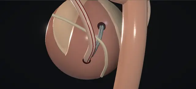
UrOops3D, danger-zone atlas

Schematic child bladder: every dimension is an authoring estimate.

Operative animation, Politano-Leadbetter ureteric reimplantation

Ureteric reimplantation: the Politano-Leadbetter suprahiatal operation

Indications

  • Persistent high-grade vesicoureteric reflux.
  • Recurrent upper tract infection despite antibiotic prophylaxis, or where prophylaxis is not taken.
  • A bladder of normal capacity: in a small or neurogenic bladder the cross-trigonal operation is preferred.

Alternatives

  • Continued prophylaxis and surveillance, with bladder and bowel dysfunction treated.
  • Endoscopic subureteric injection.
  • Cross-trigonal (Cohen) reimplantation, the most used open operation.
  • Infrahiatal advancement (Glenn-Anderson) for a laterally placed orifice; extravesical (Lich-Gregoir) repair.

Risks

  • Bleeding and infection.
  • Urine leak.
  • Ureteric obstruction from angulation or compression.
  • Persistent or recurrent reflux; recurrent upper tract infection; urinary retention.

Open antireflux surgery for a child whose vesicoureteric reflux has not settled, shown on the left side. Through the opened bladder the ureter is freed on a cuff of mucosa and taken out of its own hiatus.

It is brought back in through a new hiatus made higher and nearer the midline on that side, then drawn down a submucosal tunnel to open again at its original site. The new orifice therefore stays where the old one was.

The price is a pass behind the bladder, where peritoneum, bowel and the vas lie, and a ureter that will obstruct if it is left angulated at the new hiatus.

The lesson is the cleared plane behind the wall, the clamp kept on the muscle and in view, the straight entry at the new hiatus and the tunnel down to the native site.

Advanced tier FRCS paediatric urology, vesicoureteric reflux, reconstruction Interactive 3D

Operative visualisation

Schematic scene preview: Politano-Leadbetter ureteric reimplantation
Preparing the 3D anatomy…

UrOops3D · UroRef · © Nity G

How to use this module. Play the eight steps, or use Back and Next. Five times the view cuts to behind the bladder: watch the peritoneum swept off the wall, the clamp travel on the muscle, and the ureter leave its old hiatus for the new one. The triggers show an angulated ureter and a short tunnel.

Click me next Section 2 of 8 Anatomy The structures this operation works between, and the one it must not cross.

The structures in the scene

Politano-Leadbetter ureteric reimplantation: how the structures relateThe left ureter, moved from its own hiatus to a new one, and what lies round it. Left ureter, with its adventitia: enters here, in the line of the tunnel New hiatus: cephalad and medial, same side. Left ureter, with its adventitia: leaves here; the muscle is closed behind it Native hiatus: emptied, closed in muscle. Left ureter, with its adventitia: lies in it without tension, to its old site Submucosal tunnel to the native orifice. Left ureter, with its adventitia: angulates over it unless the lip is cut Inferior lip of the new hiatus (danger). Left ureter, with its adventitia: is passed in front of it, in a cleared plane Peritoneal fold and bowel behind the wall (danger). Left ureter, with its adventitia: is crossed by it; swept back before the pass Vas, between the two hiatuses in a boy (danger).Left ureter, with itsadventitiaNew hiatus: cephaladand medial, samesideNative hiatus:emptied, closed inmuscleSubmucosal tunnel tothe native orifice!Inferior lip of thenew hiatus!Peritoneal fold andbowel behind thewall!Vas, between the twohiatuses in a boyenters here, in theline of the tunnelleaves here; themuscle is closedbehind itlies in it withouttension, to its oldsiteangulates over itunless the lip iscutis passed in frontof it, in a clearedplaneis crossed by it;swept back beforethe passTargetDangerLandmarkContext
The left ureter, moved from its own hiatus to a new one, and what lies round it. Schematic, not to scale; relationships only.
  • Seen through the cystotomy, the dome is at the top, the bladder neck at the bottom and the patient’s left on the surgeon’s right.
  • The left orifice sits at the outer end of the interureteric bar.
  • The new hiatus is made above it and nearer the midline, on the left posterior wall, and the tunnel runs straight down from the one to the other.
  • Seen from behind the bladder, the ureter used to enter low and lateral; it now comes down the back wall and enters higher and more medially, in the line of the tunnel.
  • The peritoneal fold, the vas and the bowel lie behind the wall between the two hiatuses.
  • ›Native orifice and hiatus - the refluxing orifice sits lateral and gapes; its hiatus through the detrusor lies just lateral and cephalad of it. The hiatus is emptied and closed in muscle; the orifice site is where the ureter opens again.
  • ›New hiatus - a full-thickness opening made on the posterior wall, above the native orifice and nearer the midline, on the same side, cut down on to the tips of a clamp that lies behind the wall.
  • ›The tunnel - a submucosal plane from the new hiatus straight down to the native orifice site: mucosa above, detrusor beneath, never across the midline.
  • ›Adventitial sheath - carries the longitudinal vessels that keep the distal ureter alive once it is detached. The scissors divide detrusor fibres outside it.
  • ›Peritoneum behind the bladder - covers the dome and comes down the back wall to a fold near the level of the hiatus. It is swept off the back wall before the clamp is passed.
  • ›Vas deferens - in a boy it crosses over the ureter and runs down the back of the bladder medial to it, between the two hiatuses. It goes back with the peritoneum when that is swept.
  • ›Bowel - the rectum lies behind the bladder base and loops of bowel sit in the pouch above the peritoneal fold: what a clamp that leaves the bladder wall would meet.
  • ›Mucosa and detrusor - two layers with a plane between them. The new hiatus goes through both; the tunnel lives between them; the last closure is of mucosa alone.
  • ›Bladder neck - the lower limit of the cystotomy, guarded by the apex stitch.
  • ›Abdominal wall - skin, fat and Scarpa’s fascia, the rectus sheath opened in a curve, the recti parted in the midline, then the space in front of the bladder.
Click me next Section 3 of 8 Steps The operation in order, with the landmark that guards each move.

Operative steps

  1. Open the bladder The child is supine with the hips abducted and a urethral catheter in the bladder, which is filled so that the dome rises. A small curved transverse incision is made in the skin crease just above the pubis and carried down to the rectus sheath, which is opened in the same curve, clear of the inguinal canals. The recti are parted in the midline and the peritoneum is swept cephalad off the bladder. The anterior wall is lifted on Allis forceps and opened vertically in the midline. Stay stitches hold the edges, a stitch at the lower apex stops the cut extending caudally, the catheter is removed, and padded blades bring both orifices into view.
    where it bites: The inferior epigastric vessels lie under the recti, and a cystotomy that runs on reaches the bladder neck.
  2. Intubate and circumscribe A fine feeding tube is passed up the left ureter and secured at the orifice with a purse-string traction stitch. With gentle traction on the tube the orifice is lifted and the mucosa is circumscribed, so that a cuff of mucosa stays on the end of the ureter.
    where it bites: A tube that does not run easily is not forced: it is in a fold or a false passage.
  3. Mobilise the ureter Tenotomy scissors free the inferior aspect of the ureter first and then its whole circumference from the detrusor, keeping to the muscle so that the adventitia and its vessels stay on the ureter. Traction delivers the ureter into the bladder. A pledget is then passed through the hiatus and the peritoneum is swept off the back of the bladder under direct vision, which clears the plane the clamp will use.
    where it bites: Stripped adventitia takes the blood supply of the distal ureter with it. Behind the hiatus lie the peritoneum and bowel and, in a boy, the vas.
  4. Make the new hiatus A blunt right-angled clamp is passed out through the native hiatus and up behind the bladder, close on its outer wall, until its tips tent the muscle and mucosa cephalad and medial to the native orifice. The tented wall is cut over the tips. A second right-angled clamp is passed from inside the bladder out through this new hiatus and down to the old one, where it takes the traction stitch. As it is withdrawn the ureter leaves its native hiatus, passes behind the bladder and enters through the new one.
    where it bites: This pass is the dangerous part of the operation: peritoneum, bowel and the vas lie behind the bladder. It is made in the cleared plane, on the bladder wall and in view.
  5. Relieve the lip, close the old hiatus A short cut is made in the inferior lip of the new hiatus so that the ureter turns toward the tunnel without angulation. The bladder muscle behind the native hiatus is then closed with interrupted absorbable bites. The mucosal defect at the native orifice is left open for the anastomosis.
    where it bites: A ureter left to turn sharply over the lip of the new hiatus obstructs.
  6. Tunnel down to the native site A submucosal tunnel is made by sharp and blunt dissection from the new hiatus down to the native orifice, on the same side throughout: mucosa is its roof and detrusor its floor. The feeding tube is removed and the ureter is drawn down the tunnel by its traction stitch, to lie in it comfortably and without tension. Its lie is checked before any stitch, at the hiatus and along the tunnel.
    where it bites: Thin mucosa buttonholes, and a tunnel too short for the width of the ureter leaves it refluxing.
  7. Anastomose at the native site The ureter is spatulated ventrally and its edges are trimmed to tissue that bleeds. Dorsally it is anchored to trigone muscle and bladder mucosa with interrupted absorbable bites, and the anastomosis is completed with bites at nine, twelve and three o’clock. Urine is seen to come from the new orifice. The feeding tube is then passed up it to show that there is no obstruction or kink, and removed.
    where it bites: A pale edge is trimmed further. A tube that holds up means a kink or a bite across the lumen, to be found before the bladder is closed.
  8. Close The proximal mucosal incision, over the new hiatus, is closed with interrupted bites that stay clear of the ureter beneath. A urethral catheter is placed and the cystotomy is closed in two layers, a running stitch in mucosa and interrupted bites in muscle, then tested by irrigating the catheter. A drain is left in the space in front of the bladder, away from the cystotomy, and brought out through a separate stab. The recti, the sheath, Scarpa’s fascia and the skin are closed in turn.
    where it bites: A deep bite over the new hiatus catches the ureter that lies under it.
Click me next Section 4 of 8 Danger zones Where this operation bites, and why it bites there.
Critical relationship

Danger zones

The spatial relationships the animation is built to make automatic. Before you read each one, ask yourself: at this point in the operation, what is about to go wrong, and what is my first move? The save for each sits in the Complications tab.

  • RiskBehind the bladder, between the two hiatuses - peritoneum, bowel and the vas. The clamp travels here only after the plane has been cleared, with its tips on the bladder wall.
  • RiskThe adventitia of the ureter - its vessels are the distal ureter’s supply after mobilisation; the dissection stays on the detrusor.
  • RiskThe inferior lip of the new hiatus - where the ureter turns from crossing the muscle to running under the mucosa. A tight lip angulates it; the relieving cut is made here.
  • RiskThe tunnel roof - thin mucosa; a buttonhole leaves the ureter uncovered at that point.
  • RiskThe mucosal closure over the new hiatus - the ureter lies directly beneath; bites are shallow.
  • RiskThe bladder neck - the lower end of the cystotomy; an apex stitch keeps the cut out of it.
Practise it Take me into the scene Opens the animation at a danger moment and asks you to predict the mechanism and your first move before it reveals.
Click me next Section 5 of 8 Complications The save for each one: the first move, the escalation trigger, the op-note line.

Complications and immediate saves

Trigger each of these live in the Animation tab, then read the save: the first move, the escalation trigger, and the line that goes in the op note.

Ureteric obstruction from angulation or compression

  • the ureter turns sharply at the new hiatus, or is squeezed in the hiatus, the tunnel or a closing stitch; oedema can do the same for a few days.
  • A hiatus placed too high or too lateral, on the mobile part of the bladder, kinks the ureter as the bladder fills; the new hiatus belongs on the fixed posterior wall.
The save
  1. On the table: if the ureter is angulated at the new hiatus, lengthen the cut in its inferior lip and lay the ureter again before any stitch. first move
  2. Prove drainage before closing: efflux at the new orifice and a fine tube that passes freely.
  3. After surgery, loin pain, vomiting, fever or a falling urine output prompts an ultrasound for new or worsening dilatation.
  4. Worsening dilatation with pain, infection or a rising creatinine needs drainage and senior paediatric urology review. escalation trigger
  5. Op note: where the new hiatus was made, the relieving cut, efflux seen, tube passed, whether a stent was left. document

Persistent or recurrent reflux

  • reflux continues or returns on the operated side; a tunnel too short for the ureter, or a roof that has been breached, is the technical cause.
The save
  1. On the table: if the tunnel will be short, the new hiatus is sited further from the orifice, still on the fixed posterior wall, before the ureter is drawn down. first move
  2. Follow up clinically and with ultrasound; cystography after an uncomplicated open reimplantation is a unit decision.
  3. Look for and treat bladder and bowel dysfunction, which undermines any antireflux operation.
  4. Recurrent febrile infection with reflux on imaging goes back to the paediatric urology team for a planned decision. escalation trigger
  5. Op note: the site of the new hiatus and the judged length of the tunnel. document

Urine leak

  • urine escapes from the cystotomy closure or from the reimplanted ureter into the space round the bladder, seen as drain output or a collection.
The save
  1. On the table: test the bladder closure by irrigating the catheter and add stitches where it leaks. first move
  2. After surgery, keep the catheter draining freely and check that it is not blocked or kinked.
  3. High or persistent drain output is sent for creatinine and the upper tract is imaged.
  4. A persistent leak, a collection, or a leak with fever or ileus needs senior review and a plan for drainage. escalation trigger
  5. Op note: two-layer closure, leak test, drain site and where it lies. document
Click me next Section 6 of 8 Troubleshooting When the case turns: the problem, then the next move.

Intra-operative troubleshooting

When the case turns: the problem, then the next move.

  • ActionThe clamp does not pass easily behind the bladder - it is withdrawn, and more peritoneum is swept off the back wall under vision before another attempt. The tips stay against the bladder wall the whole way.
  • ActionThe ureter looks angulated where it comes through the new hiatus - the cut in the inferior lip is lengthened and the ureter laid again before the tunnel is used.
  • ActionThe ureter will not reach the native site without tension - more length comes from freeing it further outside the bladder, on the detrusor and with the sheath intact; it does not come from pulling.
  • ActionThe feeding tube will not pass up the new orifice - something is kinked or a bite has crossed the lumen; it is found and corrected before the bladder is closed.
  • ActionThe orifice is ectopic and lies far lateral - the chapter’s alternative is the infrahiatal advancement of Glenn and Anderson.
  • ActionA small or neurogenic bladder - the cross-trigonal (Cohen) operation is preferred.
  • ActionBoth sides reflux - the same operation is done on the other side.
  • ActionA redo operation - a vertical midline abdominal incision can be used.
  • ActionStent or no stent, drain or no drain - the chapter leaves both to the surgeon.
Click me next Section 7 of 8 FRCS viva Say it out loud, then mark yourself against the model answer.

FRCS viva questions

The examiner stems this scene prepares you for. Try to answer out loud first, then reveal, and learn the discriminator, the detail that turns a pass into a strong pass.

QDescribe a Politano-Leadbetter ureteric reimplantation.reveal answer
  • It is an open intravesical antireflux operation in which the ureter is given a new hiatus above its old one and a submucosal tunnel back down to its original orifice.
  • Through a low transverse incision I part the recti, sweep the peritoneum off the bladder and open the bladder vertically in the midline.
  • I pass a fine feeding tube up the ureter, secure it with a traction stitch, circumscribe the orifice leaving a cuff of mucosa, and free the ureter from the detrusor with scissors, keeping its adventitia.
  • I then sweep the peritoneum off the back of the bladder through the hiatus under direct vision.
  • I would pass a blunt right-angled clamp out through the hiatus and up behind the bladder, on its wall, to tent the wall cephalad and medial to the native orifice, and cut down on to its tips.
  • A second clamp passed from inside through this new hiatus takes the traction stitch and brings the ureter round into the bladder.
  • I make a short relieving cut in the inferior lip of the new hiatus, close the muscle of the old hiatus, and make a submucosal tunnel from the new hiatus down to the native orifice.
  • I remove the tube, draw the ureter down the tunnel without tension, spatulate it, anchor it dorsally to trigone muscle and mucosa and complete the anastomosis with interrupted bites.
  • I confirm efflux and the free passage of a tube, close the mucosa over the new hiatus, close the bladder in two layers and leave a catheter and a drain.
Strong pass

Say all three

  • Saying that the peritoneum is swept off the back of the bladder before the clamp is passed
  • That the new hiatus is higher and nearer the midline on the same side
  • That the tunnel runs back down to the native orifice
Examiner note
Saying that the peritoneum is swept off the back of the bladder before the clamp is passed, that the new hiatus is higher and nearer the midline on the same side, and that the tunnel runs back down to the native orifice.
QWhy is the pass behind the bladder the step people fear, and how do you make it safe?reveal answer
  • Because it is the one part of an intravesical operation that leaves the bladder.
  • Behind the posterior wall lie the peritoneal fold and, beyond it, bowel; in a boy the vas crosses there on its way down the back of the bladder.
  • A clamp pushed through the hiatus and up toward the new site without preparation can go through peritoneum or into one of those structures.
  • I would make it safe in three ways.
  • First, I clear the plane: with the ureter freed, I pass a pledget through the hiatus and sweep the peritoneum off the back of the bladder under direct vision, as the operative text describes, until the wall is bare beyond where the new hiatus will be.
  • Second, I keep the tips of a blunt clamp on the bladder muscle for the whole pass, so the instrument cannot wander away from the wall.
  • Third, I make the new opening by cutting down on to the tips from inside, where I can see them tenting the wall, and I do not push the clamp through.
  • The same care applies to the second clamp on its way down to the old hiatus.
Strong pass

Say all three

  • Naming what lies behind the wall (peritoneum, bowel, the vas in a boy) and the three safeguards: a plane cleared under vision
  • Tips kept on the muscle
  • A cut made down on to the tips
Examiner note
Naming what lies behind the wall (peritoneum, bowel, the vas in a boy) and the three safeguards: a plane cleared under vision, tips kept on the muscle, and a cut made down on to the tips.
QHow does this operation differ from the Cohen operation, and when would you choose each?reveal answer
  • Both lengthen the submucosal ureter through the opened bladder; they differ in where the ureter goes.
  • In the cross-trigonal operation the ureter keeps its own hiatus and is tunnelled across the trigone to open on the opposite side.
  • In the suprahiatal operation the ureter is given a new hiatus higher on its own side and is tunnelled down to its original orifice, so the hiatus moves and the orifice does not.
  • The European guideline calls the cross-trigonal operation the most popular and reliable open procedure and names difficulty with later endoscopic access to the ureter as its main concern;
  • it lists the suprahiatal operation as an alternative.
  • Because the orifice stays at its native site and the terminal ureter runs down toward it, I would expect later retrograde access to be more straightforward after this operation;
  • that is my reasoning from the anatomy, and I am not quoting a comparison.
  • Against it are the pass behind the bladder and the risk of angulation at the new hiatus.
  • The operative text I have followed prefers the cross-trigonal operation in a small or neurogenic bladder.
  • In practice I would do the operation my unit does well, and I would be able to explain both.
Strong pass

Say all three

  • Stating that here the hiatus moves and the orifice stays
  • The reverse of the cross-trigonal operation
  • Being honest that easier later access is an inference from the anatomy
Examiner note
Stating that here the hiatus moves and the orifice stays, the reverse of the cross-trigonal operation, and being honest that easier later access is an inference from the anatomy.
QAfter drawing the ureter down the tunnel, how do you check it, and what if it is angulated at the new hiatus?reveal answer
  • I check it before I place a stitch.
  • I look at the ureter where it comes through the muscle: it should come down through the new hiatus in the line of the tunnel, with no hook over the inferior lip.
  • I look along the tunnel for a twist and I check that it lies slack.
  • If it is angulated at the hiatus I would not accept it, because a ureter that turns sharply there obstructs.
  • I take it back up, lengthen the relieving cut in the inferior lip of the hiatus so that the opening is longer in the line of the tunnel, and lay the ureter again.
  • If the hiatus itself is in the wrong place I would rather remake it than leave a kink.
  • Once the anastomosis is done I want two proofs before the bladder is closed: urine seen to come from the new orifice, and a fine feeding tube that passes up it without holding up.
  • I record both, with the site of the hiatus, in the operation note.
Strong pass

Say all three

  • Locating the angulation at the inferior lip of the new hiatus
  • Correcting it by lengthening the relieving cut
  • Quoting efflux and a tube that passes as the on-table proofs
Examiner note
Locating the angulation at the inferior lip of the new hiatus, correcting it by lengthening the relieving cut, and quoting efflux and a tube that passes as the on-table proofs.
QWhat complications of this operation would you discuss with the family?reveal answer
  • I would go through the complications the operative text lists and say which belong to this operation in particular.
  • Bleeding and infection can follow any open bladder operation, and haematuria and bladder spasm are expected for the first days.
  • Urine can leak from the bladder closure or the reimplanted ureter, which is why a catheter and often a drain are left.
  • Reflux can persist or recur.
  • The ureter can obstruct, from angulation where it enters the bladder or from compression; early on this may be swelling that settles, and later it means a stricture or a kink that needs treatment.
  • Urinary retention and further upper tract infection are also listed.
  • I would add that the route behind the bladder puts the structures there at risk, and that this is the reason the operation is done with that plane cleared and in view.
  • I would not quote figures I could not source, and I would explain the alternatives, including continued prophylaxis, endoscopic injection and the other open operations.
Strong pass

Say all three

  • Giving the listed complications in order of relevance
  • Singling out obstruction from angulation as the one particular to the suprahiatal route
  • Declining to invent rates
Examiner note
Giving the listed complications in order of relevance, singling out obstruction from angulation as the one particular to the suprahiatal route, and declining to invent rates.
Click me next Section 8 of 8 Evidence The guidance each of these answers is built on.

Evidence and guidance

Operative Dictations in Urologic Surgery
Armenakas NA, Fracchia JA, Golan R. Ureteral reimplantation: suprahiatal transvesical advancement (Politano-Leadbetter procedure). Chapter 124. Wiley, 2019
Read for this build, 13 October 2026. Source of the indications, the order of the steps, the instruments named (feeding tube with a purse-string traction suture, tenotomy scissors, a pledget to sweep the peritoneum under direct vision, two right-angle clamps, the relieving incision, closure of the muscle behind the original hiatus, the tunnel from the new hiatus to the native orifice, the order of the anastomosis, efflux and the tube check, the proximal mucosal closure, two-layer bladder closure with a leak test, the drain), the variations and the complication list. Its numbers are not on this page. The chapter does not say what lies behind the bladder; the peritoneum, bowel and vas there, and every first-move, escalation and documentation line, are general surgical anatomy and practice.
Open guideline
EAU Guidelines
Paediatric Urology, chapter on vesicoureteric reflux: open surgical techniques and the recommendations
Read 13 October 2026. The guideline says the cross-trigonal reimplantation described by Cohen is the most popular and reliable open procedure and that its main concern is difficulty reaching the ureters endoscopically later; it names suprahiatal (Politano-Leadbetter) and infrahiatal (Glenn-Anderson) reimplantation as alternatives; it recommends offering reimplantation for persistent high-grade reflux. It gives no operative detail for the suprahiatal operation. The statement on this page that later retrograde access should be easier after this operation is an inference from the anatomy, not the guideline’s.
Open guideline
The Journal of Urology
Politano VA, Leadbetter WF. An operative technique for the correction of vesicoureteral reflux. J Urol 1958;79(6):932-941
The original description, cited for the name and the date only. The PubMed record carries no abstract and the paper itself was not read for this build; nothing on this page is taken from it.
Open guideline

Curriculum. Supports learning relevant to the ISCP Urology Curriculum syllabus topic Paediatric urology: vesicoureteric reflux. Not a workplace-based assessment and not evidence of competence.

Two minutes

Close the lap: recall it before you leave.

Say it aloud, then reveal. Reading is not remembering.

  1. 01 The relationship Say the critical spatial relationship out loud, then reveal. reveal
    Memory frame

    Seen through the cystotomy, the dome is at the top, the bladder neck at the bottom and the patient’s left on the surgeon’s right.

    Why?
    The left orifice sits at the outer end of the interureteric bar. The new hiatus is made above it and nearer the midline, on the left posterior wall, and the tunnel runs straight down from the one to the other. Seen from behind the bladder, the ureter used to enter low and lateral; it now comes down the back wall and enters higher and more medially, in the line of the tunnel. The peritoneal fold, the vas and the bowel lie behind the wall between the two hiatuses.
  2. 02 The first move Ureteric obstruction from angulation or compression. Your first move? reveal
    First move

    On the table: if the ureter is angulated at the new hiatus, lengthen the cut in its inferior lip and lay the ureter again before any stitch.

  3. 03 The escalation trigger Ureteric obstruction from angulation or compression. What makes you call for help? reveal
    Escalate when

    Worsening dilatation with pain, infection or a rising creatinine needs drainage and senior paediatric urology review.

  4. 04 The discriminator What turns a pass into a strong pass? reveal
    Strong pass

    Say all three

    • Saying that the peritoneum is swept off the back of the bladder before the clamp is passed
    • That the new hiatus is higher and nearer the midline on the same side
    • That the tunnel runs back down to the native orifice
    Examiner note
    Saying that the peritoneum is swept off the back of the bladder before the clamp is passed, that the new hiatus is higher and nearer the midline on the same side, and that the tunnel runs back down to the native orifice.
  5. 05 The pitfall Where does this operation bite first? reveal
    Pitfall

    Behind the bladder, between the two hiatuses

    Why?
    peritoneum, bowel and the vas. The clamp travels here only after the plane has been cleared, with its tips on the bladder wall.

Revision practice with self-marked answers, not an assessment of clinical competence. Kept in this browser only; it does not sync between devices.