
UrOops3D, danger-zone atlas
Operative animation, Continent cutaneous diversion

Continent cutaneous diversion: the Indiana pouch

No bag and no urethra: a reservoir made from the right colon, emptied every four hours through a small stoma with a catheter the patient carries.

The continence comes from the ileocaecal valve, reinforced, and a tapered terminal ileum; the price is a lifetime of catheterising, irrigating and watching for stones. This page is who chooses it, the pouch, the valve, the stoma and the ward after.

Advanced tier FRCS uro-oncology and reconstruction, urinary diversion Interactive 3D

Operative visualisation

Schematic scene preview: Continent cutaneous diversion
Preparing the 3D anatomy…

UrOops3D · UroRef · © Nity G

Click me next Section 2 of 8 Anatomy The structures this operation works between, and the one it must not cross.

The structures in the scene

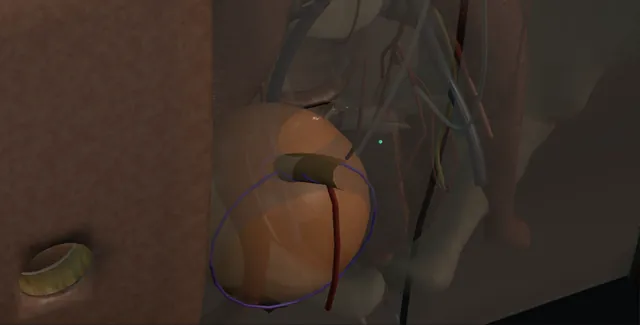
Continent cutaneous diversion: how the structures relateOpen, the right colon pouch low in the right pelvis. Right colon reservoir: sole supply to pouch, valve and channel Ileocolic pedicle (danger). Right colon reservoir: too loose it leaks, too tight it will not pass Ileocaecal valve and tapered ileum (danger). Right colon reservoir: the channel runs up through the right rectus Flush stoma, right lower quadrant. Right colon reservoir: enter from the brim; tight joins stricture Ureters into the posterior wall (danger). Right colon reservoir: leaves through the front wall; drains it Pouch tube.Right colon reservoir!Ileocolic pedicleIleocaecal valve andtapered ileumFlush stoma, rightlower quadrant!Ureters into theposterior wallPouch tubesole supply topouch, valve andchanneltoo loose it leaks,too tight it willnot passthe channel runs upthrough the rightrectusenter from thebrim; tight joinsstrictureleaves through thefront wall; drainsitTargetDangerLandmarkContext
Open, the right colon pouch low in the right pelvis. Schematic, not to scale; relationships only.
  • The right colon is opened along its taenia and folded down onto itself to make a reservoir that sits low in the right pelvis on the ileocolic pedicle.
  • At its lower right corner the ileocaecal valve leads into the tapered terminal ileum, which runs straight up through the right rectus to a flush stoma in the right lower quadrant.
  • The ureters enter the back of the reservoir from the pelvic brim, and the pouch tube leaves through its front wall.
  • ›Caecum and ascending colon - twenty-five to thirty centimetres, mobilised along the white line of Toldt; opened along the taenia and folded into the reservoir.
  • ›The ileocolic pedicle - the single blood supply of pouch, valve and channel; transilluminated and kept.
  • ›The ileocaecal valve - the native valve reinforced with rows of non-absorbable Lembert sutures; the first part of the continence mechanism.
  • ›The terminal ileum - ten to twelve centimetres tapered over a 12 or 14 French catheter on its antimesenteric side; the channel to the skin.
  • ›The ureters - implanted into the posterior wall of the colonic reservoir over single-J stents, the left brought under the sigmoid mesentery.
  • ›The stoma site - right lower quadrant or umbilicus; flush, not spouted, through a straight two-finger opening in the rectus.
  • ›The pouch tube - 20 to 24 French through the anterior pouch wall and a separate stab; the drain and the irrigation route for three weeks.
Click me next Section 3 of 8 Steps The operation in order, with the landmark that guards each move.

Operative steps

  1. Who chooses it The patient having a cystectomy who cannot have a neobladder, because the urethra has tumour at the margin, the sphincter is already weak or the urethra has been taken, but who does not want a bag; and the patient with a devastated outlet or a neuropathic bladder that is being taken out. They need the hands, the eyesight and the will to pass a catheter into a stoma every four hours by day and once at night for the rest of their life, and to irrigate the pouch for mucus. Renal function must be good enough for a large absorptive surface, the right colon must be free of disease and previous radiation, and the alternative, an ileal conduit, is explained without disparaging it, because it is simpler and fails less.
  2. The right colon and terminal ileum Through the midline incision, after the cystectomy, the caecum and ascending colon are mobilised along the white line of Toldt up to the hepatic flexure, the duodenum and the right ureter swept away behind. A segment of about twenty-five to thirty centimetres of caecum and ascending colon with ten to twelve centimetres of terminal ileum is isolated on the ileocolic pedicle, transilluminated so the vessel is seen and preserved. The colon is divided at its proximal end with a linear stapler and the ileum divided the same way; bowel continuity is restored with a stapled ileocolic anastomosis and the mesenteric window closed. The segment is irrigated until the effluent is clear.
    where it bites: the ileocolic artery is the whole blood supply of the reservoir and the valve; it is seen against the light and never divided.
  3. Opening the colon along its taenia The colonic segment is laid out and opened along its antimesenteric taenia with the diathermy from the stapled proximal end down to the caecum, stopping short of the ileocaecal valve so that the valve and the caecal cuff around it are not disturbed. The purpose is the same as for any bowel reservoir: intact colon contracts in mass movements that raise the pressure to a level the valve cannot hold, and an opened plate does not. The staple line is excised, the mucosa wiped of stool and mucus, and the edge made haemostatic with the bipolar. The appendix, if present, is removed at its base.
  4. Folding and closing the reservoir The opened plate is folded on itself, the free proximal edge brought down toward the caecum so that the segment is folded in half, and the adjacent edges sewn together with a running 3-0 Vicryl, mucosa to mucosa, in the manner of a Heineke-Mikulicz closure: a longitudinal opening closed transversely. The reservoir that results is wider than it is long, sits low in the right side of the pelvis on its pedicle, and holds 400 to 500 millilitres at low pressure once it has stretched. The closure is inspected along its length and a second seromuscular layer added where the first looks thin.
  5. The continence mechanism: valve and tapered ileum The ileocaecal valve is reinforced from outside with two or three rows of interrupted non-absorbable Lembert sutures, 3-0 silk or Prolene, placed across the ileocaecal junction to imbricate the caecum around the terminal ileum and narrow the valve so it holds against the filled pouch. The terminal ileum is then tapered: a 12 or 14 French catheter is passed through it into the pouch and the antimesenteric side of the ileum is excised alongside the catheter with a linear stapler fired two or three times, or trimmed and oversewn, leaving a tube that admits the catheter and no more. The staple line faces away from the mesentery. The catheter is passed and withdrawn several times to confirm it goes easily.
    where it bites: the taper is made on the antimesenteric side and the mesentery left untouched; a channel tapered too tight, or with its blood supply compromised, is a channel that cannot be catheterised.
  6. The ureters into the reservoir The left ureter is brought under the sigmoid mesentery through a generous window and both ureters are spatulated on their lateral wall with the adventitia preserved. Each is implanted into the posterior wall of the colonic reservoir through a separate small colotomy, mucosa to mucosa with interrupted 4-0 or 5-0 PDS, over a single-J stent passed up to the renal pelvis; a submucosal tunnel for antireflux is used by some and omitted by others because of stricture. The stents are led into the pouch and out through the pouch wall. The ureters are laid without a kink or a twist and are not on tension when the packs are removed.
    where it bites: the ureteric supply is in the adventitia, medial above the brim and lateral below it; the implant is spatulated, stented and never tensioned.
  7. The stoma The efferent ileum is brought through the abdominal wall at a site marked before surgery, in the right lower quadrant or at the umbilicus, through a two-finger opening in the rectus made as for any stoma. Only enough ileum is brought out to reach the skin, the rest trimmed, and the stoma is matured flush with the skin, not as a spout, with interrupted absorbable sutures taking skin and the full thickness of bowel, because a flush stoma needs no appliance and is hidden by a dressing. The route through the wall is straight and the pouch is fixed to the anterior abdominal wall beside the exit with a few sutures so that the channel does not angulate as the pouch fills.
    where it bites: the channel must run straight from skin to pouch; an angulated or redundant channel is a false passage waiting for the first difficult catheter.
  8. Catheters, stents, drains, irrigation A 20 to 24 French Malecot or Foley pouch tube is brought through the anterior wall of the reservoir and out through a separate stab to drain the pouch, and a 12 to 14 French catheter is left through the stoma and channel. The stents come out through the pouch wall beside the tube. A pelvic drain sits beside the ureteric implants. The pouch is filled with 100 millilitres of saline and the suture lines inspected. The abdomen is closed as a mass closure. From the first day the pouch is irrigated through the tube with 30 to 50 millilitres of saline every four to six hours, because colonic mucus is thick and a blocked pouch strains every line in it.
  9. After: teaching the stoma and the lifetime At about three weeks a pouchogram confirms no leak, the stents and tube come out and the patient is taught to catheterise the stoma: a 14 French catheter with gel, every two to three hours to begin with, then every four hours by day and once at night as the pouch stretches to 400 to 500 millilitres, with a weekly irrigation for mucus. The catheter is never forced; a channel that will not admit it is reviewed the same day. Lifelong: bicarbonate, creatinine and B12, ultrasound of the kidneys for the silent stricture, imaging for stones in the pouch, and a plain warning that the pouch that cannot be emptied is an emergency.
Click me next Section 4 of 8 Danger zones Where this operation bites, and why it bites there.
Critical relationship

Danger zones

The spatial relationships the animation is built to make automatic. Before you read each one, ask yourself: at this point in the operation, what is about to go wrong, and what is my first move? The save for each sits in the Complications tab.

  • RiskThe ileocolic pedicle - the only supply to reservoir, valve and channel; an ischaemic pouch leaks and an ischaemic channel strictures.
  • RiskThe valve and the taper - too loose it leaks, too tight or angulated it cannot be catheterised; the catheter is passed and withdrawn in theatre until it goes easily.
  • RiskThe uretero-colonic joins - stripped, tensioned or tunnelled too tight they stricture silently; the upper tracts are imaged for life.
  • RiskThe full pouch that cannot be emptied - a blocked or lost channel with 500 millilitres of urine behind it is a rupture in waiting.
Practise it Take me into the scene Opens the animation at a danger moment and asks you to predict the mechanism and your first move before it reveals.
Click me next Section 5 of 8 Complications The save for each one: the first move, the escalation trigger, the op-note line.

Complications and immediate saves

Trigger each of these live in the Animation tab, then read the save: the first move, the escalation trigger, and the line that goes in the op note.

Difficult catheterisation and stomal stenosis

  • the catheter that will not go, at the skin from stenosis of the flush stoma or deeper from an angulated channel; the commonest late problem, in about one in five to one in ten over the years.
The save
  1. Stop. Gel, a smaller catheter and a change of the patient's position; never force the catheter, which makes a false passage. first move
  2. If the pouch is full and nothing will pass, a coudé catheter or a guidewire under fluoroscopy or through a flexible scope passed via the stoma.
  3. A skin-level stenosis is dilated and, if it recurs, revised with a small skin flap.
  4. A full pouch that cannot be reached: the pouch is drained percutaneously under ultrasound before it ruptures, then the channel is revised. escalation trigger
  5. Note: the level of the obstruction, the catheter that finally passed, and the revision plan; teach the patient the number to ring. document

Pouch incontinence

  • leakage from the stoma, early from a valve that was not tight enough or late from a pouch that generates pressure or is overfilled.
The save
  1. Check the emptying: a pouch that is catheterised too seldom or that has a residual leaks by overflow first. first move
  2. A pouchogram and a pressure study of the pouch: high pressure waves mean an incompletely detubularised or a small reservoir.
  3. Anticholinergics and more frequent emptying for a high-pressure pouch; a bulking agent at the valve for a mild leak.
  4. A valve that has failed: revision of the continence mechanism, or conversion to a conduit if the patient has had enough. escalation trigger
  5. Note: the pad use, the catheterisation interval and the pouch pressures found. document

Urine leak or pouch rupture

  • urine in the drain in the first weeks from a suture line or a ureteric implant, and years later the pouch that bursts because it was full and could not be emptied.
The save
  1. Early: confirm the pouch tube and the catheter drain, flush for mucus, and send drain fluid for creatinine. first move
  2. Keep all drains and stents and image with a pouchogram or CT; most early leaks close with drainage.
  3. Late abdominal pain in a pouch patient, however vague, is a rupture until proved otherwise: catheterise the pouch, then CT with contrast through it.
  4. Peritonitis, sepsis or a rupture on imaging: laparotomy, washout and repair the same day. escalation trigger
  5. Note: the site, the repair, and the catheterisation schedule repeated to the patient and carers. document

Uretero-colonic stricture

  • hydronephrosis on the surveillance ultrasound, silent, from an implant that was ischaemic, tensioned or tunnelled too tight.
The save
  1. Ultrasound of the kidneys on a schedule for life; a new hydronephrosis is investigated with a renogram. first move
  2. Nephrostomy and an antegrade study to define the length; exclude recurrence at the join.
  3. A short stricture is dilated or incised antegrade or through the pouch with a flexible scope.
  4. A long or recurrent stricture, or a kidney losing function: open or robotic reimplantation. escalation trigger
  5. Note: the side, the length, the treatment and the interval to the next scan. document

Stones, mucus, infection and acidosis

  • colon in the urinary tract makes mucus, harbours bacteria, absorbs chloride and ammonium and loses bicarbonate; stones form in the pouch in about one in ten.
The save
  1. Irrigation for mucus, weekly at least and daily when it is thick; the frequency is set by the patient, not the clinic. first move
  2. Symptomatic infection is cultured and treated; bacteriuria without symptoms is left alone.
  3. Bicarbonate, chloride and creatinine at each visit, oral bicarbonate for acidosis; B12 yearly from five years.
  4. Recurrent infection, haematuria or a catheter that catches: a pouchoscopy through the stoma and stones cleared endoscopically or through a small open pouchotomy. escalation trigger
  5. Note: the irrigation regime, the bloods, and the stone imaging interval. document
Click me next Section 6 of 8 Troubleshooting When the case turns: the problem, then the next move.

Intra-operative troubleshooting

When the case turns: the problem, then the next move.

  • ActionThe catheter will not pass the taper in theatre - the taper is too tight or the staple line has caught the lumen; take down the last firing and redo it over the catheter with a wider margin.
  • ActionThe channel is too long to reach the skin without redundancy - trim the ileum; a short straight channel beats a long one that kinks in the wall.
  • ActionThe ureter will not reach the reservoir - mobilise it higher with its adventitia intact, or bring the pouch up to it; a tensioned join is a stricture.
  • ActionLeak on the test fill - find the point and close it with a single interrupted suture; add a seromuscular layer where the closure looks thin.
  • ActionThe pouch tube is blocked on the ward - flush 50 millilitres of saline and aspirate; if the tube will not clear, drain through the stoma catheter; a blocked pouch strains every line.
  • ActionThe patient cannot learn to catheterise - the stoma nurse, a mirror, a different catheter; if it still fails, the pouch tube stays until they can, and conversion to a conduit is discussed honestly.
Click me next Section 7 of 8 FRCS viva Say it out loud, then mark yourself against the model answer.

FRCS viva questions

The examiner stems this scene prepares you for. Try to answer out loud first, then reveal, and learn the discriminator, the detail that turns a pass into a strong pass.

QWho would you offer a continent cutaneous diversion to, and why not a neobladder or a conduit?reveal answer
  • I would offer it to a patient who needs a cystectomy or a diversion, who cannot have an orthotopic neobladder and does not want an incontinent stoma.
  • The reasons a neobladder is excluded are oncological, tumour at the urethral margin, the prostatic urethra or the bladder neck, or functional, a urethra that is absent, strictured or incontinent, or a pelvic floor that has been irradiated.
  • The patient must be able and willing to catheterise a stoma every four hours by day and once at night for life, with the dexterity, vision and cognition to do it and to irrigate for mucus, and their renal function must tolerate a large absorptive surface, so a creatinine above about 150 or an eGFR below about 50 argues against it.
  • The right colon must be free of disease and previous radiation.
  • Against the conduit I would be honest: the conduit is shorter, has fewer early complications and fewer revisions, and the continent pouch buys freedom from an appliance at the cost of catheterisation, a higher revision rate, stones and metabolic follow-up.
  • The patient chooses with the stoma nurse, having met patients living with each.
Strong pass

Say both

  • Separating the oncological from the functional reasons a neobladder is excluded
  • Comparing the pouch with the conduit honestly rather than presenting it as the better operation
Examiner note
Separating the oncological from the functional reasons a neobladder is excluded, and comparing the pouch with the conduit honestly rather than presenting it as the better operation.
QDescribe how you would construct an Indiana pouch.reveal answer
  • After the cystectomy I would mobilise the right colon along the white line of Toldt and isolate about twenty-five to thirty centimetres of caecum and ascending colon with ten to twelve centimetres of terminal ileum on the ileocolic pedicle, restoring bowel continuity with a stapled ileocolic anastomosis and closing the mesenteric window.
  • I would open the colon along its antimesenteric taenia to the caecum, sparing the ileocaecal valve, and fold the plate on itself, closing it transversely with a running absorbable suture to make a low-pressure reservoir.
  • For the continence mechanism I would reinforce the ileocaecal valve with two or three rows of interrupted non-absorbable Lembert sutures and taper the terminal ileum over a 12 or 14 French catheter with a linear stapler along its antimesenteric border, checking the catheter passes freely.
  • I would spatulate the ureters and implant them into the posterior wall of the reservoir over single-J stents, mucosa to mucosa, bringing the left under the sigmoid mesentery.
  • I would bring the efferent ileum straight through the rectus to a flush stoma in the right lower quadrant or umbilicus, fix the pouch to the abdominal wall, place a pouch tube, a stomal catheter and a drain, test the closure, and irrigate the pouch from the first day.
Strong pass

Say all four

  • The taenia as the line of opening with the valve spared
  • The continence mechanism given as two parts
  • Imbrication and taper
  • The flush stoma with the pouch fixed to the wall
Examiner note
The taenia as the line of opening with the valve spared, the continence mechanism given as two parts, imbrication and taper, and the flush stoma with the pouch fixed to the wall.
QWhere does the continence of an Indiana pouch come from, and how does it fail?reveal answer
  • The continence comes from two elements in series and from the reservoir behind them.
  • The ileocaecal valve is the natural barrier, reinforced by imbricating the caecum around the terminal ileum with rows of non-absorbable sutures so that it closes against the pressure of the filled pouch.
  • The tapered terminal ileum then provides a narrow channel of a length that generates outflow resistance, the same principle as a Mitrofanoff channel, and the fixation of the pouch to the abdominal wall keeps that channel straight.
  • Behind them the reservoir must be low pressure: the colon is detubularised along its taenia and folded so that it cannot contract in mass movements, and is large enough that the pressure stays below the resistance of the valve until it is emptied.
  • Failure is on either side of that balance.
  • Leakage follows a valve that was not imbricated tightly enough, a taper that was too generous, or a pouch that generates pressure because it was incompletely opened, is too small, or is overfilled by a patient who catheterises too seldom;
  • overflow from a residual is the first thing to exclude.
  • Obstruction follows a taper that is too tight or a channel that angulates, causing difficult catheterisation and false passage.
Strong pass

Say all three

  • Continence explained as valve
  • Taper and low-pressure reservoir in series
  • Failure described as a balance that can be lost in either direction
Examiner note
Continence explained as valve, taper and low-pressure reservoir in series, and failure described as a balance that can be lost in either direction.
QWhat is the long-term care of a patient with a continent cutaneous reservoir?reveal answer
  • I would organise it around emptying, metabolism, structure and cancer.
  • Emptying is by clean intermittent catheterisation every four hours by day and once at night, with weekly or more frequent irrigation for mucus;
  • the patient is told that a pouch that cannot be emptied is an emergency and is given a direct line to the unit, because rupture follows overdistension.
  • Metabolism: the colon absorbs chloride and ammonium and loses bicarbonate, so bicarbonate, chloride and creatinine are checked at each visit and oral bicarbonate started early;
  • B12 is measured annually from about five years because terminal ileum has been used;
  • and the loss of the ileocaecal valve can cause diarrhoea and bile salt malabsorption.
  • Structure: ultrasound of the upper tracts on a schedule for the silent uretero-colonic stricture, imaging for pouch stones, which occur in about one in ten and are treated endoscopically through the stoma, and a low threshold for pouchoscopy for haematuria or recurrent infection.
  • Cancer: adenocarcinoma has been reported in bowel exposed to urine after a decade or more, so new symptoms after ten years are investigated with endoscopy, and the oncological surveillance of the original cancer continues alongside.
Strong pass

Say both

  • Naming the loss of the ileocaecal valve as a specific metabolic consequence
  • The emptying emergency as a standing instruction rather than a clinic finding
Examiner note
Naming the loss of the ileocaecal valve as a specific metabolic consequence, and the emptying emergency as a standing instruction rather than a clinic finding.
Click me next Section 8 of 8 Evidence The guidance each of these answers is built on.

Evidence and guidance

Two minutes

Close the lap: recall it before you leave.

Say it aloud, then reveal. Reading is not remembering.

  1. 01 The relationship Say the critical spatial relationship out loud, then reveal. reveal
    Memory frame

    The right colon is opened along its taenia and folded down onto itself to make a reservoir that sits low in the right pelvis on the ileocolic pedicle.

    Why?
    At its lower right corner the ileocaecal valve leads into the tapered terminal ileum, which runs straight up through the right rectus to a flush stoma in the right lower quadrant. The ureters enter the back of the reservoir from the pelvic brim, and the pouch tube leaves through its front wall.
  2. 02 The first move Difficult catheterisation and stomal stenosis. Your first move? reveal
    First move

    Stop.

    More detail
    Gel, a smaller catheter and a change of the patient's position; never force the catheter, which makes a false passage.
  3. 03 The escalation trigger Difficult catheterisation and stomal stenosis. What makes you call for help? reveal
    Escalate when

    A full pouch that cannot be reached: the pouch is drained percutaneously under ultrasound before it ruptures, then the channel is revised.

  4. 04 The discriminator What turns a pass into a strong pass? reveal
    Strong pass

    Say both

    • Separating the oncological from the functional reasons a neobladder is excluded
    • Comparing the pouch with the conduit honestly rather than presenting it as the better operation
    Examiner note
    Separating the oncological from the functional reasons a neobladder is excluded, and comparing the pouch with the conduit honestly rather than presenting it as the better operation.
  5. 05 The pitfall Where does this operation bite first? reveal
    Pitfall

    The ileocolic pedicle

    Why?
    the only supply to reservoir, valve and channel; an ischaemic pouch leaks and an ischaemic channel strictures.

Revision practice with self-marked answers, not an assessment of clinical competence. Kept in this browser only; it does not sync between devices.