
UrOops3D, danger-zone atlas
Operative animation, Distal ureteric reconstruction

Ureteric reimplantation, psoas hitch and Boari flap

The lower ureter that has been cut, tied, burnt, irradiated or resected for a tumour, and cannot be joined to itself: the bladder is brought to the ureter instead.

A short gap is a direct reimplant, a longer one is the bladder freed and hitched to the psoas, and a longer one still is a flap of bladder rolled into a tube.

This page is the measuring, the ureter handled by its adventitia, the bladder mobilised on one pedicle, the hitch beside the genitofemoral nerve, the anastomosis over a stent, and the flap.

Advanced tier FRCS reconstructive urology, upper tract reconstruction and trauma Interactive 3D

Operative visualisation

Schematic scene preview: Distal ureteric reconstruction
Preparing the 3D anatomy…

UrOops3D · UroRef · © Nity G

Click me next Section 2 of 8 Anatomy The structures this operation works between, and the one it must not cross.

The structures in the scene

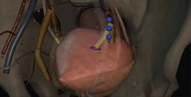
Distal ureteric reconstruction: how the structures relateFrom the operator's side, the ureter over the brim to the hitched bladder. Ureter joined to the hitched bladder: lifted with the ureter; stripped, it strictures Ureteric adventitia and blood supply (danger). Ureter joined to the hitched bladder: hitch sutures shallow, medial to the nerve Psoas and genitofemoral nerve (danger). Ureter joined to the hitched bladder: pulled to the midline; seen inside first Contralateral orifice (danger). Ureter joined to the hitched bladder: contralateral divided, ipsilateral kept Superior vesical pedicles. Ureter joined to the hitched bladder: under the hitch; the ureter not pressed on them External iliac vessels. Ureter joined to the hitched bladder: the ureter arrives straight over it JJ stent across the join.Ureter joined to thehitched bladder!Ureteric adventitiaand blood supply!Psoas andgenitofemoral nerve!ContralateralorificeSuperior vesicalpediclesExternal iliacvesselsJJ stent across thejoinlifted with theureter; stripped,it strictureshitch suturesshallow, medial tothe nervepulled to themidline; seeninside firstcontralateraldivided,ipsilateral keptunder the hitch;the ureter notpressed on themthe ureter arrivesstraight over itTargetDangerLandmarkContext
From the operator's side, the ureter over the brim to the hitched bladder. Schematic, not to scale; relationships only.
  • From the operator's side the ureter is seen coming over the pelvic brim on the iliac vessels and running down the sidewall toward a bladder that has been freed on the opposite side and pulled up until its dome lies against the psoas above the vessels.
  • The hitch sutures sit on the muscle beside the genitofemoral nerve, the ureter arrives at the raised bladder wall in a straight line over the stent, and the contralateral orifice has moved toward the midline inside.
  • When a flap is needed it rises from the front of the bladder on its posterolateral pedicle and rolls into a tube that reaches the ureter above the vessels.
  • ›The ureter at the pelvic brim - crossing the common iliac bifurcation on the medial leaf of peritoneum; found here by peristalsis and followed down.
  • ›The ureteric adventitia and its blood supply - longitudinal vessels entering medially above the brim and laterally below; lifted with the ureter and never stripped.
  • ›The superior vesical pedicles - the contralateral one divided to let the bladder swing; the ipsilateral one kept as the supply of the reimplant and the flap.
  • ›The psoas and the genitofemoral nerve - the hitch sutures go into the tendon or muscle parallel to the fibres, shallow and medial to the nerve on its surface.
  • ›The contralateral orifice - drawn toward the midline as the bladder is pulled up; seen from inside before the hitch and the flap.
  • ›The external iliac vessels - under the hitched bladder and beside the flap; the ureter crosses them and must not be compressed against them.
  • ›The stent and the catheter - a 6 French JJ across the join for four to six weeks; a urethral catheter for seven to ten days with a cystogram if the closure was long.
Click me next Section 3 of 8 Steps The operation in order, with the landmark that guards each move.

Operative steps

  1. Who: the distal ureter that cannot be joined end to end The ureter below the pelvic brim, injured at hysterectomy, colorectal resection or ureteroscopy, strictured after a stone, radiotherapy or a previous repair, resected for a distal urothelial tumour with a kidney worth keeping, or refluxing or obstructed at the orifice in a child or an adult with a megaureter. Below the brim a ureter-to-ureter join is a poor operation: the blood supply there comes from the pelvic vessels and is disrupted by the injury, the ends are fixed and the anastomosis is under tension. The bladder is mobile, well supplied and forgiving, so it is brought up to the ureter; ureteroureterostomy is for the mid and upper ureter. The kidney must be worth it: a split function under about fifteen to twenty per cent is a nephrectomy discussion.
  2. Planning: length, bladder and kidney The gap decides the operation, and it is measured before the incision, not guessed. An antegrade study through a nephrostomy or a CT urogram defines the upper end of healthy ureter and a retrograde study the lower; the length between them, corrected for magnification, is the defect. A MAG3 renogram gives the split function. The bladder is assessed at cystoscopy and, if operated on or irradiated, by a cystogram for capacity and urodynamics for compliance. A direct reimplant bridges two to three centimetres; a psoas hitch adds about five; a Boari flap adds ten to fifteen and reaches the mid ureter, and the two are combined. A small, irradiated or neuropathic bladder rules out the flap and often the hitch; ileum or autotransplantation are then discussed.
  3. Exposure and finding the ureter at the brim Open through a lower midline or a Gibson incision, extraperitoneal where the field is virgin and transperitoneal where it is not; robotic through the peritoneum with the same steps. The ureter is found where it is reliable: at the pelvic brim crossing the common iliac bifurcation, adherent to the medial leaf of the peritoneum, identified by its peristalsis when it is stroked. In the scarred pelvis it is found here and followed down, never sought first in the scar. A vessel loop is passed around it and it is traced toward the bladder to the injury or stricture, and the level of the healthy end confirmed against the imaging. The stricture or the injured segment is excised back to ureter that bleeds and is soft.
  4. Mobilising the ureter with its adventitia The ureter is lifted on the vessel loop and the peritoneum and areolar tissue are taken with it, because the blood supply runs longitudinally in the adventitia: from the renal artery and aorta entering medially above the brim, and from the iliac, vesical and uterine vessels entering laterally below it. The dissection stays a centimetre from the wall on the side the vessels come from, and the ureter is never held with forceps, only by its adventitia or the loop. It is mobilised as far up as is needed to reach the bladder without tension and no further, since every centimetre mobilised depends on the intrinsic supply alone. The end is spatulated on its anterior or lateral wall for one to one and a half centimetres.
    where it bites: a stripped ureter looks white and does not bleed at the cut edge; it strictures at the join, and the correct response is to cut it back until it bleeds and make up the length with the bladder.
  5. Mobilising the bladder The bladder has to travel, and it does so on one side. The peritoneum is taken off the dome and the urachus divided; the contralateral superior vesical pedicle is identified lateral to the bladder and ligated and divided, and the bladder is freed from the pelvic sidewall on that side, which lets the dome swing toward the ureter. The ipsilateral pedicle stays intact because it supplies the reimplant and any flap. The bladder is filled through the catheter with 200 to 300 millilitres and the dome is pushed up toward the psoas with a finger to see how far it reaches; that reach, without tension, decides between a direct reimplant, a hitch and a flap. In a woman the round ligament can be divided for more reach.
    where it bites: the contralateral orifice is drawn toward the midline as the bladder is pulled up; its position is checked from inside before the hitch sutures and the flap are placed.
  6. The psoas hitch The posterolateral bladder wall on the side of the ureter is drawn up onto the psoas minor tendon or psoas muscle above the iliac vessels and fixed with two or three interrupted 2-0 PDS or non-absorbable sutures through detrusor and tendon, the bites parallel to the muscle fibres, shallow, and medial to the genitofemoral nerve where it lies on the surface of the psoas. The femoral nerve runs deep in the muscle lateral to this and is injured by a deep bite. The fixation takes the tension off the anastomosis for good and stops the bladder dropping back. Many open the bladder first, through an anterior incision at right angles to the line of pull, and place the hitch sutures with a finger inside to feel the depth.
    where it bites: the genitofemoral nerve lies on the psoas and the femoral nerve within it; a hitch suture placed deep or lateral leaves groin pain or a numb, weak thigh.
  7. The reimplant over a stent The ureter is brought to the fixed bladder through an opening made where it arrives without a bend, because a kink at the entry is a stricture. In an adult a direct refluxing anastomosis is acceptable and usual after trauma or tumour: the spatulated ureter is sewn mucosa to mucosa with interrupted 4-0 or 5-0 PDS over a 6 French JJ stent passed to the renal pelvis, and the adventitia tacked to the bladder to take any pull. Where reflux matters, in a child or an adult with a good bladder, the ureter is passed under a submucosal tunnel of about five times its diameter before the opening is sewn as before. The stent stays four to six weeks and the catheter seven to ten days.
  8. The Boari flap for the longer gap When the hitched bladder still does not reach, a flap is raised from the anterior bladder wall, based posterolaterally on the ipsilateral superior vesical pedicle, with a base at least four centimetres wide and a length no more than three times that, wider than the ureter needs so that it tubularises to a comfortable calibre. The flap is turned up, the ureter laid on it and sewn end to end at the tip or passed under a short mucosal tunnel over a stent, the flap rolled into a tube with a running 3-0 Vicryl and the bladder defect closed in two layers. The hitch is placed as well to take the tension. It needs a bladder of about 300 millilitres or more, because the flap takes capacity from it.
    where it bites: a flap with a narrow base, a long length or an ischaemic tip does not survive; the base is wide, the pedicle is on it, and the ipsilateral superior vesical artery is never divided.
  9. Closure, drains and after The bladder is closed in two layers of 2-0 or 3-0 Vicryl, filled to check the closure and the anastomosis, and a tube drain left beside the reimplant. A urethral catheter for seven to ten days, with a cystogram before it comes out where a flap was used; the drain out when dry. The stent out at four to six weeks. An ultrasound at three months and a MAG3 at three to six months to show the kidney draining, then ultrasound at a year and at intervals after, because a stricture at the join is silent. The patient is told that the reimplant refluxes if it was made to, and that a flap bladder may hold less and void more often.
Click me next Section 4 of 8 Danger zones Where this operation bites, and why it bites there.
Critical relationship

Danger zones

The spatial relationships the animation is built to make automatic. Before you read each one, ask yourself: at this point in the operation, what is about to go wrong, and what is my first move? The save for each sits in the Complications tab.

  • RiskThe ureteric blood supply - longitudinal in the adventitia, medial above the brim and lateral below; a stripped, tensioned or kinked ureter strictures at the join.
  • RiskThe contralateral ureteric orifice - pulled toward the midline by the hitch and toward the flap by the bladder incision; obstructed by a suture placed without looking inside.
  • RiskThe genitofemoral and femoral nerves - on and within the psoas; a deep or lateral hitch suture leaves groin pain, thigh numbness or a weak quadriceps.
  • RiskThe flap base and the bladder capacity - a narrow base or a divided ipsilateral pedicle is a dead flap; a long flap from a small bladder is a small bladder that leaks and voids by the hour.
Practise it Take me into the scene Opens the animation at a danger moment and asks you to predict the mechanism and your first move before it reveals.
Click me next Section 5 of 8 Complications The save for each one: the first move, the escalation trigger, the op-note line.

Complications and immediate saves

Trigger each of these live in the Animation tab, then read the save: the first move, the escalation trigger, and the line that goes in the op note.

Urine leak and pelvic urinoma

  • urine in the drain or a collection on the scan in the first week from the anastomosis, the flap tube or the bladder closure, most often behind a blocked catheter.
The save
  1. Check that the catheter drains and flush it; a full bladder pushes urine through the fresh suture line. first move
  2. Drain fluid creatinine to confirm urine; keep the drain, the catheter and the stent, and check on a plain film that the stent has not moved.
  3. A cystogram or CT urogram to find the leak; a small leak with good drainage closes with time and a longer catheter.
  4. Sepsis, an enlarging collection or a leak that persists beyond two to three weeks: percutaneous drainage, a nephrostomy to divert the kidney, and a return to theatre if the join has given way. escalation trigger
  5. Op note: the type of reimplant, the stent size and removal date, the closure layers, and the cystogram date before the catheter comes out. document

Anastomotic stricture and recurrent obstruction

  • the silent kidney months later: hydronephrosis on the surveillance scan from a join that was ischaemic, tensioned, kinked or tunnelled too tight.
The save
  1. Ultrasound at three months and at intervals for at least two years; a new hydronephrosis after the stent is out is a stricture until proved otherwise. first move
  2. A MAG3 to confirm obstruction and measure function; exclude tumour recurrence at the join where the indication was cancer.
  3. A nephrostomy and antegrade study; a short stricture may be dilated or incised endoscopically and stented, once.
  4. A stricture that recurs, a long one, or a kidney losing function: redo reimplant with a flap or a hitch it did not have, ileal interposition, or nephrectomy if the kidney is not worth it. escalation trigger
  5. Note: the imaging schedule with dates, the split function at baseline and after, and the appearance of the join at any endoscopy. document

Contralateral orifice injury

  • loin pain or a rising creatinine on the other side after a hitch or a flap: the opposite orifice drawn to the midline and caught in a suture or the flap incision.
The save
  1. Look at the trigone from inside before the hitch sutures and the flap are placed, and again after; catheterise the contralateral orifice if it lies within a centimetre of any incision. first move
  2. A suspected injury at the time: cut the suture, catheterise the orifice with a 5 French catheter, and stent that side.
  3. Found after: ultrasound of the opposite kidney, a nephrostomy if obstructed, and an antegrade study.
  4. An orifice that will not open antegrade: reimplantation of the second ureter, with senior help, once the first has healed. escalation trigger
  5. Op note: the position of both orifices seen from inside, the distance from the sutures, and whether the contralateral ureter was catheterised. document

Genitofemoral or femoral nerve injury

  • pain or numbness in the groin and anterior thigh, or a weak quadriceps on standing, from a hitch suture placed deep or lateral into the psoas.
The save
  1. Place the hitch sutures shallow, parallel to the fibres, and medial to the genitofemoral nerve seen on the muscle; two or three sutures, no more. first move
  2. Groin or thigh pain on waking: examine the sensation and the quadriceps power and hip flexion before the patient mobilises.
  3. Sensory symptoms alone: analgesia and time; most settle over weeks and are explained to the patient.
  4. Quadriceps weakness or a foot that will not clear the ground: early return to theatre to remove or replace the offending suture, and a neurology review. escalation trigger
  5. Op note: the number and depth of the hitch sutures and their relation to the nerve; the ward note records the neurological examination on the first day. document

Flap ischaemia, reduced capacity and bladder dysfunction

  • a Boari flap that goes dusky or leaks, a bladder that now holds 150 millilitres, or a bladder that will not empty after bilateral or extensive dissection.
The save
  1. A wide base on the ipsilateral pedicle, the pedicle never divided, and a flap no longer than it needs to be; a bladder capacity checked before the flap is planned. first move
  2. A leak from the flap in the first week: catheter and drain kept, imaging, and a nephrostomy if the leak is from the tube.
  3. Frequency and small volumes after: a bladder diary and a residual; time and anticholinergics help most, and the capacity recovers partly.
  4. A necrotic flap, a bladder that will not empty with a large residual, or a capacity that stays small: revision, intermittent catheterisation, or augmentation. escalation trigger
  5. Note: the flap dimensions and base, the preoperative capacity, and the residual and diary at each review. document
Click me next Section 6 of 8 Troubleshooting When the case turns: the problem, then the next move.

Intra-operative troubleshooting

When the case turns: the problem, then the next move.

  • ActionThe ureter cannot be found in the scarred pelvis - go to the brim and find it on the iliac bifurcation by peristalsis, or pass a ureteric catheter or a lit stent from below and feel for it; never dig in the scar first.
  • ActionThe cut end of the ureter is white and does not bleed - cut it back until it bleeds and make up the length with the bladder; a hitch or a flap costs less than a stricture.
  • ActionThe bladder will not reach the ureter after the hitch - divide the contralateral pedicle if it has not been, take the peritoneum off the dome and the round ligament in a woman, fill the bladder and try again; if it still will not reach, a Boari flap, and the ureter is not pulled down to meet it.
  • ActionThe contralateral orifice sits under the hitch or in the line of the flap - catheterise it from inside, move the flap incision or the sutures away from it, and stent it if any bite comes close.
  • ActionThe stent will not pass to the kidney through the anastomosis - the ureter is kinked or twisted at the entry; take the anastomosis down, lay the ureter straight, and pass the stent over a wire before the sutures are replaced.
  • ActionLeak on the test fill - find the point, one interrupted full-thickness suture, refill; a leak from the ureteric join itself is not tested by pressure but is covered with adventitia and omentum.
Click me next Section 7 of 8 FRCS viva Say it out loud, then mark yourself against the model answer.

FRCS viva questions

The examiner stems this scene prepares you for. Try to answer out loud first, then reveal, and learn the discriminator, the detail that turns a pass into a strong pass.

QA ureter is injured 4 cm above the bladder during a hysterectomy and you are called to theatre. What are your options?reveal answer
  • I would first define the injury: its level and length, whether it is a crush, a ligature, a transection or a diathermy burn, and whether the patient is stable.
  • A ligature recognised at once is removed and the ureter stented if it looks well; a crush or a burn is excised to healthy ends, because thermal injury extends beyond what is seen.
  • For a transection four centimetres above the bladder an end to end join is a poor choice, because the distal ureter has a lateral pelvic blood supply that the hysterectomy has already disrupted, so I would bring the bladder to the ureter.
  • If the gap after trimming to bleeding ends is two to three centimetres, a direct refluxing reimplant over a stent;
  • if longer, I would divide the contralateral superior vesical pedicle, hitch the bladder to the psoas with shallow sutures medial to the genitofemoral nerve, and reimplant;
  • a Boari flap if the gap is longer still and the bladder has the capacity.
  • If the patient is unstable, the field is contaminated or I am not equipped, I would place a stent or a nephrostomy, drain the ureter, and reconstruct later.
  • Every option is over a stent with a drain and a catheter, and the injury and the repair are documented and explained to the patient.
Strong pass

Say both

  • Choosing the bladder over an end to end join below the brim with the blood supply as the reason
  • Naming damage control when the circumstances demand it
Examiner note
Choosing the bladder over an end to end join below the brim with the blood supply as the reason, and naming damage control when the circumstances demand it.
QDescribe a psoas hitch and ureteric reimplantation.reveal answer
  • Through a lower midline or Gibson incision I would find the ureter at the pelvic brim, loop it and follow it to the injury, which I would excise to soft, bleeding ureter, mobilising it with its adventitia intact and only as far as needed.
  • I would mobilise the bladder by taking the peritoneum off the dome and ligating the contralateral superior vesical pedicle, keeping the ipsilateral one, and fill it to see how far it reaches.
  • I would open the bladder anteriorly at right angles to the line of pull and inspect both orifices from inside, catheterising the contralateral one if it lies near the planned sutures.
  • I would draw the posterolateral bladder wall onto the psoas minor tendon above the iliac vessels with two or three shallow interrupted 2-0 PDS sutures, parallel to the fibres and medial to the genitofemoral nerve.
  • I would bring the spatulated ureter through the bladder wall where it arrives in a straight line, sew it mucosa to mucosa with interrupted 4-0 or 5-0 sutures over a 6 French JJ stent, refluxing in an adult or through a submucosal tunnel where reflux matters, tack the adventitia to the bladder, close the bladder in two layers, test it, leave a drain, and keep the catheter seven to ten days and the stent four to six weeks.
Strong pass

Say all four

  • The contralateral pedicle divided and the ipsilateral kept
  • The orifices seen from inside before the hitch
  • The sutures described as shallow
  • Parallel and medial to the nerve
Examiner note
The contralateral pedicle divided and the ipsilateral kept, the orifices seen from inside before the hitch, and the sutures described as shallow, parallel and medial to the nerve.
QWhen would you use a Boari flap, and what are its limits?reveal answer
  • I would use a Boari flap when the defect of the lower ureter is too long for a psoas hitch alone, typically more than about six to eight centimetres or reaching the mid ureter, and where the bladder can give a flap: a capacity of about 300 millilitres or more, a compliant detrusor, and no radiotherapy or extensive previous surgery.
  • I would raise the flap from the anterior wall, based posterolaterally on the ipsilateral superior vesical pedicle, with a base of at least four centimetres and a length no more than about three times the base, wider than the ureter needs, and combine it with a psoas hitch so that the flap carries no tension.
  • The ureter is anastomosed to the tip or passed under a short mucosal tunnel over a stent, the flap is rolled into a tube and the bladder closed in two layers.
  • Its limits are the blood supply of the flap, which fails if the base is narrow or the pedicle compromised;
  • the capacity of the bladder, which the flap reduces;
  • and the length it can replace, since it should not be asked to reach above the mid ureter.
  • Beyond those limits the options are ileal interposition, transureteroureterostomy in selected cases, autotransplantation, or nephrectomy.
Strong pass

Say all three

  • The flap dimensions and pedicle given as numbers with the reasons
  • The hitch added to protect the flap
  • The small or irradiated bladder named as the contraindication
Examiner note
The flap dimensions and pedicle given as numbers with the reasons, the hitch added to protect the flap, and the small or irradiated bladder named as the contraindication.
QWhat are the complications of distal ureteric reconstruction and how do you follow these patients up?reveal answer
  • Early: urine leak from the anastomosis, the flap or the bladder closure, usually behind a blocked catheter, managed by drainage, imaging and time, with nephrostomy or re-exploration for the leak that persists;
  • infection;
  • ileus and wound problems;
  • and injury to the contralateral orifice by a hitch suture or the flap incision, prevented by looking at the trigone from inside.
  • Nerve injury from the hitch: genitofemoral neuralgia in the groin or, with a deep suture, femoral neuropathy with a weak quadriceps, a reason to return to theatre early.
  • Late: stricture at the join from ischaemia, tension, kinking or a tight tunnel, silent and found only by scheduled imaging;
  • reflux, expected in a refluxing adult reimplant and usually harmless, with recurrent infection where it is not;
  • reduced capacity and frequency after a flap;
  • retention or bladder dysfunction after extensive or bilateral dissection;
  • and stent problems, including the stent forgotten.
  • I would follow them with a cystogram before the catheter comes out if the closure was long, stent removal at four to six weeks on a written date, ultrasound at three months and a MAG3 at three to six months to confirm drainage and function, and ultrasound at intervals for at least two years, longer where the indication was cancer, with the patient told why the imaging matters when they feel well.
Strong pass

Say both

  • The silent stricture found only by scheduled imaging
  • Femoral neuropathy separated from genitofemoral neuralgia by its management
Examiner note
The silent stricture found only by scheduled imaging, and femoral neuropathy separated from genitofemoral neuralgia by its management.
Click me next Section 8 of 8 Evidence The guidance each of these answers is built on.

Evidence and guidance

Two minutes

Close the lap: recall it before you leave.

Say it aloud, then reveal. Reading is not remembering.

  1. 01 The relationship Say the critical spatial relationship out loud, then reveal. reveal
    Memory frame

    From the operator's side the ureter is seen coming over the pelvic brim on the iliac vessels and running down the sidewall toward a bladder that has been freed on the opposite side and pulled up until its dome lies against the psoas above the vessels.

    Why?
    The hitch sutures sit on the muscle beside the genitofemoral nerve, the ureter arrives at the raised bladder wall in a straight line over the stent, and the contralateral orifice has moved toward the midline inside. When a flap is needed it rises from the front of the bladder on its posterolateral pedicle and rolls into a tube that reaches the ureter above the vessels.
  2. 02 The first move Urine leak and pelvic urinoma. Your first move? reveal
    First move

    Check that the catheter drains and flush it; a full bladder pushes urine through the fresh suture line.

  3. 03 The escalation trigger Urine leak and pelvic urinoma. What makes you call for help? reveal
    Escalate when

    Sepsis, an enlarging collection or a leak that persists beyond two to three weeks

    More detail
    Sepsis, an enlarging collection or a leak that persists beyond two to three weeks: percutaneous drainage, a nephrostomy to divert the kidney, and a return to theatre if the join has given way.
  4. 04 The discriminator What turns a pass into a strong pass? reveal
    Strong pass

    Say both

    • Choosing the bladder over an end to end join below the brim with the blood supply as the reason
    • Naming damage control when the circumstances demand it
    Examiner note
    Choosing the bladder over an end to end join below the brim with the blood supply as the reason, and naming damage control when the circumstances demand it.
  5. 05 The pitfall Where does this operation bite first? reveal
    Pitfall

    The ureteric blood supply

    Why?
    longitudinal in the adventitia, medial above the brim and lateral below; a stripped, tensioned or kinked ureter strictures at the join.

Revision practice with self-marked answers, not an assessment of clinical competence. Kept in this browser only; it does not sync between devices.