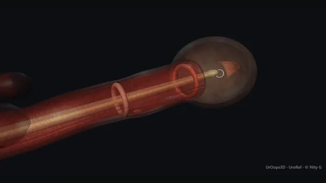
A 30-degree lens run down the urethra until the lumen pinholes, a guidewire across under vision, then a cold-knife cut at 12 o’clock where the spongiosum is thinnest.
The case is won by respecting the clock face and the depth of the cut, and by saying the word recurrence to the patient before consent.
Core tierFRCS urethra, stricture diseaseInteractive 3D
Along the male urethra on its curve, meatus to bladder. Schematic, not to scale; relationships only.
A sagittal view of the male urethra on its curve: from the meatus the scope rises over the bulbar bend where the stricture and the spongiosum sit, passes the membranous urethra and sphincter at the pelvic floor, and climbs through the prostate to the bladder.
The clock face is read against the spongiosum: 12 o’clock is the thin dorsal line, 6 o’clock is the bulk.
›Bulbar stricture - a fibrotic ring inside the spongiosum: the lumen pinholes and the surrounding tissue is pale and unyielding.
›Corpus spongiosum - envelops the bulbar urethra; thinnest dorsally at 12 o’clock, thickest ventrally with the supply entering laterally.
›Membranous urethra and sphincter - at the pelvic floor, proximal to the bulbar segment; the limit for instrument passage and incision.
›Corpora cavernosa - paired cylinders diverging anteriorly from the bulbar segment; the lateral no-go zone of the incision.
Cystoscopy to the strictureThe 30-degree lens runs from the meatus over the natural bulbar curve under continuous irrigation, reading the urethra as it goes until the lumen pinholes to a point. The scope stops there, and the pinhole position, appearance and distance from the meatus are noted: nothing crosses a stricture blind. The pale, unyielding ring around the narrowed lumen is what distinguishes true fibrosis from under-irrigation or spasm. Forcing the scope past a pinhole lumen makes a false passage, not a stricture, the problem. where it bites: forcing the scope past a pinhole lumen makes a false passage, not a stricture, the problem.
Wire through the lumenA guidewire is advanced under direct vision through the pinhole, threaded gently until it runs freely and coiled in the bladder, where its arrival is confirmed rather than assumed. The wire is the spine of the case: knife, scope and catheter are all railroaded over it, so access once gained is never surrendered. It should glide centrally through the pinhole without resistance, and its progress at the stricture is watched the whole way. A wire that stops or wanders off-axis is in the spongiosum, not the lumen. where it bites: a wire that stops or wanders off-axis is in the spongiosum, not the lumen.
Cold knife at 12 o’clockThe Sachse cold knife is mounted over the wire with its blade set to 12 o'clock before any cut is made, the one position where the spongiosum is thinnest and the lateral bulbar vessels are furthest away. The blade is advanced along the wire and the fibrotic ring divided in one controlled motion at the marked depth. The clock face is chosen on anatomy rather than convention: dorsally the blade meets fibrosis, laterally and ventrally it meets vascularised erectile bulk. A cut at 3 or 9 o'clock, or a dorsal cut driven too deep, opens the spongiosum and the view fills with blood. where it bites: cutting at 3 or 9 o’clock, or cutting too deep, opens the vascularised spongiosum.
Open the lumenThe incision is deepened cut by cut until the fibrotic ring springs open and the lumen visibly widens, then the scope is passed freely over the wire into the bladder to confirm the whole length to the bladder neck. The endpoint is a ring that gapes, not a channel that merely accepts the instrument. One clean cut at the right depth is the aim, not several ragged ones stacked in different planes. Over-deep division converts a stricture into a bleeding spongiosal bed and takes away the very view the next decision depends on. where it bites: over-deep division converts a stricture into a bleeding spongiosal bed.
CatheterA catheter is railroaded over the wire with the wire held taut, and the balloon is inflated only once the catheter is confirmed in the bladder, by seeing it coil under vision or by free return of urine. The catheter provides drainage, splints the incised urethral segment and can provide local compression through its shaft; the balloon is a retention balloon and remains in the bladder. Irrigation should run in and return freely, which is the final check before the case ends. Inflating a balloon not confirmed in the bladder, especially after a difficult wire passage, is the error this confirmation exists to prevent. where it bites: inflating a balloon that is not confirmed in the bladder, especially after a difficult wire passage.
Trial without catheterThe catheter comes out after a day or two, once the urine has cleared, and the first void is timed and measured rather than merely observed. That first flow rate, with a post-void residual, becomes the baseline against which every later flow is compared, anchoring follow-up to numbers from the first visit. The patient leaves with the plan and with the recurrence conversation already had: it happens at consent, not at re-presentation. A weakening stream then brings the patient back early, which is when a recurrence is most usefully treated.
The spatial relationships the animation is built to make automatic. Before you read each one, ask yourself: at this point in the operation, what is about to go wrong, and what is my first move? The save for each sits in the Complications tab.
Risk12 o’clock and the spongiosum - the only clock position where the blade meets fibrosis rather than vascularised bulk.
RiskMembranous urethra and external sphincter - the proximal limit of the bulbar segment; instrument torque and over-long incision threaten continence.
RiskDepth of incision - deep enough to spring the ring open, never so deep as to exit the spongiosum.
Trigger each of these live in the Animation tab, then read the save: the first move, the escalation trigger, and the line that goes in the op note.
Recurrence
the fibrotic process can outlive the incision; risk varies with length, site and prior treatment.
Selected primary, single, short, non-obliterative bulbar strictures have the best durable patency, while repetitive treatment increases complexity.
The save
Recognise it early: symptom review and flow rates from the first follow-up visit, not wait-and-see.first move
Re-stage the stricture before choosing treatment: length, calibre, site, prior intervention and fitness for reconstruction all change the answer.
Avoid repetitive DVIU or dilatation with curative intent when urethroplasty is viable; more than two endoluminal treatments increase stricture complexity and delay definitive repair.
A recurrent, long or obliterated stricture should be discussed with a reconstructive urethral service rather than automatically re-cut.
Retention with a known stricture: catheter over a wire under vision in theatre, or suprapubic drainage; never force a catheter.
Fistula, abscess or failed instrumentation → suprapubic drainage and specialist referral.escalation trigger
Op note: stricture length and site, number of endoscopic treatments to date, plan for urethroplasty referral.document
Corpus spongiosal bleeding
an incision at 3 or 9 o’clock, or a dorsal cut driven too deep, opens the vascularised erectile bulk around the urethra.
The save
Stop incising the moment the field fills; preserve the wire and restore controlled drainage.first move
Never deepen a lateral cut blind; if vision holds, complete only the 12 o’clock line.
Railroad the catheter over the wire, inflate its retention balloon in the bladder, and use the catheter shaft for local urethral compression while drainage is observed.
Persistent bleeding or an expanding perineal haematoma → return to theatre, imaging, and rarely open spongioplasty.escalation trigger
Op note: clock position and depth of the cut, bleeding encountered, catheter size and drainage plan, irrigation volumes.document
False passage
the wire or scope leaves the pinhole and tracks through the spongiosum beside the true lumen.
The save
Stop at the first resistance with off-axis progress: that combination is the false passage announcing itself.first move
Do not dilate, do not railroad, and do not repeat the same blind movement.
Withdraw to the last true view and re-find the lumen under vision, or image before any further passage.
Confirm wire position in the bladder before the balloon is inflated: a catheter balloon in a false passage destroys the reconstruction options.escalation trigger
Lumen not found → stop, suprapubic catheter, retrograde study and a planned definitive procedure.escalation trigger
Op note: instrument that left the lumen, direction of the track, how position was confirmed, drainage plan.document
When the case turns: the problem, then the next move.
ActionWire will not cross the pinhole - stop pushing; try a hydrophilic-tipped wire or flexible-scope assistance, and if it still will not cross, suprapubic drainage and imaging on a planned list.
ActionBleeding takes the view during the cut - pause, irrigate, preserve wire access and use the catheter shaft for local urethral compression; return to cutting only when the field is clear.
ActionCatheter will not railroad over the wire - check the wire is still coiled in the bladder under vision; re-position over a stiffer wire rather than pushing against resistance.
ActionLumen pinholes sooner than expected - the stricture is longer than the imaging suggested: stop and complete a retrograde urethrogram before cutting.
The examiner stems this scene prepares you for. Try to answer out loud first, then reveal, and learn the discriminator, the detail that turns a pass into a strong pass.
QDVIU or urethroplasty for a bulbar stricture?reveal answer
The decision is made on length, site, calibre and previous treatment.
Endoscopic urethrotomy is a reasonable first treatment for a primary, single, short, non-obliterative bulbar stricture.
In that selected group the EAU guideline reports a five-year stricture-free rate of up to 77%, so recurrence is a material risk but not an inevitable outcome.
DVIU should not be used as solitary treatment for a long stricture or a penile stricture.
If disease recurs, I would re-stage it and discuss urethroplasty rather than enter a cycle of repeated endoluminal treatment: more than two DVIU or dilatations should not be performed when urethroplasty is viable because repetition increases length and complexity and delays definitive repair.
In a frail patient who is unsuitable for reconstruction, a palliative endoluminal strategy may still be proportionate if its aim and follow-up are explicit.
I would map the stricture with flow assessment, retrograde imaging and endoscopy as appropriate before promising either option, and document the recurrence discussion in terms that match that individual stricture.
Strong pass
Selecting DVIU for a primary, single, short, non-obliterative bulbar stricture, and avoiding more than two endoluminal treatments when urethroplasty is viable.
QWhy does the Sachse knife cut at 12 o’clock?reveal answer
The clock face exists because the urethra is not symmetrical inside the corpus spongiosum.
Dorsally, at 12 o’clock, the spongiosum is at its thinnest and relatively avascular; ventrally and laterally the erectile bulk is thick and the bulbar supply enters at the lateral positions.
Cutting at 12 divides fibrosis with the least risk of bleeding into the spongiosum, and keeps the blade away from the corpora cavernosa and their neurovascular supply anteriorly.
The same logic dictates depth: the incision is carried through the fibrotic ring until it springs open, one clean cut, without exiting the spongiosum.
A lateral cut, or a dorsal cut driven too deep, opens vascularised erectile tissue: minor bleeding may settle with drainage and local compression from the catheter shaft, while persistent bleeding, fistula or infection requires escalation.
Position and depth are the whole operation; everything else is plumbing.
Strong pass
Say both
Linking 12 o’clock to spongiosal thickness and the lateral arterial supply
Not to convention alone
Examiner note
Linking 12 o’clock to spongiosal thickness and the lateral arterial supply, not to convention alone.
QA ward call: failed catheter insertion in a man with known stricture disease. Take me through it.reveal answer
A ward attempt that meets an obstruction at the bulbar urethra stops.
The first move is not to repeat blind passes, because each one tracks a false passage through the spongiosum and converts a catheter problem into a reconstruction problem.
I would ask about previous stricture disease, instrumentation or trauma, examine for a palpable bladder, and move to a controlled setting: flexible cystoscopy to the stricture, a guidewire through the true lumen under vision, and a catheter railroaded over the wire.
If the lumen cannot be cannulated, a suprapubic catheter under ultrasound control is the safe answer, not a stronger hand.
The patient then gets a stricture work-up with flow rates and a retrograde study, and a plan for definitive treatment rather than repeated attempts.
I would document the passes already made, because an established false passage changes both the imaging findings and the surgical plan.
Strong pass
Say both
Wire-over-vision or suprapubic as the only two options after one failed pass
No repeat blind attempts
Examiner note
Wire-over-vision or suprapubic as the only two options after one failed pass, and no repeat blind attempts.
QHow do you follow up a patient after an uncomplicated DVIU?reveal answer
The catheter drains and splints the incised segment, with its retention balloon in the bladder, and after an uncomplicated DVIU the EAU guideline advises removal within 72 hours rather than prolonged catheterisation.
The first void is timed and measured, and that flow rate is the baseline everything later is judged against.
Follow-up is symptom-led but objective: a flow rate and post-void residual at the first review, repeated if the stream weakens, because recurrence often declares itself within the first year.
At consent I explain that recurrence risk depends on stricture length, site and previous treatment, so a weakening stream brings the patient back early rather than late.
A symptomatic recurrence is re-staged and discussed with a reconstructive service; repeated DVIU is not a default pathway when urethroplasty is viable.
If retention recurs in the community, the plan written in the notes is a controlled wire-guided catheter or suprapubic drainage, never repeated blind passages.
Strong pass
Say all three
Catheter removal within 72 hours after an uncomplicated DVIU
An objective baseline flow
Re-staging rather than automatic repeat cutting if symptoms recur
Examiner note
Catheter removal within 72 hours after an uncomplicated DVIU, an objective baseline flow, and re-staging rather than automatic repeat cutting if symptoms recur.
Say it aloud, then reveal. Reading is not remembering.
01The relationshipSay the critical spatial relationship out loud, then reveal.reveal
Memory frame
A sagittal view of the male urethra on its curve
Why?
A sagittal view of the male urethra on its curve: from the meatus the scope rises over the bulbar bend where the stricture and the spongiosum sit, passes the membranous urethra and sphincter at the pelvic floor, and climbs through the prostate to the bladder. The clock face is read against the spongiosum: 12 o’clock is the thin dorsal line, 6 o’clock is the bulk.
02The first moveRecurrence. Your first move?reveal
First move
Recognise it early: symptom review and flow rates from the first follow-up visit, not wait-and-see.
03The escalation triggerRecurrence. What makes you call for help?reveal
Escalate when
Fistula, abscess or failed instrumentation → suprapubic drainage and specialist referral.
04The discriminatorWhat turns a pass into a strong pass?reveal
Strong pass
Selecting DVIU for a primary, single, short, non-obliterative bulbar stricture, and avoiding more than two endoluminal treatments when urethroplasty is viable.
05The pitfallWhere does this operation bite first?reveal
Pitfall
12 o’clock and the spongiosum
Why?
the only clock position where the blade meets fibrosis rather than vascularised bulk.
Revision practice with self-marked answers, not an assessment of clinical competence. Kept in
this browser only; it does not sync between devices.