
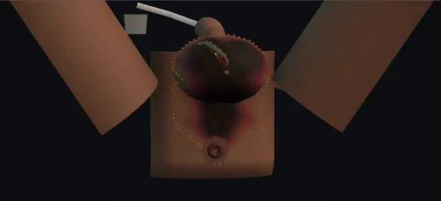
UrOops3D, danger-zone atlas

Schematic model.

Operative animation, Fournier gangrene debridement

Fournier gangrene: radical debridement, the second look and the reconstruction pathway

Emergency source control for necrotising fasciitis of the perineum and genitalia. Resuscitation and broad-spectrum antibiotics start at once, and the patient goes to theatre without waiting for scans.

The first incision goes over the worst area, and the excision is carried through every layer of dead tissue to bleeding, healthy tissue in every direction. The testes, urethra and anal sphincter are kept, the wound is packed open, and the patient returns for a second look.

Reconstruction waits for a clean, granulating wound. The lesson is that the fascia is always worse than the skin.

Advanced tier FRCS emergency urology, necrotising soft tissue infection and sepsis Interactive 3D

Operative visualisation

Schematic scene preview: Fournier gangrene: debridement
Preparing the 3D anatomy…

UrOops3D · UroRef · © Nity G

How to use this module. Play through the nine steps, or use Back and Next. Watch the grey fascia show beyond the black skin, the margin marked, the excision spread to bleeding edges, and the testes kept. The triggers show a margin that creeps on, a dead testis, a urethral tear and septic shock.

Click me next Section 2 of 8 Anatomy The structures this operation works between, and the one it must not cross.

The structures in the scene

Fournier gangrene debridement: how the structures relateThe superficial fascial plane of the perineum, and what lies round it. Colles, dartos and Scarpa fascia: dead beyond the visible skin change Skin: shows less than the fascia. Colles, dartos and Scarpa fascia: stripped off them; their supply is the cord Testes on their spermatic cords (danger). Colles, dartos and Scarpa fascia: over it in the perineum; the catheter marks it Bulbar urethra under bulbospongiosus (danger). Colles, dartos and Scarpa fascia: ends at its edge; the sphincter is kept Anal sphincter and rectum (danger). Colles, dartos and Scarpa fascia: superficial to them at the lateral margin Femoral vessels in the groin (danger). Colles, dartos and Scarpa fascia: continues onto it; healthy tissue for repair Medial thigh: pouches and graft donor.Colles, dartos andScarpa fasciaSkin: shows lessthan the fascia!Testes on theirspermatic cords!Bulbar urethra underbulbospongiosus!Anal sphincter andrectum!Femoral vessels inthe groinMedial thigh:pouches and graftdonordead beyond thevisible skin changestripped off them;their supply is thecordover it in theperineum; thecatheter marks itends at its edge;the sphincter iskeptsuperficial to themat the lateralmargincontinues onto it;healthy tissue forrepairTargetDangerLandmarkContext
The superficial fascial plane of the perineum, and what lies round it. Schematic, not to scale; relationships only.
  • Seen from the foot of a patient in lithotomy, the scrotum lies in front of the perineum, with the anus at the back of the field and the penis lying up on the abdomen.
  • Infection runs in the superficial fascia: Colles fascia in the perineum, the dartos of the scrotum and penis, and Scarpa fascia on the abdominal wall.
  • The deep fascia, the testes on their cords, the bulbar urethra and the anal sphincter lie under or beside that plane, which is why the excision follows the dead plane outward and keeps them.
  • ›Scrotal and perineal skin - shows only part of the disease: black, dusky or red over a much wider area of dead fascia.
  • ›Dartos and Colles fascia - the superficial perineal fascia in which the infection spreads; continuous with the dartos of the scrotum and penis.
  • ›Scarpa fascia - the same plane on the lower abdominal wall; the route of spread toward the groin and abdomen.
  • ›Testes and spermatic cords - supplied from above through the cord, so they usually survive the loss of the scrotal skin.
  • ›Bulbospongiosus and bulbar urethra - deep in the perineal wound in the midline, with the catheter inside the urethra.
  • ›Anal sphincter and rectum - at the back of the perineal wound; involvement suggests an anorectal source.
  • ›Femoral vessels - deep in the groin at the lateral edge of the excision.
  • ›Medial thighs - the site of the thigh pouches and a donor site for the split skin graft.
Click me next Section 3 of 8 Steps The operation in order, with the landmark that guards each move.

Operative steps

  1. The septic patient: resuscitate and decide A man, often diabetic, with pain out of proportion to the signs, fever, tachycardia and hypotension. The scrotum and perineum are swollen, tense and dusky, with black skin and crepitus. Blood cultures, broad-spectrum intravenous antibiotics and fluids start at once; theatre is booked without waiting for imaging.
    where it bites: the skin understates the disease; delay to theatre is the avoidable cause of death.
  2. Catheter, then the first incision over the worst area Under general anaesthesia in lithotomy, a urethral catheter is passed first: it drains the bladder, monitors output and marks the urethra in the perineum. The first incision goes straight over the worst area, through skin and swollen subcutaneous tissue down to the fascia.
    where it bites: a catheter that will not pass is a reason for a suprapubic catheter, not for force.
  3. The finger test and the margin A finger passes along the plane above the deep fascia with no resistance. The tissue is grey, the fluid thin, grey and foul (dishwater), and nothing bleeds. The skin looks better than the fascia beneath it, so the margin is marked where tissue stops giving way and bleeds. Pus and tissue go to microbiology.
    where it bites: judging the excision by the skin leaves dead fascia behind.
  4. Radical excision to bleeding tissue All dead skin, fat and fascia are excised, starting at the incision and working outward along Colles fascia onto the perineum and Scarpa fascia toward the groin and lower abdomen. The excision stops only where the edge bleeds and the fascia is healthy and adherent, in every direction.
    where it bites: the femoral vessels lie deep in the groin and the urethra deep in the perineum; the excision follows the dead plane, not a depth.
  5. Keep the testes, urethra and sphincter The testes are exposed but viable: their blood supply comes down the spermatic cord, not through the scrotal skin, so they are kept. In the perineum the bulbospongiosus lies over the urethra with the catheter inside it. The anal sphincter is preserved. Bleeding points are controlled with diathermy.
    where it bites: the spermatic cords, the bulbar urethra and the anal sphincter are the structures the excision must not take.
  6. Packed open; the second look booked The wound is washed out and packed open with moist gauze, the testes wrapped in it. The patient goes to intensive care. A second look in theatre is planned at 24 to 48 hours, and sooner if he deteriorates or the wound looks worse.
    where it bites: closure at the first operation traps residual infection.
  7. The second look In theatre again, the packs are removed and every edge is inspected. A residual corner of grey fascia is trimmed back to bleeding tissue, and the wound is packed again. Debridement is repeated as often as the wound needs, guided by the tissue, the patient and the culture results.
    where it bites: a margin that has moved on means the first excision was not enough or the infection is still spreading.
  8. Granulation and thigh pouches Once the sepsis has settled and the wound is clean and granulating, reconstruction is planned. When too little scrotal skin remains to cover them, each testis is tunnelled into a pouch under the skin of the upper medial thigh, where it lies covered and protected.
    where it bites: reconstruction on a wound that is still infected fails.
  9. Split skin graft, diversion only when needed A split skin graft taken from the thigh, meshed, is laid over the granulating wound; smaller scrotal defects close directly, and larger ones may need a flap. A suprapubic catheter or a defunctioning colostomy is kept for urethral or anorectal involvement or a wound that cannot be kept clean, not used by default.
    where it bites: graft loss follows infection, shear and a wound not ready for it.
Click me next Section 4 of 8 Danger zones Where this operation bites, and why it bites there.
Critical relationship

Danger zones

The spatial relationships the animation is built to make automatic. Before you read each one, ask yourself: at this point in the operation, what is about to go wrong, and what is my first move? The save for each sits in the Complications tab.

  • RiskUrethra and corpora beneath infected tissues - the bulbar urethra under the bulbospongiosus in the perineal wound; the catheter marks it.
  • RiskRectum and anal sphincter - at the back of the perineal wound; the sphincter is kept.
  • RiskSpermatic cords and testicular blood supply - at the upper edge of the scrotal excision; the testes live on them.
  • RiskMajor femoral and perineal vessels - deep in the groin laterally and in the perineum; the excision follows the dead plane, not a depth.
  • RiskThe margin itself - dead fascia extends beyond the skin change; the margin is where the tissue bleeds.
Practise it Take me into the scene Opens the animation at a danger moment and asks you to predict the mechanism and your first move before it reveals.
Click me next Section 5 of 8 Complications The save for each one: the first move, the escalation trigger, the op-note line.

Complications and immediate saves

Trigger each of these live in the Animation tab, then read the save: the first move, the escalation trigger, and the line that goes in the op note.

Inadequate first debridement

  • at the second look the necrosis has crept past the first edge: dusky skin and grey fascia beyond it, and the patient is not improving.
The save
  1. Back to theatre now and excise to a new bleeding, healthy margin in every direction. first move
  2. Send fresh tissue for culture and review the antibiotics with microbiology.
  3. Image the pelvis and abdomen for deeper, pararectal or retroperitoneal spread if the source is unclear.
  4. A margin that keeps moving, or spread onto the abdominal wall or into the pelvis, is a senior and multidisciplinary decision today. escalation trigger
  5. Op note: the extent of new necrosis, what was excised, specimens sent and the plan for the next look. document

Non-viable testis

  • the testis is black and its cord dark.
  • Because the testes live on their cords, this suggests infection reaching the cord from above.
The save
  1. Confirm it: the testis does not bleed when its tunica is incised. first move
  2. Remove only the non-viable testis; the viable one is kept.
  3. Look for a source above: retroperitoneal or intra-abdominal spread along the cord, by imaging and with the general surgeons. escalation trigger
  4. Record the finding and the orchidectomy, and tell the patient and family when he can hear it. document

Urethral injury

  • deep over the bulb, the catheter shows through a tear in the urethra, from the disease or from the excision.
The save
  1. Leave the catheter in: it is now the stent. Do not pass anything blindly. first move
  2. Divert the urine with a suprapubic catheter so the perineal wound stays dry.
  3. Formal repair waits for a clean wound and a reconstructive urologist; a small clean tear may be closed over the catheter. escalation trigger
  4. Op note: where the tear is, how long, what was done and the catheter plan. document

Systemic collapse

  • during the excision the blood pressure falls, the pulse and lactate rise and the skin mottles: septic shock.
The save
  1. Tell the anaesthetist and keep going with source control; the dead tissue is the cause. first move
  2. Complete the sepsis six: oxygen, blood cultures, intravenous antibiotics, fluids, lactate and urine output.
  3. Vasopressors through the anaesthetist; shorten the operation to what controls the source, pack and plan an earlier look.
  4. Call intensive care now; he goes there from theatre. escalation trigger
  5. Record the timing of antibiotics, fluids and vasopressors, and what was left for the next look. document
Click me next Section 6 of 8 Troubleshooting When the case turns: the problem, then the next move.

Intra-operative troubleshooting

When the case turns: the problem, then the next move.

  • ActionThe catheter will not pass - stop and place a suprapubic catheter; a false passage in an infected perineum adds a urethral injury to the disease.
  • ActionPerianal or pararectal spread - image to define it; the EAU guideline notes that pararectal involvement may need bowel diversion. Otherwise a faecal management system keeps the wound clean.
  • ActionSpread onto the abdominal wall - follow Scarpa fascia up as far as it is dead; extensive abdominal or pelvic involvement needs general surgery in theatre.
  • ActionNot sure where the margin is - use the finger: tissue that separates easily and does not bleed is dead. Healthy fascia is adherent and bleeds.
  • ActionAdjuncts: hyperbaric oxygen, negative pressure, honey - the EAU guideline advises against adjunctive treatments outside trials; negative pressure dressings are used by many units once the wound is clean.
  • ActionMost of the scrotum is gone - thigh pouches protect the testes while the wound granulates; a scrotal defect of under half may close directly, larger ones need a graft or flap.
Click me next Section 7 of 8 FRCS viva Say it out loud, then mark yourself against the model answer.

FRCS viva questions

The examiner stems this scene prepares you for. Try to answer out loud first, then reveal, and learn the discriminator, the detail that turns a pass into a strong pass.

QA diabetic man has a painful, swollen scrotum and a small black patch on the perineum. How do you make the diagnosis, and how do you judge his severity?reveal answer
  • I would make it clinically.
  • The features are pain out of proportion to the signs, swelling and erythema of the scrotum or perineum, dusky or black skin, crepitus and a foul discharge, in a patient who is often diabetic, obese or immunocompromised.
  • The EAU guideline warns that in up to 40 per cent of cases the onset is insidious, so I keep a high index of suspicion.
  • If he is unwell and the signs are there, I take him to theatre; imaging must not delay that, although CT is useful to define deep or pararectal spread when it can be done without delay.
  • For severity I would use the Fournier gangrene severity index, which Laor and colleagues built from the physiological and biochemical variables of APACHE II.
  • In their series a score above 9 carried a 75 per cent probability of death, and a score of 9 or less a 78 per cent probability of survival.
  • It describes how sick he is and helps prognosis and counselling; it does not decide whether he needs surgery.
Strong pass

Say both

  • Saying that the score predicts and the clinical diagnosis decides
  • Not waiting for a scan
Examiner note
Saying that the score predicts and the clinical diagnosis decides, and not waiting for a scan.
QWhat organisms do you expect, and what antibiotics do you start?reveal answer
  • It is a polymicrobial synergistic infection: aerobic Gram-negative bacilli such as E.
  • coli, streptococci and staphylococci, and anaerobes such as Bacteroides and clostridia.
  • The source is usually the urinary tract, the anorectum or the skin.
  • I would take blood cultures and then start broad-spectrum intravenous antibiotics at once, as the EAU guideline recommends strongly, covering all the probable organisms.
  • Its suggested combinations include a broad-spectrum penicillin or a third-generation cephalosporin with gentamicin and metronidazole or clindamycin, or piperacillin-tazobactam with vancomycin, or a carbapenem, following local policy.
  • Clindamycin is often included for its effect on toxin production.
  • I would refine the regimen with microbiology once the tissue cultures from theatre are back.
  • But antibiotics do not penetrate dead tissue, so they support the operation and never replace it.
Strong pass

Say both

  • Calling it polymicrobial synergistic infection
  • Saying that the antibiotics do not reach dead tissue
Examiner note
Calling it polymicrobial synergistic infection, and saying that the antibiotics do not reach dead tissue.
QHe is hypotensive and his lactate is raised. In what order do you do things, and how far do you debride?reveal answer
  • Resuscitation and surgery run together, not one after the other.
  • I start the sepsis six, give antibiotics and fluids, involve the anaesthetist and intensive care early, and take him to theatre as soon as possible: the EAU guideline recommends debridement within 24 hours and says it must be complete.
  • In theatre I place a catheter, make the first incision over the worst area and use my finger: dead fascia separates easily, yields grey dishwater fluid and does not bleed.
  • The internal necrosis is usually far greater than the skin suggests, so I excise all dead skin, fat and fascia in every direction along Colles and Scarpa fascia until every edge bleeds.
  • I keep the testes, which live on their cords, the urethra and the sphincter.
  • I pack the wound open and plan a second look at 24 to 48 hours, sooner if he worsens, and I keep going back until there is no dead tissue.
Strong pass

Say both

  • Excising to bleeding tissue rather than to the skin change
  • Planning the return before leaving theatre
Examiner note
Excising to bleeding tissue rather than to the skin change, and planning the return before leaving theatre.
QWhen would you divert the urine or the faeces?reveal answer
  • Not routinely.
  • A urethral catheter is enough for most patients: it drains the bladder, lets me measure output and marks the urethra.
  • I would place a suprapubic catheter if a urethral catheter cannot be passed, if the urethra is injured or is the source, as with a stricture or a periurethral abscess, or if there is extensive penile or perineal involvement.
  • The EAU guideline names suprapubic catheterisation as the route for urinary diversion.
  • For the bowel, the guideline says that pararectal involvement may need diversion.
  • I would consider a defunctioning colostomy with the general surgeons when the anal sphincter or rectum is involved, or faecal soiling of a large perineal wound cannot be controlled.
  • Short of that, a faecal management system can keep the wound clean without a stoma.
Strong pass

Say all three

  • Diverting for a reason
  • The urethra or the anorectum
  • Naming the less invasive options first
Examiner note
Diverting for a reason, the urethra or the anorectum, and naming the less invasive options first.
QWhat is his chance of survival, and how will the wound be reconstructed?reveal answer
  • Case series from referral centres report mortality of 20 to 40 per cent, but the population figures are lower.
  • In the State Inpatient Databases study by Sorensen and colleagues, 1,641 men, the case fatality rate was 7.5 per cent, and the EAU guideline summarises three registry studies with mortality of 10, 7.5 and 5 per cent.
  • Mortality rises with age, comorbidity, renal failure and coagulopathy, and in that study hospitals treating more than one case a year had lower mortality, which supports care in experienced centres.
  • Reconstruction waits for a clean, granulating wound.
  • The EAU guideline recommends closure, primary or secondary, for scrotal defects up to 50 per cent, and flaps or skin grafts for larger defects or those beyond the scrotum.
  • When the testes are exposed they can be placed in thigh pouches and the wound covered with a meshed split skin graft.
  • I would also plan for the late effects on sexual and urinary function and his psychological recovery.
Strong pass

Say both

  • Quoting the population mortality with its source rather than the case-series figures alone
  • Staging the reconstruction
Examiner note
Quoting the population mortality with its source rather than the case-series figures alone, and staging the reconstruction.
Click me next Section 8 of 8 Evidence The guidance each of these answers is built on.

Evidence and guidance

Operative Dictations in Urologic Surgery
Armenakas NA, Fracchia JA, Golan R. Operative Dictations in Urologic Surgery. Wiley, 2019
The book has no chapter on Fournier gangrene; the owner's PDF was searched for Fournier and necrotising fasciitis on 19 October 2026 with no result. Nothing on this page follows the book. The order of the steps is the hub sequence, and the clinical statements come from the EAU guideline and the papers below. General practice, from none of them: the urethral catheter first, the finger test, the testes kept on their cords, a second look at 24 to 48 hours, thigh pouches and a meshed graft, and every first, escalate and document step.
Open guideline
EAU
European Association of Urology. Guidelines on Urological Infections, 2026. Section 3.13, Fournier's gangrene, read 19 October 2026
An aggressive, frequently fatal polymicrobial infection of the perineum, perianal region and external genitalia, a subcategory of necrotising fasciitis. Three US registry studies of 650, 1,641 and 9,249 cases reported mortality of 10, 7.5 and 5 per cent. Onset is insidious in up to 40 per cent. Internal necrosis is usually far greater than the external signs suggest. Debridement early (within 24 hours), complete and repeated (strong); broad-spectrum antibiotics on presentation, refined by culture (strong). Suprapubic catheterisation for urinary diversion; bowel diversion may be needed for pararectal involvement. Closure for scrotal defects up to 50 per cent; flaps or grafts beyond that. Adjunctive treatments only within trials (weak).
Open guideline
The Journal of Urology
Laor E, Palmer LS, Tolia BM, Reid RE, Winter HI. Outcome prediction in patients with Fournier's gangrene. J Urol 1995;154:89-92
Abstract read 19 October 2026. 30 patients over 15 years, 13 deaths. APACHE II modified into the Fournier's gangrene severity index. Mean index 6.9 in survivors and 13.5 in non-survivors. A score above 9 carried a 75 per cent probability of death; 9 or less a 78 per cent probability of survival. Physiological derangement, not the extent of disease or the number of debridements, predicted outcome.
Open guideline
The Journal of Urology
Sorensen MD, Krieger JN, Rivara FP, et al. Fournier's gangrene: population based epidemiology and outcomes. J Urol 2009;181:2120-2126
Abstract read 19 October 2026. US State Inpatient Databases: 1,641 males and 39 females. Incidence 1.6 per 100,000 males, peaking at 3.3 per 100,000 between 50 and 79 years. Case fatality 7.5 per cent, lower than the 20 to 40 per cent of case series from referral centres. Most hospitals treat fewer than one case a year.
Open guideline
The Journal of Urology
Sorensen MD, Krieger JN, Rivara FP, Klein MB, Wessells H. Fournier's gangrene: management and mortality predictors in a population based study. J Urol 2009;182:2742-2747
Abstract read 19 October 2026. 1,641 men at 593 hospitals. Mortality rose with age, comorbidity, congestive heart failure, renal failure, coagulopathy and admission by transfer. Hospitals treating more than one case a year had lower mortality after adjustment, supporting regional care.
Open guideline
Therapeutic Advances in Urology
Chennamsetty A, Khourdaji I, Burks F, Killinger KA. Contemporary diagnosis and management of Fournier's gangrene. Ther Adv Urol 2015;7:203-215
Abstract read 19 October 2026. Obliterative endarteritis of the subcutaneous arteries with gangrene of the overlying skin; a synergistic polymicrobial infection from the genitourinary tract, lower gastrointestinal tract or skin. Diagnosis is clinical; the FGSI grades severity. Rapid radical debridement, resuscitation and broad-spectrum antibiotics; colostomy or a faecal management system for severe perineal involvement; reconstruction for the large defects that follow.
Open guideline

Curriculum. Supports learning relevant to the ISCP Urology Curriculum critical condition Fournier's Gangrene. Not a workplace-based assessment and not evidence of competence.

Two minutes

Close the lap: recall it before you leave.

Say it aloud, then reveal. Reading is not remembering.

  1. 01 The relationship Say the critical spatial relationship out loud, then reveal. reveal
    Memory frame

    Seen from the foot of a patient in lithotomy, the scrotum lies in front of the perineum, with the anus at the back of the field and the penis lying up on the abdomen.

    Why?
    Infection runs in the superficial fascia: Colles fascia in the perineum, the dartos of the scrotum and penis, and Scarpa fascia on the abdominal wall. The deep fascia, the testes on their cords, the bulbar urethra and the anal sphincter lie under or beside that plane, which is why the excision follows the dead plane outward and keeps them.
  2. 02 The first move Inadequate first debridement. Your first move? reveal
    First move

    Back to theatre now and excise to a new bleeding, healthy margin in every direction.

  3. 03 The escalation trigger Inadequate first debridement. What makes you call for help? reveal
    Escalate when

    A margin that keeps moving, or spread onto the abdominal wall or into the pelvis, is a senior and multidisciplinary decision today.

  4. 04 The discriminator What turns a pass into a strong pass? reveal
    Strong pass

    Say both

    • Saying that the score predicts and the clinical diagnosis decides
    • Not waiting for a scan
    Examiner note
    Saying that the score predicts and the clinical diagnosis decides, and not waiting for a scan.
  5. 05 The pitfall Where does this operation bite first? reveal
    Pitfall

    Urethra and corpora beneath infected tissues

    Why?
    the bulbar urethra under the bulbospongiosus in the perineal wound; the catheter marks it.

Revision practice with self-marked answers, not an assessment of clinical competence. Kept in this browser only; it does not sync between devices.