
UrOops3D, danger-zone atlas
Operative animation, GreenLight laser vaporisation

GreenLight photoselective vaporisation of the prostate

The laser that boils the adenoma away instead of cutting it: a 532 nanometre beam absorbed by haemoglobin, fired sideways from a fibre held a millimetre or two off the tissue, in saline, with almost no bleeding.

This page is for the man on anticoagulants and the trainee on the fibre: the goggles, the distance, the bubbles, the sequence, and the endpoint that is the same as the loop's.

Advanced tier FRCS endourology, BPH / LUTS and the alternatives to TURP Interactive 3D

Operative visualisation

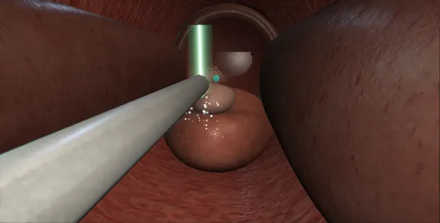
Schematic scene preview: GreenLight laser vaporisation
Preparing the 3D anatomy…

UrOops3D · UroRef · © Nity G

Click me next Section 2 of 8 Anatomy The structures this operation works between, and the one it must not cross.

The structures in the scene

GreenLight laser vaporisation: how the structures relateAlong the prostatic urethra with the side-firing fibre, apex to bladder. Sphincter, beyond the veru: never fire beyond the veru Verumontanum (danger). Verumontanum: every apical sweep stops at the veru Median and lateral lobes. Median and lateral lobes: no more bubbles: the endpoint Surgical capsule: pale fibrous floor. Median and lateral lobes: a median lobe puts the orifices in the beam Bladder neck and the orifices (danger).Distal: penile endProximal: bladder!Sphincter, beyond the veruVerumontanumMedian and lateral lobesSurgical capsule:pale fibrousfloor!Bladder neck and theorificesnever fire beyond the veruevery apical sweep stops atthe veruno more bubbles:the endpointa median lobe puts theorifices in the beamTargetDangerLandmark
Along the prostatic urethra with the side-firing fibre, apex to bladder. Schematic, not to scale; relationships only.
  • The fibre sits in the working channel just ahead of the lens and fires its beam sideways, so the tissue being vaporised is at the edge of the endoscopic view at the clock position the fibre has been turned to.
  • The median lobe is taken from the neck toward the veru, each lateral lobe from the neck to the apex, the apex last, each sweep drawn back to the veru;
  • the bubbles show the beam is at the right distance and the pale fibrous floor shows it has reached the capsule.
  • ›The fibre window and the aiming beam - the beam fires sideways at an angle to the shaft; the red spot shows where it will land.
  • ›The distance - one to two millimetres off the tissue vaporises; touching chars; too far coagulates.
  • ›Verumontanum and sphincter - the distal limit and the structure beyond it; every apical sweep stops at the veru.
  • ›Surgical capsule - a pale fibrous floor with no more bubbles: the endpoint.
  • ›Bladder neck and the orifices - the dangerous end when a median lobe reaches the trigone.
  • ›The coagulated bed - a thin layer that sloughs over weeks: the reason for the dysuria and the pink urine.
  • ›The laser room - goggles on everyone, the sign on the door, standby with the foot off the pedal.
Click me next Section 3 of 8 Steps The operation in order, with the landmark that guards each move.

Operative steps

  1. Who gets a laser The man on anticoagulants or antiplatelets who cannot safely stop them, the poor heart that cannot take blood loss or fluid absorption, the gland up to about eighty grams, and the man who wants to go home the same day. Vaporisation bleeds less than a loop, runs in saline so there is no TUR syndrome, and needs a shorter catheter time. The trade-offs are no tissue for histology, so the PSA and examination must be reassuring first; storage symptoms and dysuria for some weeks as the coagulated bed sloughs; and a longer operation for a large gland, where enucleation is the better laser.
  2. Consent and laser safety The consent names retrograde ejaculation, storage symptoms and dysuria for weeks, haematuria as the bed sloughs, infection, retention after the catheter comes out, the small risks of incontinence and stricture, no histology, and retreatment in a proportion over the years. Laser safety before anything else: the sign on the door, wavelength-specific goggles on everyone in the room including the patient, the fibre never fired outside the body or at the telescope, the laser on standby whenever the foot is off the pedal, and a named laser safety officer.
  3. The stack and the fibre A 23 or 26 French continuous-flow laser cystoscope with a working channel, a 30 degree telescope, saline irrigation by gravity, and the 180 watt lithium triborate laser with its side-firing fibre. The fibre has a window near its tip that fires the beam at an angle to the shaft, and a red aiming beam that shows where the treatment beam will land. The generator has a vaporisation setting and a coagulation setting at lower power; the fibre is fed through the channel and its window orientation is checked on the screen before the pedal is touched.
  4. Cystoscopy: the landmarks The sheath goes in under vision. The sphincter, the verumontanum as the distal limit, the lobes, the bladder neck, and in the bladder both ureteric orifices, measured against the fibre: a median lobe that reaches back to the trigone makes the neck the dangerous end of the operation, because the beam swept across the neck reaches the orifice. Any tumour or stone in the bladder is dealt with or noted before the laser starts.
    where it bites: a median lobe that sits against the trigone puts the orifices in the path of the beam at the neck.
  5. The distance The whole technique is the distance between the fibre window and the tissue. At one to two millimetres the beam vaporises: the tissue disappears with a cloud of bubbles and a smooth bed is left. Touching the tissue chars the fibre tip and coagulates rather than vaporises, and the char then absorbs the beam and destroys the fibre. Too far away the energy is spread and coagulates a thick layer that sloughs later, with more storage symptoms and no channel. The fibre is swept side to side and the bubbles are watched, not the power display. The beam is fired only at tissue, never into water or at the scope.
    where it bites: touching chars and bleeds; too far coagulates and removes nothing; the bubbles are the feedback.
  6. The median lobe and the working space The median lobe goes first, from the bladder neck toward the veru, swept in layers so that the vaporisation front moves as a plane and no trench is dug. Creating working space at the neck first lets the scope move and the irrigant flow. The bed is levelled toward the veru until the capsule shows as a pale fibrous floor with no more bubbles: the endpoint is the same as the loop's, and it is recognised the same way. The neck fibres are respected and the orifices kept out of the sweep.
  7. The lateral lobes The right lobe from the neck to the apex, the fibre rotated so the window faces the tissue at nine and ten o'clock, sweeping and retracting in layers; then the left at two and three. Each lobe is taken until the capsule shows across the sector. A vessel that bleeds is coagulated by pulling the fibre back a few millimetres so the beam defocuses, or by changing to the coagulation setting, and never by pressing the fibre onto it. Large glands take time: an hour of laser for eighty grams is usual, and the fibre may need changing.
  8. The apex The apex is last and at the lowest power. Every sweep is drawn back toward the verumontanum and stops at it, never firing distal to the veru. The roof at twelve o'clock, where the two lobes met, is taken in short sweeps toward the veru. Then the scope is withdrawn into the bulbar urethra and, still looking forward, the sphincter ring is seen to close ahead with the veru intact beyond it. A small apical remnant is accepted; the sphincter is not touched.
    where it bites: the beam fired distal to the veru enters the sphincter; every apical sweep is drawn back toward the veru and stops at it.
  9. The end, the catheter and the note The irrigation is slowed and the cavity inspected: a dry, pale, coagulated bed with no chips to evacuate, both orifices seen again with their jets. A 20 French catheter goes in, usually without irrigation, and comes out the next morning or the same day. The note records the total energy in kilojoules, the fibre used and whether it was changed, the lasing time, the landmarks seen intact, the catheter and the plan, and the patient is warned about the weeks of dysuria and the sloughing that brings pink urine.
Click me next Section 4 of 8 Danger zones Where this operation bites, and why it bites there.
Critical relationship

Danger zones

The spatial relationships the animation is built to make automatic. Before you read each one, ask yourself: at this point in the operation, what is about to go wrong, and what is my first move? The save for each sits in the Complications tab.

  • RiskThe orifices behind a median lobe - in the path of the beam when the neck is swept; measured before the laser starts.
  • RiskDistal to the veru - the sphincter; every apical sweep is drawn back toward the veru and never beyond it.
  • RiskThe fibre touching the tissue - char, a destroyed fibre, and a vessel opened that the beam then cannot see through.
  • RiskThe beam fired at nothing - into water or at the telescope: a burnt scope, a fire, an eye.
Practise it Take me into the scene Opens the animation at a danger moment and asks you to predict the mechanism and your first move before it reveals.
Click me next Section 5 of 8 Complications The save for each one: the first move, the escalation trigger, the op-note line.

Complications and immediate saves

Trigger each of these live in the Animation tab, then read the save: the first move, the escalation trigger, and the line that goes in the op note.

Bleeding from a touched vessel

  • the fibre touched the tissue and opened a vessel; the field is red and the beam cannot see through it.
The save
  1. Pull the fibre back a few millimetres and slow the irrigation until the point can be seen. first move
  2. Coagulate with the beam defocused or on the coagulation setting; never press the fibre onto the vessel.
  3. A field that will not clear: change to a resectoscope with a bipolar loop or button and coagulate under vision.
  4. Bleeding that continues: catheter and traction, blood, senior help. escalation trigger
  5. Note: the point, its control, and the catheter plan. document

Orifice in the beam

  • the median lobe taken back to the neck and the beam swept across the right orifice.
The save
  1. Stop firing at the neck. first move
  2. Find the orifice; cannulate and stent it for four to six weeks if it can be found.
  3. If it cannot be found: ultrasound at two weeks and three months for obstruction.
  4. Loin pain, fever or a rising creatinine: image and drain by nephrostomy. escalation trigger
  5. Note: which orifice, stented or not, and the imaging plan. document

Fired beyond the veru

  • the apical sweep carried distal to the verumontanum into the sphincter.
The save
  1. Stop. Withdraw into the bulbar urethra and, looking forward, check the sphincter ring. first move
  2. No further energy at the apex in that sector; accept the remnant.
  3. Catheter as planned; tell the patient before the trial without catheter; pelvic floor physiotherapy from the start.
  4. Incontinence persisting beyond six to twelve months: urodynamics and a continence surgeon. escalation trigger
  5. Note: the sector, the ring seen afterwards, and the plan for review. document

Capsular perforation

  • the beam held too long in one place has gone through the capsule: fat, and irrigant leaving.
The save
  1. Stop in that sector; keep the scope still and re-orient. first move
  2. Haemostasis at the breach with the beam defocused.
  3. A small extraperitoneal perforation: complete or curtail, catheter for a week.
  4. A large deficit, distension or peritonism: imaging, a drain, senior help. escalation trigger
  5. Note: the site, the deficit, the catheter and drainage plan. document

Retention after the catheter comes out

  • oedema of the coagulated bed: a failed trial without catheter on the first day.
The save
  1. Recatheterise; it is the bed, not the operation, that has failed for now. first move
  2. An alpha-blocker, and a trial without catheter in a week as the bed sloughs.
  3. A second failure: flexible cystoscopy for a residual lobe, a clot or a stricture. escalation trigger
  4. Note: the residual, the plan, and the date of the next trial. document
Click me next Section 6 of 8 Troubleshooting When the case turns: the problem, then the next move.

Intra-operative troubleshooting

When the case turns: the problem, then the next move.

  • ActionNo bubbles - too far from the tissue or the window facing the wrong way: check the aiming beam and close the distance.
  • ActionThe tip is black - the fibre has touched tissue and charred: pull back, clean or change the fibre; do not keep firing through char.
  • ActionThe vaporisation is slow - a fibrous or previously treated gland, a degraded fibre, or the power turned down for the apex; check each.
  • ActionThe field is red - a touched vessel: defocus and coagulate; if it will not clear, change to a resectoscope.
  • ActionLost orientation - back to the veru with the laser on standby; nothing is fired until the landmarks are seen.
  • ActionThe gland is bigger than the ultrasound said - accept a longer operation with a fibre change, or convert to enucleation or resection with a senior decision.
Click me next Section 7 of 8 FRCS viva Say it out loud, then mark yourself against the model answer.

FRCS viva questions

The examiner stems this scene prepares you for. Try to answer out loud first, then reveal, and learn the discriminator, the detail that turns a pass into a strong pass.

QA 78-year-old man on warfarin for a mechanical valve has retention with a 60 g prostate. Which operation and why?reveal answer
  • The problem is a man who needs outlet surgery but cannot safely stop anticoagulation, so the operation chosen should bleed as little as possible and should run in saline.
  • I would offer photoselective vaporisation with the GreenLight laser, which can be done with anticoagulation continued or bridged, has a low transfusion rate compared with TURP, uses saline so there is no TUR syndrome, and suits a 60 g gland.
  • Holmium or thulium enucleation is the alternative for a large gland and is also haemostatic, and bipolar TURP in saline is possible with bridging but bleeds more.
  • I would coordinate the anticoagulation with the cardiologist and haematologist, with a bridging plan around the valve rather than a long interruption.
  • I would counsel him about no histology, so I would want a reassuring PSA and examination first, about storage symptoms and dysuria for some weeks as the bed sloughs, retrograde ejaculation, the risk of retention at the trial without catheter, and retreatment in a proportion over the years.
  • Catheter time is short and most go home within a day.
Strong pass

Say all three

  • Choosing the laser for its haemostasis and saline
  • Coordinating the anticoagulation with cardiology rather than stopping it
  • Naming the no-histology caveat
Examiner note
Choosing the laser for its haemostasis and saline, coordinating the anticoagulation with cardiology rather than stopping it, and naming the no-histology caveat.
QExplain how the GreenLight laser works and what determines whether it vaporises or coagulates.reveal answer
  • GreenLight is a 532 nanometre laser, produced by frequency-doubling a neodymium or lithium triborate source, which is strongly absorbed by oxyhaemoglobin and poorly absorbed by water.
  • The energy is therefore deposited in the vascular prostatic tissue rather than the irrigant, and at high power density it heats the intracellular water above boiling point instantly, vaporising the tissue with a visible cloud of bubbles and leaving a thin coagulated layer of about one to two millimetres that gives haemostasis.
  • The power density depends on the power setting and, above all, on the distance from the fibre window to the tissue: at one to two millimetres the beam is concentrated and vaporises;
  • further away the same energy is spread over a larger area, the power density falls below the vaporisation threshold, and the tissue is coagulated in depth instead, which removes nothing and sloughs later.
  • Touching the tissue chars the fibre tip; the char absorbs the beam and the fibre degrades.
  • So the surgeon controls the effect with distance and sweep, watching the bubbles as the feedback, and uses a lower power or a deliberately defocused beam for coagulation.
Strong pass

Say both

  • Absorption by haemoglobin not water
  • Power density set by distance as the switch between vaporisation and coagulation
Examiner note
Absorption by haemoglobin not water, and power density set by distance as the switch between vaporisation and coagulation.
QWhat is your laser safety protocol in theatre?reveal answer
  • Laser safety is a named responsibility, with a laser protection adviser for the hospital and a laser safety officer in the room.
  • Before the case the sign goes on the door and access is controlled, the windows are covered, and everyone in the room, including the patient, wears goggles specific to the wavelength;
  • for 532 nanometres those are orange filters and they are checked for the marking.
  • The laser is kept on standby whenever the foot is off the pedal, is only made ready when the fibre is in the patient and the aiming beam is seen on tissue, and is never fired outside the body, into air, at the telescope or at a drape.
  • The fibre is inspected for damage and changed if the tip is charred, and it is handled so it cannot break in the channel.
  • Flammable preparations are avoided near the field.
  • Any injury or near miss is reported.
  • I would also confirm before starting that the irrigant is saline, the generator settings are correct for the fibre, and that the whole team has done the laser safety training.
Strong pass

Say all four

  • Naming the safety officer
  • Wavelength-specific goggles including the patient
  • Standby with the foot off the pedal
  • Never firing outside tissue
Examiner note
Naming the safety officer, wavelength-specific goggles including the patient, standby with the foot off the pedal, and never firing outside tissue.
QHow do you deal with bleeding during a GreenLight vaporisation?reveal answer
  • Bleeding during vaporisation usually means the fibre has touched the tissue or a larger vessel has been opened faster than the beam can seal it.
  • I would first pull the fibre back a few millimetres and slow the irrigation so the point can be seen, since a red field absorbs the beam before it reaches the vessel.
  • I would coagulate by defocusing: holding the fibre further from the tissue so the power density falls into the coagulation range, or switching the generator to its coagulation setting, and I would never press the fibre onto the vessel, which chars it.
  • Most bleeding stops this way.
  • If the field cannot be cleared, I would exchange the laser scope for a resectoscope and coagulate with a bipolar loop or button under direct vision, and if bleeding still continued I would place a three-way catheter with traction and irrigation, check the haemoglobin and clotting, and seek senior help, with embolisation as the further option.
  • I would document the bleeding point and its control.
Strong pass

Say both

  • Defocus rather than contact for coagulation
  • The willingness to change to a resectoscope when the laser cannot see the point
Examiner note
Defocus rather than contact for coagulation, and the willingness to change to a resectoscope when the laser cannot see the point.
Click me next Section 8 of 8 Evidence The guidance each of these answers is built on.

Evidence and guidance

Two minutes

Close the lap: recall it before you leave.

Say it aloud, then reveal. Reading is not remembering.

  1. 01 The relationship Say the critical spatial relationship out loud, then reveal. reveal
    Memory frame

    The fibre sits in the working channel just ahead of the lens and fires its beam sideways, so the tissue being vaporised is at the edge of the endoscopic view at the clock position the fibre has been turned to.

    Why?
    The median lobe is taken from the neck toward the veru, each lateral lobe from the neck to the apex, the apex last, each sweep drawn back to the veru; the bubbles show the beam is at the right distance and the pale fibrous floor shows it has reached the capsule.
  2. 02 The first move Bleeding from a touched vessel. Your first move? reveal
    First move

    Pull the fibre back a few millimetres and slow the irrigation until the point can be seen.

  3. 03 The escalation trigger Bleeding from a touched vessel. What makes you call for help? reveal
    Escalate when

    Bleeding that continues: catheter and traction, blood, senior help.

  4. 04 The discriminator What turns a pass into a strong pass? reveal
    Strong pass

    Say all three

    • Choosing the laser for its haemostasis and saline
    • Coordinating the anticoagulation with cardiology rather than stopping it
    • Naming the no-histology caveat
    Examiner note
    Choosing the laser for its haemostasis and saline, coordinating the anticoagulation with cardiology rather than stopping it, and naming the no-histology caveat.
  5. 05 The pitfall Where does this operation bite first? reveal
    Pitfall

    The orifices behind a median lobe

    Why?
    in the path of the beam when the neck is swept; measured before the laser starts.

Revision practice with self-marked answers, not an assessment of clinical competence. Kept in this browser only; it does not sync between devices.