UrOops3D, danger-zone atlas

Schematic infant anatomy: every dimension is an authoring estimate.

Operative animation, MAGPI hypospadias repair

Hypospadias repair: meatal advancement and glanuloplasty (MAGPI)

Indications

  • Distal hypospadias with the meatus at the corona.
  • No chordee on the artificial erection.
  • A glanular groove that is not deep and wide.

Alternatives

  • Tubularised incised plate repair: the usual choice for distal hypospadias today.
  • Glans approximation procedure, where the glanular groove is deep and wide.
  • Other distal repairs, the Mathieu repair among them.

Risks

  • Bleeding; infection.
  • Dehiscence of the glans closure.
  • Meatal stenosis.
  • Retraction of the meatus toward the corona.
  • Urethral stricture.

A repair for the boy whose meatus opens at the corona on a straight penis. No urethra is built and nothing is tubularised. A vertical cut through the bridge of glans tissue beyond the meatus is closed transversely, which carries the dorsal lip toward the tip.

The ventral lip is then lifted on a hook and the glans wings are closed under it in three layers. The result depends on choosing the right glans and on a solid glans closure: the failures that belong to this operation are a meatus that slips back and a meatus that narrows.

Advanced tier FRCS paediatric urology, hypospadias, genital reconstruction Interactive 3D

Operative visualisation

Schematic scene preview: MAGPI hypospadias repair
Preparing the 3D anatomy…

UrOops3D · UroRef · © Nity G

How to use this module. Play the eight steps, or use Back and Next. Follow the meatus: its dorsal lip travels as the vertical cut is sewn across, its ventral lip when the hook lifts it, and the glans then closes under it, layer by layer. The triggers show stenosis, retraction and dehiscence.

Click me next Section 2 of 8 Anatomy The structures this operation works between, and the one it must not cross.

The structures in the scene

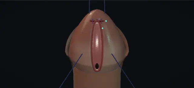
MAGPI hypospadias repair: how the structures relateThe ventral midline of the glans, and what the meatus is moved past. Meatus at the corona: just distal: cut vertically, closed transversely Bridge of glans tissue (dorsal web). Meatus at the corona: the dorsal lip is advanced into it Glanular groove, running to the tip. Meatus at the corona: closed under its lifted ventral edge, three layers Glans wings, one each side. Meatus at the corona: runs up to it directly beneath skin and dartos Urethra under the ventral skin (danger). Meatus at the corona: on the dorsal side; excised or reconstructed Dorsal hood of prepuce. Meatus at the corona: shown straight by the artificial erection Corpora cavernosa.Meatus at the coronaBridge of glanstissue (dorsal web)Glanular groove,running to the tipGlans wings, oneeach side!Urethra under theventral skinDorsal hood ofprepuceCorpora cavernosajust distal: cutvertically, closedtransverselythe dorsal lip isadvanced into itclosed under itslifted ventraledge, three layersruns up to itdirectly beneathskin and dartoson the dorsal side;excised orreconstructedshown straight bythe artificialerectionTargetDangerLandmarkContext
The ventral midline of the glans, and what the meatus is moved past. Schematic, not to scale; relationships only.
  • Seen from the ventral side with the glans uppermost, everything lies on one midline.
  • From below upward: the ventral shaft skin with the urethra under it, the coronal sulcus, the meatus at the corona, the bridge of glans tissue, the glanular groove and the tip.
  • A glans wing lies on each side of that line, and the hood of prepuce lies behind on the dorsum.
  • The operation moves the meatus up the line in two halves.
  • The dorsal lip goes first, when the bridge is cut along the midline and sewn across it; the ventral lip follows on a hook, and the wings are closed below it.
  • The patient’s left is on the right of the picture.
  • ›Coronal meatus - opens on the ventral glans at the corona; the first half of the operation moves its dorsal lip, the second its ventral lip.
  • ›Bridge of glans tissue (dorsal web) - the bar between the dorsal lip of the meatus and the glanular groove; it is incised along the midline and sutured across it.
  • ›Glanular groove - the shallow furrow from the bridge to the tip; the cut ends at its distal end and the advanced lip is sewn into it.
  • ›Glans wings - the ventral glans on each side of the midline; drawn together as the ventral edge is lifted, then sutured to each other.
  • ›Ventral meatal edge - the proximal lip of the meatus; lifted on the hook, it stays up only if the glans is closed soundly under it.
  • ›Dorsal hood of prepuce - redundant dorsally and deficient ventrally; excised at the end, or used to reconstruct the foreskin.
  • ›Ventral shaft skin and dartos - thin, with the urethra directly beneath; incised transversely a short way below the sulcus.
  • ›Corpora cavernosa - filled with saline under a tourniquet to show that the shaft is straight.
Click me next Section 3 of 8 Steps The operation in order, with the landmark that guards each move.

Operative steps

  1. Assess, stay and infiltrate Under general anaesthesia the boy lies supine and the genitalia are prepared. The findings are confirmed: a meatus at the corona, a bridge of glans tissue between its dorsal lip and the glanular groove, and a dorsal hood of prepuce. A stay stitch is placed through the mid glans for traction. The corona and the dorsal web of glans tissue are infiltrated with local anaesthetic containing adrenaline, to help haemostasis. A further fine stitch is placed ventrolaterally in each glans wing to help the dissection.
    where it bites: A deep and wide glanular groove calls for a glans approximation procedure.
  2. Ventral skin incision With loupe magnification a transverse incision is made in the ventral penile skin a short distance below the coronal sulcus. It is taken down beneath the dartos fascia and developed toward the penoscrotal junction. The urethra lies immediately under this skin.
    where it bites: The urethral wall is directly beneath the ventral skin and is not to be injured.
  3. Artificial erection A tourniquet is placed round the base of the penis and sterile saline is injected through a butterfly needle into one corpus cavernosum. The erect shaft is inspected for chordee. None is seen, and the tourniquet is removed.
    where it bites: The test is there to exclude chordee before the glans is touched.
  4. Incise the bridge vertically A vertical incision is made from the dorsal edge of the meatus to the distal end of the glanular groove, through the bridge of glans tissue that lies between them. The cut opens into a V, which widens the meatus.
    where it bites: The cut runs the whole way from the meatus to the distal end of the groove.
  5. Close it transversely The widened V is sutured to the glanular groove with interrupted fine absorbable stitches. A vertical cut is thereby closed transversely, on the Heineke-Mikulicz principle, and the meatus is advanced as far distally as it will go.
    where it bites: A closure under tension narrows the new meatus.
  6. Lift the ventral edge The proximal edge of the meatus is lifted with a skin hook toward the tip of the glans. Lifting it draws the two glans wings together.
    where it bites: The edge has to reach the tip without tension.
  7. Glans wings in three layers Several interrupted stitches close the deep glans tissue. Buried subcutaneous stitches are added to minimise tension. The two midline edges are then closed with vertical mattress stitches, which leaves the new meatus in its normal place at the tip of the glans.
    where it bites: A glans closure that does not hold lets the ventral wall of the meatus fall back.
  8. Excise the prepuce and close The excess preputial skin is excised and haemostasis is secured with diathermy. The penile skin is closed with interrupted fine absorbable stitches. A sterile adhesive dressing is applied to the penis and a double nappy is put on. No catheter is left. A fine infant feeding tube is an option where urinary retention is a concern.
    where it bites: Neither dressing nor nappy may cover the meatus.
Click me next Section 4 of 8 Danger zones Where this operation bites, and why it bites there.
Critical relationship

Danger zones

The spatial relationships the animation is built to make automatic. Before you read each one, ask yourself: at this point in the operation, what is about to go wrong, and what is my first move? The save for each sits in the Complications tab.

  • RiskThe urethra under the ventral skin - it lies immediately beneath skin and dartos where the transverse incision is made; the dissection stays off its wall.
  • RiskThe dorsal cut - from the dorsal edge of the meatus to the distal end of the groove; a cut that is short or shallow leaves the bridge and a tight meatus.
  • RiskThe transverse closure - tension on this line narrows the new meatus.
  • RiskThe glans closure - it is what holds the ventral wall forward; if it parts, the meatus falls back to the corona.
  • RiskThe meatus at the end - left uncovered by the dressing and the nappy so that the child can void.
Practise it Take me into the scene Opens the animation at a danger moment and asks you to predict the mechanism and your first move before it reveals.
Click me next Section 5 of 8 Complications The save for each one: the first move, the escalation trigger, the op-note line.

Complications and immediate saves

Trigger each of these live in the Animation tab, then read the save: the first move, the escalation trigger, and the line that goes in the op note.

Meatal stenosis

  • the new meatus narrows as it heals: a fine, forceful or deviated stream, and straining.
  • A shallow dorsal cut or a closure under tension is the technical cause.
The save
  1. Watch the stream and look at the meatus: the diagnosis is made by inspection. first move
  2. Calibrate the meatus gently where there is doubt.
  3. Dilatation is a temporising measure and no more.
  4. A narrowing that persists is treated by a planned meatotomy or meatoplasty.
  5. Retention, urinary infection or upper tract dilatation brings review and treatment forward. escalation trigger
  6. Note: the stream, the calibre found and the plan. document

Meatal retraction

  • the meatus slips back toward the corona because the ventral wall was not held forward by the glans.
  • The stream is deflected downward.
The save
  1. At follow-up, look at where the meatus lies and watch the child void. first move
  2. A minor regression with a straight stream may need review alone.
  3. Wait for the tissues to soften before planning anything further.
  4. A meatus back at the corona is revised: by a repeat advancement where the fault was technical, or by another distal repair.
  5. A meatus that has regressed below the corona, or a second failure, goes to a surgeon who revises hypospadias regularly. escalation trigger
  6. Note: the position of the meatus at the end of the operation and at each review. document

Glans dehiscence

  • the glans closure parts in the first days, from tension, swelling or infection, and the wings lie open again with the meatus sitting low between them.
The save
  1. Recognise it at the first dressing change: the wings apart, the stitches cut through. first move
  2. Do not re-suture into swollen tissue.
  3. Keep the wound clean and treat infection.
  4. Let it heal, then reassess the meatus and the stream once the tissues have settled.
  5. A complete breakdown is planned for revision by a surgeon who revises hypospadias regularly. escalation trigger
  6. Note: the layers used at closure, what has parted and the plan. document
Click me next Section 6 of 8 Troubleshooting When the case turns: the problem, then the next move.

Intra-operative troubleshooting

When the case turns: the problem, then the next move.

  • ActionThe glanular groove is deep and wide - consider a glans approximation procedure in place of this repair.
  • ActionThe artificial erection shows chordee - this repair does not correct it; the plan changes to one that does.
  • ActionThe dorsal lip will not advance - check that the vertical cut reaches the distal end of the groove before any stitch is asked to carry tension.
  • ActionThe ventral edge will not reach the tip on the hook - it is not forced up, because a meatus held there by tension is the one that slips back; another distal repair is chosen.
  • ActionUrinary retention is a concern - a fine infant feeding tube may be left in the bladder for drainage.
  • ActionThe family wishes to keep the foreskin - the skin closure can be completed by reconstructing the foreskin in place of a circumcision.
  • ActionOozing along the suture lines - a haemostatic sealant such as fibrin glue can be used with the stitches to reinforce the repair.
Click me next Section 7 of 8 FRCS viva Say it out loud, then mark yourself against the model answer.

FRCS viva questions

The examiner stems this scene prepares you for. Try to answer out loud first, then reveal, and learn the discriminator, the detail that turns a pass into a strong pass.

QDescribe a MAGPI repair and say which boy it suits.reveal answer
  • MAGPI is meatal advancement and glanuloplasty, a repair for distal hypospadias in which the meatus lies at the corona.
  • I would offer it only where there is no chordee and where the glanular groove is not deep and wide.
  • Under general anaesthesia I place a stay stitch through the glans and infiltrate the corona and the dorsal web of glans tissue with local anaesthetic and adrenaline.
  • I make a transverse incision in the ventral penile skin below the sulcus and develop the plane beneath the dartos, keeping off the urethral wall.
  • I then induce an artificial erection with a tourniquet and saline to exclude chordee.
  • The meatal advancement comes next.
  • I make a vertical incision from the dorsal edge of the meatus to the distal end of the glanular groove and close it transversely with interrupted absorbable stitches: a Heineke-Mikulicz closure, which carries the dorsal lip distally.
  • For the glanuloplasty I lift the proximal edge of the meatus toward the tip with a skin hook, which draws the glans wings together, and I close them in three layers: deep glans tissue, buried subcutaneous stitches, and vertical mattress stitches at the edges.
  • I finish with a circumcision or a foreskin reconstruction, close the skin and apply a dressing that leaves the meatus clear.
Strong pass

Say both

  • Naming both halves and how each is done: the vertical cut closed transversely for the dorsal lip
  • The glans closed in layers under a lifted ventral lip
Examiner note
Naming both halves and how each is done: the vertical cut closed transversely for the dorsal lip, and the glans closed in layers under a lifted ventral lip.
QWhere does MAGPI stand today against the tubularised incised plate repair?reveal answer
  • It has a narrow place.
  • The European paediatric urology guideline describes the tubularised incised plate repair as the treatment of choice in distal and mid-penile hypospadias.
  • It says a wide range of other techniques is available for distal forms; MAGPI appears in its algorithm as one of the options for distal forms, and the guideline does not single it out.
  • So I would describe MAGPI as an option for a selected glanular or coronal meatus and would not call it the standard distal repair.
  • Its attraction is that no urethra is constructed, and Duckett and Snyder reported doing it as an outpatient operation without urinary diversion.
  • What it cannot do is correct chordee or bring forward a meatus that is too proximal.
  • The same authors named the cases to avoid: excessively thin or rigid ventral skin beside the meatus, and a meatus that is too proximal or too wide.
  • If I had any doubt on the table, after the artificial erection or when I tried the ventral edge on the hook, I would change to a tubularised incised plate repair before committing to the advancement.
Strong pass

Say all three

  • Quoting the guideline accurately: the incised plate repair as the treatment of choice for distal forms
  • MAGPI as one option in its algorithm for distal forms
  • Selection as the whole of its indication
Examiner note
Quoting the guideline accurately: the incised plate repair as the treatment of choice for distal forms, MAGPI as one option in its algorithm for distal forms, and selection as the whole of its indication.
QA year after a MAGPI the meatus has slipped back to the corona. Why, and what do you do?reveal answer
  • This is meatal regression, the late failure that belongs to this operation.
  • The ventral wall of the advanced meatus is held forward only by the glans closed under it, so if that closure is thin, tight or breaks down, the ventral lip falls back.
  • Duckett and Snyder made the point that preventing a retrusive meatus needs a solid approximation of glans tissue in two layers.
  • Selection matters as much.
  • Issa and Gearhart described eight boys with regression after MAGPI among 142 patients with hypospadias seen over two years: five they put down to technical failure and three, with the meatus back in a subcoronal position, to poor selection.
  • None had chordee.
  • How often it happens depends on the cases chosen and on who is counting.
  • Duckett and Snyder reported a secondary procedure in 1.2 per cent of 1,111 cases.
  • Unlüer and colleagues re-examined 41 boys at a mean of 29 months and found partial regression in 22 per cent and complete regression in 15 per cent, with excellent cosmetic results in every glanular case, 65 per cent of the coronal and 15 per cent of the subcoronal.
  • I would examine the boy, watch him void and wait until the tissues were mature.
  • A glanular meatus with a straight stream I would leave alone.
  • If it needed revision I would repeat the advancement where the first failure was technical and use another distal repair where the meatus now lay below the corona;
  • in the series of Issa and Gearhart five were revised by a repeat MAGPI and three needed a Mathieu repair.
Strong pass

Say both

  • Explaining regression by loss of glans support under the ventral lip
  • Giving the reported rates with their authors and case mix in place of a single figure
Examiner note
Explaining regression by loss of glans support under the ventral lip, and giving the reported rates with their authors and case mix in place of a single figure.
QHow do you avoid meatal stenosis in this repair, and how would you manage it?reveal answer
  • Stenosis comes from the two suture lines that make the new meatus.
  • Dorsally the vertical cut has to be adequate: Duckett and Snyder attributed the avoidance of stenosis to a sufficiently deep Heineke-Mikulicz rearrangement, with the incision carried from within the meatus distally into the glanular groove.
  • Ventrally the glans has to come together without tension.
  • The European guideline gives meatal stenosis as 5 to 15 per cent, particularly when the meatus is reconstructed under tension; that figure is for hypospadias repair as a whole and is not specific to this operation.
  • So I would cut the bridge fully, close it without tension, use the buried stitches to take the load off the glans edges, and look at the calibre of the meatus before I finish.
  • Afterwards I would ask about the stream at every review.
  • A boy with a fine, forceful stream or with straining is examined and the meatus calibrated.
  • I would treat dilatation as a temporising measure and plan a meatotomy or meatoplasty for a narrowing that persists, sooner if there were retention or infection.
Strong pass

Say both

  • Placing the cause at the dorsal cut and at tension in the closure
  • Saying that the guideline figure is for hypospadias repair in general
Examiner note
Placing the cause at the dorsal cut and at tension in the closure, and saying that the guideline figure is for hypospadias repair in general.
QWhat do you tell the parents before a MAGPI?reveal answer
  • I would explain that their son’s opening lies a little short of the tip, that the penis is straight, and that the aim is to bring the opening to the tip and give the glans a normal shape.
  • I would say that primary repair is usually done between 6 and 18 months of age, the range the European guideline gives, under a general anaesthetic.
  • I would tell them that it is one of several operations for this degree of hypospadias and why I think it suits him.
  • I would ask whether they wish the foreskin removed or reconstructed, since the operation can end either way.
  • I would name the risks: bleeding, infection, breakdown of the glans closure, narrowing of the new opening, and the opening slipping back toward where it started, any of which may mean a second operation.
  • I would explain that a catheter is not usually left, that a fine tube is sometimes placed to drain the bladder, and that the dressing and nappies are arranged so that he can pass urine.
  • I would arrange review and ask them to tell me about a fine or deflected stream.
Strong pass

Say both

  • Offering the choice between circumcision and foreskin reconstruction
  • Naming retraction and stenosis as the particular late problems
Examiner note
Offering the choice between circumcision and foreskin reconstruction, and naming retraction and stenosis as the particular late problems.
Click me next Section 8 of 8 Evidence The guidance each of these answers is built on.

Evidence and guidance

Operative Dictations in Urologic Surgery
Armenakas NA, Fracchia JA, Golan R. Meatal advancement and glanuloplasty (MAGPI repair). Chapter 117. Wiley, 2019
Read 13 October 2026. The source of the operation as drawn and of the order of its steps: the indication (meatus at the corona); stay stitch, infiltration of the corona and dorsal web, a stitch in each glans wing; transverse ventral skin incision taken beneath the dartos; artificial erection to exclude chordee; vertical incision from the dorsal edge of the meatus to the distal end of the glanular groove, closed transversely; the proximal meatal edge lifted on a skin hook; glans wings in three layers; excision of the excess prepuce or foreskin reconstruction; dressing. Its variations are used in the troubleshooting: glans approximation for a deep and wide groove, an infant feeding tube for drainage, a haemostatic sealant. Its complication list: bleeding, infection, dehiscence, meatal stenosis or retraction, urethral stricture. Its sizes, suture gauges and drug concentration are kept off the page. The management of each complication, and the danger lines on steps 4 to 7, are written from general practice and the papers below.
Open guideline
EAU Guidelines
Paediatric Urology, chapter 9 Hypospadias: sections 9.5.3, 9.5.4, 9.5.5, 9.5.7, 9.5.8 and 9.6.2
Read 13 October 2026; the 2026 edition by its figure and file names. Primary repair is usually at 6 to 18 months. The tubularised incised plate repair is described as the treatment of choice in distal and mid-penile hypospadias; a wide range of other techniques is said to be available for distal forms, and MAGPI appears in the algorithm figure as one of the options for distal forms; the guideline does not single it out. An erection test during the repair assesses curvature. Preputial reconstruction can be considered where circumcision is not routine. No drainage after distal repair is an option. Meatal stenosis is given as 5 to 15 per cent, particularly with reconstruction under tension, and glans and wound dehiscence as 9 to 17 per cent: figures for hypospadias repair in general, none of them specific to MAGPI.
Open guideline
The Journal of Urology
Duckett JW, Snyder HM. Meatal advancement and glanuloplasty hypospadias repair after 1,000 cases: avoidance of meatal stenosis and regression. J Urol 1992;147(3):665-669
Abstract read on PubMed, 13 October 2026. Case selection is critical: excessively thin or rigid ventral parameatal skin, and a meatus that is too proximal or too wide, are to be avoided. A solid approximation of glans tissue in two layers stops the ventral meatal wall falling back; an adequately deep dorsal Heineke-Mikulicz rearrangement avoids stenosis. Done as an outpatient operation without urinary diversion. A secondary procedure was required in 1.2 per cent of 1,111 cases over 12 years. A single-centre series from the surgeons who devised the operation.
Open guideline
British Journal of Urology
Issa MM, Gearhart JP. The failed MAGPI: management and prevention. Br J Urol 1989;64(2):169-171
Abstract read on PubMed, 13 October 2026. Eight boys with meatal regression after MAGPI among 142 patients with hypospadias seen over two years: five attributed to technical failure, three with severe regression to a subcoronal position attributed to poor selection; none had chordee on artificial erection. Five were treated by a repeat MAGPI and three required a Mathieu repair. The 142 are all the patients with hypospadias seen, so this is not a failure rate for the operation.
Open guideline
International Urology and Nephrology
Unlüer ES, Miroglu C, Ozdiler E, Ozturk R. Long-term follow-up results of the MAGPI operations in distal hypospadias. Int Urol Nephrol 1991;23(6):581-587
Abstract read on PubMed, 13 October 2026. Of 54 boys, 41 were re-examined at 6 to 40 months (mean 29): partial regression in 22 per cent and complete regression in 15 per cent. Excellent cosmetic results in all glanular cases, 65 per cent of coronal and 15 per cent of subcoronal cases; the authors judged the operation unsuitable for subcoronal hypospadias.
Open guideline
Urologic Clinics of North America
Duckett JW. MAGPI (meatoplasty and glanuloplasty): a procedure for subcoronal hypospadias. Urol Clin North Am 1981;8(3):513-519
Abstract read on PubMed, 13 October 2026. The original description. Its author found the operation difficult to teach except by demonstration and advised watching it done by a surgeon who has mastered it before attempting it.
Open guideline

Curriculum. Supports learning relevant to the ISCP Urology Curriculum syllabus topic Paediatric urology: hypospadias. Not a workplace-based assessment and not evidence of competence.

Two minutes

Close the lap: recall it before you leave.

Say it aloud, then reveal. Reading is not remembering.

  1. 01 The relationship Say the critical spatial relationship out loud, then reveal. reveal
    Memory frame

    Seen from the ventral side with the glans uppermost, everything lies on one midline.

    Why?
    From below upward: the ventral shaft skin with the urethra under it, the coronal sulcus, the meatus at the corona, the bridge of glans tissue, the glanular groove and the tip. A glans wing lies on each side of that line, and the hood of prepuce lies behind on the dorsum. The operation moves the meatus up the line in two halves. The dorsal lip goes first, when the bridge is cut along the midline and sewn across it; the ventral lip follows on a hook, and the wings are closed below it. The patient’s left is on the right of the picture.
  2. 02 The first move Meatal stenosis. Your first move? reveal
    First move

    Watch the stream and look at the meatus: the diagnosis is made by inspection.

  3. 03 The escalation trigger Meatal stenosis. What makes you call for help? reveal
    Escalate when

    Retention, urinary infection or upper tract dilatation brings review and treatment forward.

  4. 04 The discriminator What turns a pass into a strong pass? reveal
    Strong pass

    Say both

    • Naming both halves and how each is done: the vertical cut closed transversely for the dorsal lip
    • The glans closed in layers under a lifted ventral lip
    Examiner note
    Naming both halves and how each is done: the vertical cut closed transversely for the dorsal lip, and the glans closed in layers under a lifted ventral lip.
  5. 05 The pitfall Where does this operation bite first? reveal
    Pitfall

    The urethra under the ventral skin

    Why?
    it lies immediately beneath skin and dartos where the transverse incision is made; the dissection stays off its wall.

Revision practice with self-marked answers, not an assessment of clinical competence. Kept in this browser only; it does not sync between devices.