UrOops3D, danger-zone atlas

Schematic model.

Operative animation, Hypospadias: onlay island flap

Hypospadias repair: the transverse preputial onlay island flap

Indications

  • Mid-shaft hypospadias, the indication the operative text gives.
  • A healthy urethral plate that is to be kept but is not suited to tubularisation.
  • A straight penis on the erection test once the skin has been released.
  • Enough inner prepuce for an island of the length the plate needs.

Alternatives

  • Tubularised incised plate repair, the guideline’s choice for distal and mid-penile forms.
  • A tubular preputial flap, or the split prepuce technique.
  • A staged repair, preferred for proximal hypospadias.
  • Dorsal plication first where a significant bend remains on the erection test.

Risks

  • Bleeding and infection.
  • Urethrocutaneous fistula, usually needing a planned closure.
  • Dehiscence of the glans or the wound.
  • Meatal stenosis or retraction; urethral stricture.

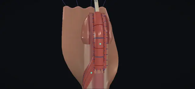
A one-stage repair for mid-shaft hypospadias in which the urethral plate is kept as the dorsal wall of the new urethra and a strip of inner prepuce, carried on its own dartos pedicle, is laid on it as the ventral wall.

The plate is neither incised in the midline nor rolled into a tube. The island is raised across the dorsal hood, brought round one side of the shaft, turned skin surface down on the plate, and sewn to its two borders and to the spatulated meatus over a stent.

The lesson is the pedicle: which plane it is found in, how far it is freed, and how it lies once the flap is in place.

Advanced tier FRCS paediatric urology, hypospadias Interactive 3D

Operative visualisation

Schematic scene preview: Hypospadias: onlay island flap
Preparing the 3D anatomy…

UrOops3D · UroRef · © Nity G

How to use this module. Play the ten steps, or use Back and Next. Watch the hood spread into its two layers, the island lift off the outer skin, the pedicle follow it round the shaft, and the flap close over the stent. The triggers show a fistula, a stricture at the join and a tight meatus.

Click me next Section 2 of 8 Anatomy The structures this operation works between, and the one it must not cross.

The structures in the scene

Hypospadias: onlay island flap: how the structures relateThe island of inner prepuce and what it is raised from, carried on and sewn to. Island of inner prepuce: the ventral wall: lives on it; no twist or tension Dartos pedicle: its only blood supply (danger). Island of inner prepuce: the ventral wall: dissected off it, under the plexus Outer preputial skin and its plexus (danger). Island of inner prepuce: the ventral wall: sewn to both borders, skin to lumen Urethral plate: kept whole, the dorsal wall. Island of inner prepuce: the ventral wall: proximal edge joined to it widely Hypospadiac meatus, spatulated. Island of inner prepuce: the ventral wall: covered by them at the glans Glans wings, closed over the repair. Island of inner prepuce: the ventral wall: closed over it on the far line Stent through the new urethra.Island of innerprepuce: the ventralwall!Dartos pedicle: itsonly blood supply!Outer preputial skinand its plexusUrethral plate: keptwhole, the dorsalwallHypospadiac meatus,spatulatedGlans wings, closedover the repairStent through thenew urethralives on it; notwist or tensiondissected off it,under the plexussewn to bothborders, skin tolumenproximal edgejoined to it widelycovered by them atthe glansclosed over it onthe far lineTargetDangerLandmarkContext
The island of inner prepuce and what it is raised from, carried on and sewn to. Schematic, not to scale; relationships only.
  • Seen from its ventral side with the glans uppermost, the meatus lies at mid-shaft and the urethral plate runs up from it to the tip of the glans, between the two glans wings.
  • The hood is dorsal.
  • Its inner layer faces the glans and its outer layer is continuous with the shaft skin; the dartos lies between them and runs down the dorsum to the root of the penis.
  • The island is cut across the inner layer, so its long axis lies transversely there.
  • Brought round one side of the shaft on that dartos, it lies lengthways on the plate with its skin surface against it.
  • ›Urethral plate - the strip of urethral tissue from the meatus to the tip of the glans; kept whole as the dorsal wall of the new urethra.
  • ›Hypospadiac meatus - at mid-shaft; spatulated ventrally so that the flap joins it widely.
  • ›Glans wings - formed by the incisions along the plate margins; closed in two layers over the new urethra.
  • ›Dorsal hood - the prepuce, deficient ventrally: two layers that meet at a fold.
  • ›Inner prepuce - the layer of the hood that faces the glans; the island is a transverse rectangle of it.
  • ›Outer preputial skin - continuous with the shaft skin; the plane of dissection lies just beneath its subdermal plexus.
  • ›Dartos pedicle - the vascular layer between the two skins, freed to the penoscrotal junction; the island has no other supply.
  • ›Buck’s fascia - the level of the degloving and of the dissection beside the plate.
  • ›Corpora cavernosa - the butterfly needle for the erection test goes into one of them; they are not opened.
  • ›Stent - a fine feeding tube through the new urethra, secured to the glans stay.
Click me next Section 3 of 8 Steps The operation in order, with the landmark that guards each move.

Operative steps

  1. Stay, infiltration, incision The child is supine under general anaesthesia with antibiotic cover, and the genitalia, lower abdomen and perineum are prepared. A traction suture is placed through the mid-glans and the penis is put on stretch. The ventral tissue round the meatus is infiltrated with local anaesthetic containing adrenaline to help haemostasis. Under loupe magnification a circumscribing skin incision is made, passing a short distance below the meatus, and the urethral plate is preserved.
    where it bites: the plate distal to the meatus is one wall of the repair; the incision passes below the meatus and leaves it alone.
  2. Plate margins and glans wings The lateral margins of the urethral plate are marked and then incised from below the meatus to the tip of the glans. On the glans these incisions form the glans wings. The dissection is taken down to Buck’s fascia, which releases any tethering. The plate is left as one strip between the two cuts; nothing in this operation incises it in the midline or rolls it into a tube.
    where it bites: a cut that strays into the plate narrows the strip the flap is to be sewn to.
  3. Erection test and degloving An artificial erection is induced with saline through a butterfly needle placed in one corpus cavernosum, to confirm that no ventral chordee remains. The penis is then degloved to the level of Buck’s fascia by sharp and blunt dissection. The skin sleeve and the dorsal hood come back together toward the base, and the plate stays where it was on the ventral surface.
    where it bites: a bend that persists on the erection test changes the operation; it is settled before the flap is raised.
  4. Raise the island Four stay sutures are placed in the inner prepuce for traction and the hood is spread. A rectangle is marked on the inner preputial surface with its long axis across the hood. It is incised at its two ends and along its distal border, and dissected carefully off the outer skin that lies behind it.
    where it bites: the island lives on the dartos beneath it; the dissection stays on the outer skin and leaves that layer on the island.
  5. Develop the pedicle The dissection is continued proximally in the same plane, just beneath the subdermal plexus of the outer skin, as far as the penoscrotal junction. What remains attached to the island is a broad, well-vascularised dartos pedicle. Bleeding points are controlled with fine bipolar diathermy.
    where it bites: too deep and the pedicle loses its vessels; too superficial and the outer skin loses its own.
  6. Rotate and invert The island is rotated ventrally on its pedicle and inverted over the urethral plate, so that its skin surface faces the plate, and it is trimmed to fit.
    where it bites: a pedicle that is twisted or tight as it comes round has lost the supply the flap depends on.
  7. Pedicle edge, then the meatus The pedicle edge of the flap is sutured to the lateral border of the plate in one layer with a fine running absorbable suture. The hypospadiac meatus is minimally debrided and spatulated ventrally. The proximal edge of the flap is sutured to it with interrupted sutures, which makes a wide, patent anastomosis.
    where it bites: the join to the native meatus runs across the tube; the spatulation is what keeps it wide.
  8. Stent and the far edge The urethra is stented with a fine infant feeding tube. The other edge of the flap is sutured to the opposite border of the plate with a running absorbable suture in two layers. The plate is now the dorsal wall of the new urethra and the flap is its ventral wall.
    where it bites: the long suture lines are where a fistula starts; each needs a covering layer over it.
  9. Close the glans The glans wings are closed over the new urethra in two layers of interrupted absorbable sutures, so that a patent meatus lies at the tip of the glans. The stent is secured to the traction suture placed in the glans at the start.
    where it bites: wings closed tightly over the tube narrow the meatus.
  10. Skin cover and dressing Before the skin is closed the penis is inspected for bleeding and haemostasis is secured. The preputial hood is excised and the penile skin is closed with interrupted absorbable sutures. A penile dressing with a plastic adhesive cover is applied. A double nappy is fitted, taking care not to occlude the meatus, and the stent is placed within the nappies.
    where it bites: skin closed tightly over the pedicle compresses the vessels the flap lives on.
Click me next Section 4 of 8 Danger zones Where this operation bites, and why it bites there.
Critical relationship

Danger zones

The spatial relationships the animation is built to make automatic. Before you read each one, ask yourself: at this point in the operation, what is about to go wrong, and what is my first move? The save for each sits in the Complications tab.

  • RiskThe urethral plate - kept whole; the margin incisions run beside it and nothing crosses it.
  • RiskThe plane under the outer skin - just beneath the subdermal plexus: the dartos stays with the island and the skin keeps its plexus.
  • RiskThe pedicle as it comes round - it carries all the flap’s vessels; it should lie round the side of the shaft without twist or tension.
  • RiskThe join at the meatus - the proximal anastomosis; the ventral spatulation makes it wide.
  • RiskThe new meatus - the glans is closed over the tube without squeezing it.
Practise it Take me into the scene Opens the animation at a danger moment and asks you to predict the mechanism and your first move before it reveals.
Click me next Section 5 of 8 Complications The save for each one: the first move, the escalation trigger, the op-note line.

Complications and immediate saves

Trigger each of these live in the Animation tab, then read the save: the first move, the escalation trigger, and the line that goes in the op note.

Urethrocutaneous fistula

  • urine leaks through a second opening on the ventral shaft; a listed complication of this operation.
The save
  1. Watch the child pass urine: confirm the second stream and note where on the shaft it comes from. first move
  2. Examine the meatus and the stream beyond the hole; narrowing downstream keeps a fistula open and has to be found.
  3. Nothing is passed through the opening and nothing is sutured on the ward. Closure is planned for when the tissues have softened.
  4. A large fistula, more than one, or a fistula with narrowing beyond it is a revision of the repair and goes to a surgeon who does that work. escalation trigger
  5. Record: the site and size of the opening, the stream from the meatus, the interval since the repair and the plan. document

Urethral stricture at the join

  • narrowing of the new urethra, shown where the flap meets the native urethra; urethral stricture is a listed complication of this operation.
The save
  1. Take the history of the stream and measure the flow where the child is old enough: a slow, prolonged stream with straining is the pattern. first move
  2. Ultrasound of the bladder and kidneys for residual urine and for dilatation of the upper tracts.
  3. Examination under anaesthetic, with urethroscopy or calibration, defines the level and the length before anything is treated.
  4. Retention, infection with a poor stream, or a dilated upper tract needs drainage and urgent paediatric urology review. escalation trigger
  5. Record: flow, residual urine, the level and length of the narrowing, and what was done. document

Meatal stenosis

  • the new meatus heals narrow; the European guideline relates it to a meatus reconstructed under tension.
  • Meatal stenosis or retraction is a listed complication of this operation.
The save
  1. Watch the stream: fine, forceful or deflected, with straining. first move
  2. Examine the meatus directly, and calibrate it where there is doubt.
  3. A meatotomy or meatoplasty is planned once the tissue has matured.
  4. Straining with residual urine, infection or upper tract dilatation needs urgent paediatric urology review. escalation trigger
  5. Record: the stream, the calibre, the residual urine and the plan. document
Click me next Section 6 of 8 Troubleshooting When the case turns: the problem, then the next move.

Intra-operative troubleshooting

When the case turns: the problem, then the next move.

  • ActionThe erection test still shows a ventral bend after the skin has been released - Baskin and colleagues found a significant bend in 50 of 374 onlay repairs at this point and corrected it by dorsal plication of the tunica albuginea, leaving the plate intact. The EAU paediatric guideline advises correcting a curvature of more than 30 degrees, and notes that dorsal plication shortens the penis.
  • ActionThe plate cannot be used as one wall of the new urethra - the textbook names two other ways of fashioning the new urethra from the prepuce: a tubular flap, and the split prepuce technique. European guidance prefers staged repair for proximal hypospadias.
  • ActionThe island looks dusky once it has been brought round - look at the pedicle before anything is sutured. Take the island back, undo any twist, free the base of the pedicle further if it is tight, and wait for the colour to return.
  • ActionThe glans wings will not come together over the tube - the textbook describes a glans cap approach as an alternative: fine scissors, kept flat on the corporal bodies, cut a wide tunnel under the glans to its tip, excess glanular tissue is excised and a V-shaped meatus is fashioned at the tip.
  • ActionA suture line looks thin - a haemostatic sealant such as fibrin glue can be used with the sutures to reinforce the repair, as the dictation notes. The EAU guideline regards adequate covering of the reconstruction as most important.
Click me next Section 7 of 8 FRCS viva Say it out loud, then mark yourself against the model answer.

FRCS viva questions

The examiner stems this scene prepares you for. Try to answer out loud first, then reveal, and learn the discriminator, the detail that turns a pass into a strong pass.

QWhen would you choose an onlay island flap, and why keep the urethral plate?reveal answer
  • I would consider it for a child with a mid-shaft hypospadias, which is the indication the operative text gives.
  • The European guideline describes the preputial island onlay as a good option for proximal hypospadias, or if the urethral plate is unsuitable for tubularisation.
  • For distal and mid-penile forms that guideline names the tubularised incised plate repair as the treatment of choice, so I would use the onlay where the plate is healthy enough to keep but I do not want to roll it into a tube.
  • Keeping the plate rests on work from Philadelphia.
  • Baskin and colleagues reported that in most cases the plate is not the cause of the curvature: after the skin had been taken down, an artificial erection showed a significant bend in 50 of 374 onlay repairs, and that bend was corrected by dorsal plication with the plate left in place.
  • The guideline now calls preservation of the well-vascularised plate the mainstay of hypospadias repair.
  • The practical gain is that the new urethra has a native, vascular wall behind and its two long suture lines lie at the sides, with no join running round the whole circumference.
  • I would tell the family that this is a one-stage repair using the foreskin, that it carries a risk of fistula, of narrowing at the meatus or in the urethra, and of breakdown, and that a further operation is sometimes needed.
Strong pass

Say all three

  • Knowing that the plate is kept because it is seldom the cause of the bend
  • With the figure and whose series it is
  • That the guideline places the onlay with proximal forms or a plate unsuitable for tubularisation
Examiner note
Knowing that the plate is kept because it is seldom the cause of the bend, with the figure and whose series it is, and that the guideline places the onlay with proximal forms or a plate unsuitable for tubularisation.
QDescribe the operation.reveal answer
  • I place a traction suture through the glans, infiltrate round the meatus for haemostasis and, with loupes, make a circumscribing skin incision that passes a short distance below the meatus and preserves the plate.
  • I mark and incise the lateral margins of the plate from below the meatus to the tip of the glans, which forms the glans wings, and take the dissection down to Buck’s fascia to release any tethering.
  • I induce an artificial erection with saline through a butterfly needle in one corpus to confirm that the penis is straight, then deglove it to Buck’s fascia.
  • I place four stay sutures in the inner prepuce, mark a transverse rectangle on its inner surface, incise it at its ends and distally and dissect it off the outer skin.
  • I carry that plane proximally, just beneath the subdermal plexus, to the penoscrotal junction, so that the island is left on a broad dartos pedicle.
  • I rotate the island ventrally, invert it over the plate and trim it.
  • I sew its pedicle edge to one border of the plate with a running suture in one layer, spatulate the meatus ventrally and sew the proximal edge of the flap to it with interrupted sutures.
  • I pass a fine feeding tube as a stent and sew the other edge to the opposite border in two layers.
  • I close the glans wings in two layers over the new urethra, secure the stent to the glans suture, check haemostasis, excise the hood and close the skin.
Strong pass

Say all three

  • The order of the three suture lines: pedicle edge first
  • The proximal edge to the spatulated meatus
  • The stent and the far edge in two layers
Examiner note
The order of the three suture lines: pedicle edge first, then the proximal edge to the spatulated meatus, then the stent and the far edge in two layers.
QWhat keeps this flap alive, and how do you protect it?reveal answer
  • The island is skin with no attachment except its pedicle, so everything it receives comes through the dartos.
  • I would protect that in three places.
  • When I raise the island I dissect on the outer skin, just beneath its subdermal plexus, so that the whole thickness of the dartos stays on the island and the outer skin keeps its own plexus.
  • I carry that dissection all the way to the penoscrotal junction, because a pedicle freed only part of the way has to be pulled to reach the ventral side.
  • And when I bring the island round the shaft I look at the pedicle before I place a stitch: it should lie flat round the side with no twist and no tension, and the island should stay pink.
  • If it did not, I would take it back, undo the twist and free the base further.
  • At the end I close the skin without compressing the pedicle.
  • The plane and the extent of the dissection are as the chapter describes them; the checks on twist, tension and colour are general flap practice.
Strong pass

Naming the plane, just beneath the subdermal plexus of the outer skin, and the extent, to the penoscrotal junction, and checking the pedicle for twist and tension before the first stitch.

QWhat are the complications, and how often do they happen?reveal answer
  • The chapter lists bleeding, infection, dehiscence, meatal stenosis or retraction, urethrocutaneous fistula and urethral stricture.
  • For figures I would quote two kinds of source and say which is which.
  • The original reports come from one hospital.
  • Elder, Duckett and Snyder reported 50 onlay island flap repairs for mid and distal penile hypospadias with a complication rate of 6 per cent, and Baskin and colleagues later gave a fistula rate of 6 per cent for the onlay.
  • In proximal hypospadias the numbers are higher.
  • Castañón and colleagues compared 38 onlay repairs with 42 tubularised island flaps: fistula in 18.4 per cent of the onlays and 21.4 per cent of the tubes, which was not a significant difference, anastomotic stricture in 2.6 per cent against 7.1 per cent, and megaurethra only in the tube group.
  • Across all techniques the EAU guideline gives fistula rates of 5 to 50 per cent depending on the complexity of the case, and meatal stenosis in 5 to 15 per cent, particularly when the meatus is reconstructed under tension.
  • It gives urethral stricture in 8 to 13 per cent, especially after proximal repairs, and glans and wound dehiscence in 9 to 17 per cent.
  • I would tell the family that a further operation is a real possibility, and that the guideline asks for long-term follow-up.
Strong pass

Say both

  • Separating the early single-centre figures for mid-shaft repairs from the guideline’s ranges across all techniques
  • Knowing that the comparison with the tubularised flap favoured the onlay for anastomotic stricture
Examiner note
Separating the early single-centre figures for mid-shaft repairs from the guideline’s ranges across all techniques, and knowing that the comparison with the tubularised flap favoured the onlay for anastomotic stricture.
QA second stream appears on the shaft after the stent has come out. What do you do?reveal answer
  • I would watch the child void and confirm it, note the site and size of the opening, and look carefully at the stream from the meatus, because narrowing of the meatus or of the urethra beyond the hole keeps a fistula open and has to be dealt with as well.
  • I would not pass anything through it or try to suture it on the ward.
  • I would explain to the family that this is a recognised complication and plan an elective closure once the tissues have softened.
  • At that operation I would put a vascular layer between the urethral repair and the skin: the EAU guideline regards adequate covering of the reconstruction as most important, and recommends a double layer if dartos is used.
  • If the fistula were large, if there were more than one, or if the flap had broken down, I would treat it as a revision of the whole repair.
  • I would record the site, the stream and the plan.
Strong pass

Say both

  • Looking for narrowing beyond the fistula before planning its closure
  • Interposing a vascular layer at the repair
Examiner note
Looking for narrowing beyond the fistula before planning its closure, and interposing a vascular layer at the repair.
Click me next Section 8 of 8 Evidence The guidance each of these answers is built on.

Evidence and guidance

Operative Dictations in Urologic Surgery
Armenakas NA, Fracchia JA, Golan R. Transverse preputial onlay island flap. Chapter 121. Wiley, 2019
The indication (mid-shaft hypospadias), the eight essential steps, the template dictation followed step by step here, the variations (a tubular flap or the split prepuce technique; the glans cap approach; a haemostatic sealant) and the complication list: bleeding, infection, dehiscence, meatal stenosis or retraction, urethrocutaneous fistula, urethral stricture. Where the essential steps and the dictation differ in order, the dictation is followed: the plate margins are incised before the erection test. The chapter says the margin incisions separate the plate from the corporal bodies; that is read here as freeing its borders down to Buck’s fascia, with the plate left on its bed. The chapter’s sizes (the width of the flap, the spatulation, the level of the incision, the stent and the sutures) are kept off the page. The model is schematic. The chapter lists complications without their management: the first, escalate and document steps, the danger lines on steps two, three, five, six, eight and ten, and the troubleshooting line on a dusky island are general surgical practice.
Open guideline
EAU Guidelines
Paediatric Urology, 2026: Hypospadias, sections 9.5.3 to 9.5.5, 9.5.8, 9.6 and 9.8, read 13 October 2026
Preservation of the well-vascularised urethral plate is the mainstay of repair; the tubularised incised plate repair is the treatment of choice in distal and mid-penile hypospadias; the onlay technique with a preputial island flap is a good option for proximal hypospadias or if the plate is unsuitable for tubularisation; staged repairs are preferred for proximal forms. Curvature of 30 degrees or more warrants correction (recommendation: weak), and dorsal plication shortens the penis. Adequate covering of the reconstruction appears most important; a double layer is recommended if dartos is used. Fistula 5% to 50% depending on complexity; meatal stenosis 5% to 15%, particularly when the meatus is reconstructed under tension; urethral stricture 8% to 13%, especially in proximal repairs; glans and wound dehiscence 9% to 17%. Long-term follow-up is recommended (strong). These ranges are across techniques and are not figures for the onlay alone.
Open guideline
The Journal of Urology
Elder JS, Duckett JW, Snyder HM. Onlay island flap in the repair of mid and distal penile hypospadias without chordee. J Urol 1987;138:376-379
Abstract read 13 October 2026. The onlay island flap as a variation of the transverse preputial island flap; 50 patients, compared with 34 who had a meatal-based flap repair in the same period; a complication rate of 6% in each group; described as applicable particularly to mid-shaft hypospadias without chordee. A single-centre comparative series.
Open guideline
The Journal of Urology
Baskin LS, Duckett JW, Ueoka K, Seibold J, Snyder HM. Changing concepts of hypospadias curvature lead to more onlay island flap procedures. J Urol 1994;151:191-196
Abstract read 13 October 2026. Of 1,109 primary hypospadias operations from 1987 to 1992, 374 were onlay island flaps; an artificial erection after the skin had been taken down showed a significant bend in 50 of the 374, corrected by dorsal plication of the tunica albuginea; a fistula rate of 6% with the onlay. The authors conclude that the plate is usually healthy and need not be divided to straighten the penis. A retrospective single-centre series.
Open guideline
Journal of Pediatric Surgery
Castañón M, Muñoz E, Carrasco R, Rodó J, Morales L. Treatment of proximal hypospadias with a tubularized island flap urethroplasty and the onlay technique: a comparative study. J Pediatr Surg 2000;35:1453-1455
Abstract read 13 October 2026. A retrospective comparison of 42 tubularised island flaps and 38 onlay island flaps for proximal hypospadias: fistula 21.4% against 18.4% (not significant); anastomotic stricture 7.14% against 2.63% (significant); megaurethra only after the tubularised flap (4.7%). Proximal cases, so the figures do not transfer directly to a mid-shaft repair.
Open guideline

Curriculum. Supports learning relevant to the ISCP Urology Curriculum syllabus topic Paediatric urology: hypospadias. Not a workplace-based assessment and not evidence of competence.

Two minutes

Close the lap: recall it before you leave.

Say it aloud, then reveal. Reading is not remembering.

  1. 01 The relationship Say the critical spatial relationship out loud, then reveal. reveal
    Memory frame

    Seen from its ventral side with the glans uppermost, the meatus lies at mid-shaft and the urethral plate runs up from it to the tip of the glans, between the two glans wings.

    Why?
    The hood is dorsal. Its inner layer faces the glans and its outer layer is continuous with the shaft skin; the dartos lies between them and runs down the dorsum to the root of the penis. The island is cut across the inner layer, so its long axis lies transversely there. Brought round one side of the shaft on that dartos, it lies lengthways on the plate with its skin surface against it.
  2. 02 The first move Urethrocutaneous fistula. Your first move? reveal
    First move

    Watch the child pass urine: confirm the second stream and note where on the shaft it comes from.

  3. 03 The escalation trigger Urethrocutaneous fistula. What makes you call for help? reveal
    Escalate when

    A large fistula, more than one, or a fistula with narrowing beyond it is a revision of the repair and goes to a surgeon who does that work.

  4. 04 The discriminator What turns a pass into a strong pass? reveal
    Strong pass

    Say all three

    • Knowing that the plate is kept because it is seldom the cause of the bend
    • With the figure and whose series it is
    • That the guideline places the onlay with proximal forms or a plate unsuitable for tubularisation
    Examiner note
    Knowing that the plate is kept because it is seldom the cause of the bend, with the figure and whose series it is, and that the guideline places the onlay with proximal forms or a plate unsuitable for tubularisation.
  5. 05 The pitfall Where does this operation bite first? reveal
    Pitfall

    The urethral plate

    Why?
    kept whole; the margin incisions run beside it and nothing crosses it.

Revision practice with self-marked answers, not an assessment of clinical competence. Kept in this browser only; it does not sync between devices.