
UrOops3D, danger-zone atlas
Operative animation, Posterior urethral valve ablation

Posterior urethral valve ablation

The commonest cause of bladder outlet obstruction in a boy: two leaflets running from the verumontanum toward the sphincter that let a catheter in and stop urine getting out.

Drain first, define the anatomy, then divide the leaflets at five, seven and twelve o'clock in a urethra a few millimetres wide, without cutting the sphincter that sits just beyond them.

This page is the drainage, the assessment, the cuts, and the long follow-up of the valve bladder.

Advanced tier FRCS paediatric urology, obstructive uropathy Interactive 3D

Operative visualisation

Schematic scene preview: Posterior urethral valve ablation
Preparing the 3D anatomy…

UrOops3D · UroRef · © Nity G

Click me next Section 2 of 8 Anatomy The structures this operation works between, and the one it must not cross.

The structures in the scene

Posterior urethral valve ablation: how the structures relateAlong the posterior urethra from the cystoscope, sphincter to bladder. External sphincter: lies just distal; the cut stops at the wall Valve leaflets (danger). Valve leaflets: leaflets arise from its distal end Verumontanum. Valve leaflets: divided at five, seven and twelve, never resected Dilated posterior urethra. Dilated posterior urethra: thick and high, looked at and not cut Hypertrophied bladder neck. Hypertrophied bladder neck: cellules, diverticula, reflux in about half Trabeculated bladder and the orifices.Distal: penile endProximal: bladder!External sphincterValve leafletsVerumontanumDilated posterior urethraHypertrophied bladder neckTrabeculated bladder andthe orificeslies just distal; the cutstops at the wallleaflets arise fromits distal enddivided at five, seven andtwelve, never resectedthick and high, looked at andnot cutcellules, diverticula, refluxin about halfTargetDangerLandmarkContext
Along the posterior urethra from the cystoscope, sphincter to bladder. Schematic, not to scale; relationships only.
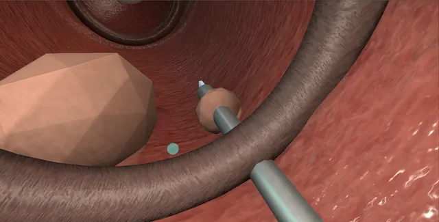
  • From inside the posterior urethra the veru sits on the floor and the two leaflets leave its distal end, sweep forward along the walls and meet at twelve o'clock, leaving only a slit in the roof.
  • The sphincter is a short distance beyond them and the thick bladder neck a short distance behind.
  • The cuts fall at five and seven where each leaflet meets the wall, and at twelve where they fuse; the atlas is adult-sized, the infant urethra is a fraction of it.
  • ›Valve leaflets - from the distal veru forward and laterally to fuse at twelve o'clock; divided at five, seven and twelve, never resected.
  • ›Verumontanum - the fixed landmark on the floor; the leaflets take origin from its distal end.
  • ›Dilated posterior urethra - long and wide above the valve on the MCUG and at cystoscopy; the sign of years of straining before birth.
  • ›External sphincter - just distal to the leaflets; a cut carried into it is lifelong incontinence.
  • ›Hypertrophied bladder neck - thick and high from working against the valve; looked at, not cut.
  • ›The trabeculated bladder and the orifices - cellules, diverticula, reflux in about half; the valve bladder that needs years of follow-up.
  • ›The atlas - adult-proportioned in this scene; the infant anatomy is the same in miniature, in a urethra a few millimetres wide.
Click me next Section 3 of 8 Steps The operation in order, with the landmark that guards each move.

Operative steps

  1. The boy Antenatal ultrasound shows bilateral hydronephrosis, a thick-walled bladder that never empties and, in the worst cases, oligohydramnios and a keyhole sign at the neck. Or the infant presents later with a dribbling stream, a palpable bladder, urosepsis or failure to thrive. Whatever the route, the first move is drainage: a 6 or 8 French feeding tube passed urethrally, never a balloon catheter, because a balloon inflated in the dilated posterior urethra or a spasming infant bladder does harm and the tube drains just as well. The tube is secured to the penis and left, and the creatinine, electrolytes and fluid balance are followed from that hour.
    where it bites: a balloon catheter in a valve infant sits in the posterior urethra or provokes the bladder; a feeding tube is the drainage.
  2. Assessment before the scope The creatinine falls on drainage and its nadir over the first days and weeks is the best early guide to what the kidneys have left; a creatinine that will not come down means the damage is renal, not obstructive, and no cut will fix it. The MCUG makes the diagnosis: a dilated, elongated posterior urethra above an abrupt narrowing, a thick trabeculated bladder, often reflux, which is present in about half, and sometimes a diverticulum. Ultrasound shows the upper tracts and the bladder wall. The nephrologist is a partner from the start: fluids, electrolytes, acidosis, blood pressure and the timing of surgery are shared decisions.
  3. The instrument A paediatric resectoscope of 8 to 11 French, or a paediatric cystoscope with a working channel, with a 0 or 12 degree telescope. The cutting tool is one of three: a cold knife on the resectoscope, a Bugbee electrode on low cutting current, or a laser fibre passed through the cystoscope. Whichever is used, the principle is a division of the leaflet, not a resection of tissue. The urethra of a small or premature infant may not take the smallest instrument, and forcing it makes a stricture; that infant keeps the tube or has a vesicostomy and is ablated when the urethra has grown.
    where it bites: an instrument that does not pass freely is the wrong size for this urethra; the alternative is a vesicostomy, not force.
  4. Cystoscopy: what a valve looks like The scope passes the bulbar urethra and the sphincter and enters the posterior urethra, which is wide and long. The verumontanum sits on the floor and from its distal end two leaflets run forward and laterally, curving up to meet at twelve o'clock, so that from below they look like a membrane with a slit at the top. They billow toward the scope on irrigation and flatten when the flow is stopped. Beyond them the bladder neck is thick and high, sometimes a ring that looks obstructive itself, and the bladder is trabeculated with cellules and diverticula. Both orifices are found, and reflux may be seen.
  5. Ablation at five and seven o'clock The leaflets are divided where they attach to the wall on either side of the veru, at five and seven o'clock, in their distal third where the tissue is thinnest and the sphincter is furthest away. The knife or electrode is placed on the leaflet close to its attachment and the cut is made toward the wall under continuous vision, with the veru watched as the fixed landmark. The leaflet falls away and the lumen opens. The cut is short, a few millimetres, and the current is the lowest that cuts; the urethra here is thin and the tissue beyond it is the sphincter and the corpus spongiosum.
    where it bites: the sphincter lies just distal to the leaflets; the cut is on the leaflet, toward the wall, and no further.
  6. Twelve o'clock and the check The anterior fusion of the leaflets at twelve o'clock is divided next, a single short cut through the membrane where the two halves meet, and this often does the most for flow. Then the irrigation is turned on again and the scope drawn back to the level of the veru: the leaflets should lie flat against the wall and no longer coapt when fluid runs past them. If they still billow into the lumen and meet, the cuts are extended a little at the attachments. The bladder neck is looked at but not cut; it is thick because it has been working against a valve and it settles once the valve is gone.
  7. No resection The temptation is to remove the leaflets rather than divide them, and it is resisted. A resected leaflet takes urethral wall with it and heals as a stricture in a urethra that has no width to spare; a deep cut at five or seven that enters the sphincter leaves a boy incontinent for life. The endpoint is a valve that no longer obstructs, not a urethra that looks clean. Slightly under-treated is recoverable at a second look; over-treated is not. The same restraint applies to the bladder neck: a bladder neck incision in an infant with valves is a specialist decision made later, if at all.
  8. The catheter and the days after A feeding tube goes back in, sized to the urethra, and stays for a day or two, longer if the urethra was tight or bled. Antibiotic prophylaxis continues, particularly with reflux. The creatinine is checked because the ablation changes nothing about the kidneys in the short term, and a post-obstructive diuresis after the first drainage is watched for. The note records the instrument, the sites divided, the sphincter and veru seen intact, the state of the bladder neck and bladder, reflux if seen, and the catheter. A check cystoscopy or MCUG at about three months confirms that the leaflets no longer obstruct.
  9. The valve bladder and the long game The obstruction is gone in an afternoon; the damage it did takes a lifetime. The bladder that grew thick against the valve is poorly compliant, later often overdistended and polyuric because the kidneys cannot concentrate, and it empties incompletely. Reflux resolves in many once the pressure falls and is watched rather than repaired. Follow-up is shared with nephrology for years: creatinine and growth, ultrasound of the upper tracts, infections, blood pressure, and from toilet training onward, continence, timed and double voiding, anticholinergics and sometimes overnight catheter drainage. A proportion progress to end-stage renal disease in childhood or adolescence, and that is said at the first consultation.
Click me next Section 4 of 8 Danger zones Where this operation bites, and why it bites there.
Critical relationship

Danger zones

The spatial relationships the animation is built to make automatic. Before you read each one, ask yourself: at this point in the operation, what is about to go wrong, and what is my first move? The save for each sits in the Complications tab.

  • RiskThe external sphincter - just distal to the leaflets; the cut stays on the leaflet and stops at the wall.
  • RiskThe small urethra - a few millimetres wide; an instrument forced through it, or a leaflet resected rather than divided, heals as a stricture.
  • RiskThe bladder neck - thick and high but not the obstruction; it is left alone at the first operation.
  • RiskThe kidneys - the creatinine that will not fall after drainage is renal dysplasia; the ablation does not change it and the family is told.
Practise it Take me into the scene Opens the animation at a danger moment and asks you to predict the mechanism and your first move before it reveals.
Click me next Section 5 of 8 Complications The save for each one: the first move, the escalation trigger, the op-note line.

Complications and immediate saves

Trigger each of these live in the Animation tab, then read the save: the first move, the escalation trigger, and the line that goes in the op note.

Urethral stricture

  • the narrow urethra scarred by a forced instrument or a resected leaflet: a poor stream returning weeks or months after a good ablation.
The save
  1. Recognise it early: a stream that worsens after it had improved, or a tube that will not pass, means the urethra, not the valve. first move
  2. Cystoscopy with the smallest instrument to define the length and site of the stricture.
  3. A short stricture is incised endoscopically once; a long or recurrent one needs a paediatric reconstructive surgeon and may need a vesicostomy while the urethra rests. escalation trigger
  4. Prevent the next one: the smallest instrument that passes freely, low current, division not resection.
  5. Note: the site and length, the instrument used, what was done and the plan for review. document

Residual valve or persistent obstruction

  • leaflets that still coapt after the cuts: a stream that did not improve, a bladder that still does not empty, hydronephrosis that does not settle.
The save
  1. A check cystoscopy or MCUG at about three months, earlier if the stream and ultrasound have not improved. first move
  2. Residual leaflets are divided again at the same sites; a second look is expected in a proportion and is not a failure.
  3. Obstruction with no residual valve is the bladder neck or the bladder itself: urodynamics and a specialist decision before any further cutting. escalation trigger
  4. Upper tracts that stay dilated with rising creatinine despite a clear urethra: nephrology, and consider drainage above the bladder.
  5. Note: what was seen at the second look, what was divided and the imaging that follows. document

Sphincter injury and incontinence

  • a cut at five or seven carried past the leaflet into the striated sphincter: a boy who is never dry.
The save
  1. Stop cutting in that sector as soon as the leaflet has fallen away; look at the sphincter ring from the bulbar side. first move
  2. Nothing more is done at the operation; incontinence declares itself only at toilet training, years later.
  3. The family is told what was seen, and the note says so.
  4. Incontinence at school age: video-urodynamics to separate sphincter injury from the valve bladder, then a paediatric continence surgeon. escalation trigger
  5. Note: the sector, the depth of the cut, the sphincter seen or not seen, and the plan for review. document

Urinary infection and sepsis

  • an obstructed, refluxing system instrumented: fever within days, or urosepsis in an infant with little reserve.
The save
  1. Cultures, intravenous antibiotics, and confirm the catheter is draining. first move
  2. Ultrasound the upper tracts: an infected obstructed kidney above a valve bladder needs draining.
  3. An infant with sepsis and a rising creatinine or acidosis: the paediatric intensive care team and nephrology the same hour. escalation trigger
  4. Prophylaxis afterwards while reflux persists; circumcision reduces infections in these boys and is discussed.
  5. Note: the organism, the imaging, the antibiotic course and the prophylaxis plan. document

Bladder dysfunction and renal decline

  • the valve bladder: poor compliance, incomplete emptying, polyuria and reflux; the kidneys that were dysplastic before the ablation.
The save
  1. Accept from the first day that the ablation treats the valve and not the bladder or the kidneys; set the follow-up accordingly. first move
  2. Creatinine, growth, blood pressure and ultrasound at fixed intervals with nephrology; urodynamics when the boy is old enough to void.
  3. Timed and double voiding, anticholinergics for poor compliance, overnight catheter drainage for the polyuric bladder that overfills.
  4. A rising creatinine, proteinuria or hypertension: nephrology leads, and transplant planning starts early because the bladder must be made safe first. escalation trigger
  5. Note: the shared follow-up schedule, who owns each part of it, and what the family has been told. document
Click me next Section 6 of 8 Troubleshooting When the case turns: the problem, then the next move.

Intra-operative troubleshooting

When the case turns: the problem, then the next move.

  • ActionThe scope will not pass the urethra - stop; the smallest instrument, or a vesicostomy and an ablation later when the urethra has grown.
  • ActionCannot see the leaflets - turn the irrigation up so they billow toward the lens, then off so they flatten; look from the veru forward.
  • ActionThe view fills with blood after a cut - the current was too high or the cut too deep; irrigate, wait, lower the current and do not chase the bleeding with more energy.
  • ActionThe leaflets still meet after five and seven - divide the fusion at twelve o'clock; then extend the lateral cuts a little at the attachments.
  • ActionThe bladder neck looks obstructive - leave it; it is hypertrophied from the valve and is reassessed months later, never cut at the first operation.
  • ActionThe creatinine does not fall after drainage - the tube is checked and the bladder scanned; if the bladder is empty the problem is the kidneys, and nephrology is called.
Click me next Section 7 of 8 FRCS viva Say it out loud, then mark yourself against the model answer.

FRCS viva questions

The examiner stems this scene prepares you for. Try to answer out loud first, then reveal, and learn the discriminator, the detail that turns a pass into a strong pass.

QA newborn boy has antenatally detected bilateral hydronephrosis and a thick-walled bladder. How would you manage him?reveal answer
  • I would treat this as posterior urethral valves until proved otherwise and my first step is drainage: a 6 or 8 French feeding tube passed urethrally and secured, not a balloon catheter, with antibiotic prophylaxis.
  • I would check the creatinine, electrolytes and acid-base status and repeat them daily, expecting the creatinine to fall as the bladder drains, and I would involve the paediatric nephrologist from the first day.
  • I would confirm the diagnosis with an ultrasound of the kidneys and bladder and a micturating cystourethrogram, looking for the dilated posterior urethra, the thick trabeculated bladder and reflux, which is present in about half.
  • Once the infant is stable, the creatinine has reached a nadir and the urethra will take a paediatric instrument, I would proceed to endoscopic valve ablation.
  • If the urethra is too small, as in a premature infant, I would keep the tube or create a cutaneous vesicostomy and ablate later.
  • I would tell the family at the outset that the ablation treats the obstruction but that bladder and kidney function need lifelong follow-up.
Strong pass

Say all three

  • A feeding tube rather than a balloon catheter
  • The creatinine nadir as the early prognostic marker
  • The vesicostomy alternative for the urethra that will not take an instrument
Examiner note
A feeding tube rather than a balloon catheter, the creatinine nadir as the early prognostic marker, and the vesicostomy alternative for the urethra that will not take an instrument.
QDescribe the endoscopic ablation of posterior urethral valves.reveal answer
  • Under general anaesthesia in lithotomy I would pass a paediatric resectoscope or cystoscope of 8 to 11 French with a 0 or 12 degree telescope, gently and without force.
  • I would identify the bulbar urethra, the sphincter, then the wide posterior urethra with the verumontanum on the floor, and the two leaflets arising from its distal end, curving forward and laterally to fuse at twelve o'clock.
  • I would inspect the bladder neck, the bladder and both orifices.
  • Using a cold knife, a Bugbee electrode on low cutting current or a laser fibre, I would divide each leaflet close to its attachment at five and seven o'clock, cutting toward the wall under vision and no deeper, then divide the anterior fusion at twelve.
  • I would restart the irrigation and confirm that the leaflets lie flat and no longer coapt.
  • I would not resect tissue and would not touch the bladder neck.
  • I would leave a feeding tube for a day or two, continue prophylaxis, document the sites and the sphincter seen intact, and arrange a check cystoscopy or MCUG at about three months.
Strong pass

Five, seven and twelve o'clock with the veru as the landmark, division rather than resection, and the sphincter named as the structure just beyond the cut.

QWhat determines the long-term outcome after valve ablation?reveal answer
  • The outcome is decided by the kidneys and the bladder, not by the ablation.
  • Renal outcome depends on the degree of dysplasia present before birth, which is fixed, and on how much further damage is avoided afterwards.
  • The creatinine nadir in the first year is the most used early marker, with a low nadir predicting preserved function and a persistently raised creatinine predicting chronic kidney disease.
  • Reflux, recurrent infection, and a bladder that stores at high pressure all add secondary damage.
  • The valve bladder is poorly compliant early, then often large and poorly emptying, with polyuria from a concentrating defect that overfills it, and it must be managed actively with timed and double voiding, anticholinergics and overnight drainage to protect the upper tracts.
  • A proportion of boys, quoted as about a third with chronic kidney disease and a smaller group reaching end-stage disease in childhood or adolescence, will need transplant planning, and the bladder must be safe before that.
  • So follow-up is lifelong and shared with nephrology.
Strong pass

Say all three

  • Separating fixed dysplasia from preventable secondary damage
  • Naming the creatinine nadir
  • Describing the valve bladder as a condition in its own right
Examiner note
Separating fixed dysplasia from preventable secondary damage, naming the creatinine nadir, and describing the valve bladder as a condition in its own right.
QWhat are the complications of valve ablation and how are they avoided?reveal answer
  • The intra-operative complications are urethral injury and stricture, from an instrument too large for the urethra or from resecting rather than dividing the leaflets, and sphincter injury from a cut at five or seven carried past the leaflet into the striated muscle, which causes lifelong incontinence.
  • Both are avoided by using the smallest instrument that passes freely, low current, short cuts made on the leaflet toward the wall with the verumontanum watched, and no resection.
  • Residual valve tissue causing persistent obstruction is common enough that a check cystoscopy or MCUG at about three months is routine, and a second division is not a failure.
  • Infection and sepsis follow instrumentation of a refluxing, obstructed system and are reduced by prophylaxis, and by circumcision in selected boys.
  • The late problems are the valve bladder with poor compliance and emptying, persistent reflux, and progressive renal decline, which are not complications of the operation but of the disease, and are managed by a long shared follow-up with nephrology.
Strong pass

Say both

  • Pairing each early complication with the technical step that prevents it
  • Separating the late problems of the disease from those of the operation
Examiner note
Pairing each early complication with the technical step that prevents it, and separating the late problems of the disease from those of the operation.
Click me next Section 8 of 8 Evidence The guidance each of these answers is built on.

Evidence and guidance

Two minutes

Close the lap: recall it before you leave.

Say it aloud, then reveal. Reading is not remembering.

  1. 01 The relationship Say the critical spatial relationship out loud, then reveal. reveal
    Memory frame

    From inside the posterior urethra the veru sits on the floor and the two leaflets leave its distal end, sweep forward along the walls and meet at twelve o'clock, leaving only a slit in the roof.

    Why?
    The sphincter is a short distance beyond them and the thick bladder neck a short distance behind. The cuts fall at five and seven where each leaflet meets the wall, and at twelve where they fuse; the atlas is adult-sized, the infant urethra is a fraction of it.
  2. 02 The first move Urethral stricture. Your first move? reveal
    First move

    Recognise it early: a stream that worsens after it had improved, or a tube that will not pass, means the urethra, not the valve.

  3. 03 The escalation trigger Urethral stricture. What makes you call for help? reveal
    Escalate when

    A short stricture is incised endoscopically once; a long or recurrent one needs a paediatric reconstructive surgeon and may need a vesicostomy while the urethra rests.

  4. 04 The discriminator What turns a pass into a strong pass? reveal
    Strong pass

    Say all three

    • A feeding tube rather than a balloon catheter
    • The creatinine nadir as the early prognostic marker
    • The vesicostomy alternative for the urethra that will not take an instrument
    Examiner note
    A feeding tube rather than a balloon catheter, the creatinine nadir as the early prognostic marker, and the vesicostomy alternative for the urethra that will not take an instrument.
  5. 05 The pitfall Where does this operation bite first? reveal
    Pitfall

    The external sphincter

    Why?
    just distal to the leaflets; the cut stays on the leaflet and stops at the wall.

Revision practice with self-marked answers, not an assessment of clinical competence. Kept in this browser only; it does not sync between devices.