
UrOops3D, danger-zone atlas
Operative animation, Infected obstructed kidney

Emergency decompression of the infected obstructed kidney

A ureteric stone, a temperature and a blood pressure that is starting to fall: pus under pressure in a closed system, draining into the blood.

The only treatment is to let it out, by nephrostomy or by stent, within hours of the diagnosis and after the antibiotics and the fluids have started, and to leave the stone entirely alone until the patient is well.

This page is the recognition, the sepsis bundle, the CT, the choice of route, the technique of each, the pus, and the definitive treatment weeks later.

Core tier FRCS emergency urology, urosepsis and upper tract obstruction Interactive 3D

Operative visualisation

Schematic scene preview: Infected obstructed kidney: emergency decompression
Preparing the 3D anatomy…

UrOops3D · UroRef · © Nity G

Click me next Section 2 of 8 Anatomy The structures this operation works between, and the one it must not cross.

The structures in the scene

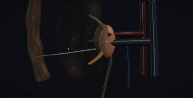
Infected obstructed kidney: how the structures relateProne-oblique, the obstructed kidney and what surrounds the needle line. Dilated collecting system: thin inflamed wall at the stone; never force Stone impacted in the ureter (danger). Dilated collecting system: puncture stays below the twelfth rib Pleura and the twelfth rib (danger). Dilated collecting system: lateral and in front; seen on CT first The colon (danger). Dilated collecting system: enter a calyx through parenchyma, not the pelvis Segmental vessels at the infundibulum (danger). Dilated collecting system: JJ drains the pelvis to an empty bladder Bladder and urethra: retrograde route.Dilated collectingsystemStone impacted inthe ureter!Pleura and thetwelfth rib!The colon!Segmental vessels atthe infundibulumBladder and urethra:retrograde routethin inflamed wallat the stone; neverforcepuncture staysbelow the twelfthriblateral and infront; seen on CTfirstenter a calyxthrough parenchyma,not the pelvisJJ drains thepelvis to an emptybladderTargetDangerLandmark
Prone-oblique, the obstructed kidney and what surrounds the needle line. Schematic, not to scale; relationships only.
  • The kidney sits high in the retroperitoneum with its upper pole under the eleventh and twelfth ribs and the pleura reflecting across the twelfth rib behind it, the colon lateral and in front, and the psoas medial.
  • The collecting system is dilated with the stone impacted in the ureter below the pelvis, and the nephrostomy needle comes from behind and below the twelfth rib, through the parenchyma of the lower pole into a posterior calyx.
  • The retrograde wire comes the other way, up the ureter from the bladder to the stone.
  • ›The dilated collecting system - pus under pressure behind the stone; entered through a posterior lower pole calyx and never distended.
  • ›The obstructing stone in the ureter - impacted in an inflamed thin ureteric wall; passed gently with a hydrophilic wire or left for the nephrostomy.
  • ›The pleura and the twelfth rib - the pleural reflection crosses the twelfth rib posteriorly; the puncture stays below it.
  • ›The colon - lateral and posterior to the kidney in the thin patient and on the left; seen on the CT and the ultrasound before the needle.
  • ›The segmental vessels at the infundibulum - the puncture through the parenchyma along the avascular plane into a calyx avoids them; a direct pelvic puncture does not.
  • ›The bladder and the urethra - the retrograde route; a catheter left so the stent drains into an empty bladder.
  • ›The nephrostomy tube and the stent - 8 French for urine, 10 to 12 for pus; 6 French JJ from pelvis to bladder; each with an owner and a removal date.
Click me next Section 3 of 8 Steps The operation in order, with the landmark that guards each move.

Operative steps

  1. Who: the stone, the fever and the falling blood pressure The patient with loin pain, a temperature above 38 or below 36, a pulse above 90 and a respiratory rate above 20, sometimes rigors, sometimes confusion in the elderly, and a systolic pressure that has come down. The urine may be clear, because the infected kidney is not draining into the bladder. The obstruction is usually a ureteric stone; it may be a stricture, a tumour, a pregnancy, a clot or a pelvi-ureteric junction. The diabetic, the immunosuppressed, the patient with a single kidney and the pregnant woman deteriorate fastest. The sepsis screen shows a raised white count or a low one, a high CRP, a rising lactate and a creatinine that may be climbing. This is a urological emergency and the registrar is told at once.
  2. Sepsis six and the clock The hour begins when sepsis is suspected. Three in: high-flow oxygen to a target saturation, intravenous fluid, a bolus of crystalloid if the pressure or the lactate demands it, and intravenous antibiotics after the cultures according to the hospital policy for urosepsis, typically a broad-spectrum agent with or without an aminoglycoside, adjusted to any previous cultures and to renal function. Three out: blood cultures, a lactate, and a catheter with hourly urine output. The early warning score is recorded and the critical care team is told about anyone with a lactate above 4, a systolic pressure that does not respond to fluid, or a rising oxygen requirement. Group and save, clotting, and nil by mouth from now, because a procedure is coming.
    where it bites: antibiotics without drainage do not treat an obstructed infected kidney; the drug does not reach the pus and the pressure keeps forcing organisms into the blood.
  3. Imaging: CT without contrast A non-contrast CT of the kidneys, ureters and bladder is done as soon as the patient has been resuscitated enough to lie on the scanner, within hours, not the next morning: it shows the stone, its size and level, the degree of hydronephrosis, perinephric stranding and fluid, gas in the collecting system or parenchyma that means emphysematous pyelonephritis, a perinephric abscess, and the anatomy for the nephrostomy, the position of the colon and the height of the pleura. In pregnancy an ultrasound is used and the CT reserved. Ultrasound alone can show hydronephrosis but not the level of the stone. The report is read for the size of the stone and the amount of parenchyma, because both decide what happens later.
  4. Nephrostomy or stent Both drain the kidney and the evidence does not favour one over the other; the choice is made by the patient, the anatomy and the hour. A nephrostomy is done under local anaesthetic in radiology, spares the shocked patient a general anaesthetic, drains reliably, gives a sample of the pus and does not disturb an impacted stone; it needs a normal clotting, a patient who can lie prone, a dilated system and a radiologist. A stent needs an anaesthetic and a theatre, may fail to pass an impacted stone, and pushes contrast and instruments into an infected system; but it is the route when the clotting is abnormal, the pelvis is not dilated, or the patient cannot lie prone. Neither includes any attempt to treat the stone.
    where it bites: a pressurised infected system that is filled with contrast or pushed against with a wire empties into the blood; every manoeuvre in either route is done at low pressure with the minimum of injection.
  5. The nephrostomy: prone, the posterior calyx, the wire Prone or prone-oblique, monitored, oxygen on, the antibiotics already running. Under ultrasound a posterior calyx of the lower pole is chosen, below the twelfth rib to avoid the pleura, with the colon seen and avoided, and the needle passed through the parenchyma along the avascular plane into the calyx, never straight into the pelvis. Urine or pus is aspirated and sent for culture, and only as much is withdrawn as is needed to make room: the system is not distended with contrast. A wire is coiled in the pelvis, the tract dilated over it, and an 8 French pigtail placed, or a 10 to 12 French if the pus is thick, fixed to the skin and on free drainage, its position confirmed with a few millilitres of contrast.
    where it bites: above the twelfth rib is the pleura, lateral and posterior on the left is the colon, and the infundibulum and the pelvis carry the segmental vessels; the puncture is through parenchyma into a posterior lower pole calyx.
  6. The retrograde stent: the alternative Lithotomy under general or spinal anaesthetic, the anaesthetist warned that the patient may deteriorate at induction and on drainage. A cystoscope, the orifice on the affected side, and a retrograde study with a little dilute contrast, enough to see the ureter below the stone. A hydrophilic guidewire is passed up to the stone and gently past it under screening; if it will not pass, an open-ended catheter is advanced to the stone for support and the wire tried again; if it still will not pass, the attempt stops and a nephrostomy is arranged. Over the wire a 6 French JJ stent is placed, upper coil in the pelvis and lower coil in the bladder. A urethral catheter is left so the bladder stays empty and the stent drains.
    where it bites: the ureteric wall at an impacted stone is inflamed and thin; a wire or a stent forced against it perforates and creates a false passage, and the attempt at ureteroscopy that follows loses the ureter.
  7. Pus, and what to do with it Frank pus from the needle or the orifice is pyonephrosis and changes three things. The sample goes straight to the laboratory for microscopy and culture, and the antibiotics are reviewed against the result. The tube stays larger and longer: a 10 or 12 French nephrostomy, not flushed on the first day and then only gently if it blocks, left until the drainage is clear and the patient afebrile. And the patient is expected to get worse before they get better: organisms released at decompression bring rigors, a fall in pressure and sometimes septic shock within an hour or two, so the patient goes to a monitored bed and critical care knows about them. Gas on the CT, or a destroyed kidney, is a nephrectomy discussion, not a drain.
  8. The stone later The stone is not touched during the acute illness. The patient is treated with antibiotics guided by the culture, the fever and the CRP are watched, and the definitive treatment is planned for after recovery, usually within a few weeks, with the nephrostomy or stent left in place in the meantime and the urine culture repeated before the operation. Ureteroscopy and laser fragmentation for the ureteric stone, or shockwave lithotripsy where the stone and the patient suit it; a percutaneous nephrolithotomy for a large renal stone through the same tract if the nephrostomy was placed with that in mind. The stent or nephrostomy is then removed or exchanged with a written plan, and the metabolic screen and the prevention advice follow at the clinic.
  9. After The temperature, the blood pressure, the lactate and the urine output over the first twelve hours, with the tube checked to be draining every hour. A nephrostomy that stops draining is checked for a kink and then gently flushed; a stented patient who spikes a temperature has a blocked stent or an undrained collection and is scanned. The antibiotics are narrowed to the culture and continued for a course. A perinephric abscess on the CT is drained separately. The creatinine is followed to see what function returns, and a renogram at about three months tells the patient what the kidney is worth. Every drain and stent has a named owner and a date, because the forgotten stent in an infected kidney is the next admission.
Click me next Section 4 of 8 Danger zones Where this operation bites, and why it bites there.
Critical relationship

Danger zones

The spatial relationships the animation is built to make automatic. Before you read each one, ask yourself: at this point in the operation, what is about to go wrong, and what is my first move? The save for each sits in the Complications tab.

  • RiskThe pressurised infected system - contrast, flushing or a wire pushed against the stone forces organisms into the circulation; every step is low pressure.
  • RiskThe ureteric wall at the stone - thin and inflamed; a wire or stent forced past perforates it and the false passage costs the ureter.
  • RiskThe pleura, the colon and the vessels - above the twelfth rib, lateral to the kidney, and at the infundibulum; the puncture is below the rib, through parenchyma, into a posterior calyx.
  • RiskThe undrained kidney - a tube that looks in place but does not drain, a kidney with two systems, or a perinephric abscess that the nephrostomy does not reach.
Practise it Take me into the scene Opens the animation at a danger moment and asks you to predict the mechanism and your first move before it reveals.
Click me next Section 5 of 8 Complications The save for each one: the first move, the escalation trigger, the op-note line.

Complications and immediate saves

Trigger each of these live in the Animation tab, then read the save: the first move, the escalation trigger, and the line that goes in the op note.

Deterioration and septic shock at drainage

  • rigors, a fall in blood pressure and a rising lactate in the hour after the needle or the stent: the bacteraemia of decompression, expected and dangerous.
The save
  1. Antibiotics and fluids running before the procedure starts, and the patient monitored throughout and for hours afterwards, not sent back to a ward bed. first move
  2. Fluid bolus, repeat lactate, oxygen, and the drain checked to be draining; the fastest way to reduce the bacteraemia is a tube that works.
  3. Repeat blood cultures and a review of the antibiotic against the pus microscopy.
  4. A systolic pressure that does not respond to fluid, a lactate above 4 or a rising oxygen requirement: critical care now for vasopressors and support. escalation trigger
  5. Note: the observations before and after drainage, the fluid given, the cultures sent and the critical care contact. document

Failure to drain

  • a nephrostomy in the perinephric fat or the wrong calyx, a stent that could not pass the stone, a kidney with a duplex system, or a perinephric abscess beside a drained pelvis: the patient still septic with a tube in place.
The save
  1. Look at the drainage every hour: a tube that drains nothing in a dilated kidney is not in the system. first move
  2. A nephrostogram with a few millilitres of contrast, or an ultrasound of the kidney with the tube in, to see where it lies and whether the pelvis is still full.
  3. A stent that failed: a nephrostomy the same day; a nephrostomy that failed: a repeat under ultrasound or a stent.
  4. A persistent fever with both routes tried, or a collection outside the collecting system: CT and a separate percutaneous drain, or a nephrectomy for a destroyed kidney. escalation trigger
  5. Note: the route, the position confirmed and by what, the drainage volumes and the plan if the drainage stops. document

Ureteric perforation and false passage

  • a wire or stent forced against an impacted stone through an inflamed wall: contrast outside the ureter, a stent in the retroperitoneum, and a ureter that will be harder to treat for good.
The save
  1. Stop pushing; withdraw the wire to below the stone and look at the screen. first move
  2. A small perforation with the wire still in the lumen: place the stent and leave it for longer; a wire outside the ureter is withdrawn.
  3. If the true lumen cannot be found, abandon the retrograde route and place a nephrostomy; an antegrade wire may later cross the stone.
  4. Extravasation with an undrained septic kidney, or a stent seen outside the ureter on imaging: nephrostomy now and a senior review of the ureter after recovery. escalation trigger
  5. Op note: the level, the wire and stent used, the appearance on screening, and that the retrograde attempt was abandoned and why. document

Bleeding and adjacent organ injury at nephrostomy

  • blood in the tube or a falling haemoglobin from a segmental vessel, a pneumothorax from a puncture above the twelfth rib, or a colon transfixed on the way to the calyx.
The save
  1. Blood-stained drainage is common and clears; brisk blood, a falling haemoglobin or a haematoma on ultrasound is a vessel. first move
  2. Clamp the nephrostomy for an hour to tamponade the tract and correct the clotting; a chest film for breathlessness after a high puncture.
  3. Faeces or gas in the tube: the colon; leave the tube as a colostomy, antibiotics, a CT, and a surgical opinion.
  4. Bleeding that does not stop with clamping and correction: CT angiography and embolisation of the segmental vessel; a pneumothorax with respiratory compromise: a chest drain. escalation trigger
  5. Note: the puncture site relative to the rib, the calyx entered, the tube size, and the bleeding or injury and its management. document

The displaced tube, the forgotten stent and the untreated stone

  • a nephrostomy that falls out on the ward, a stent that blocks or encrusts, and the patient who is discharged well and never brought back for the stone.
The save
  1. The tube stitched and dressed, the stent and nephrostomy entered on a register with a named owner and a date before the patient leaves the department. first move
  2. A nephrostomy that falls out in the first days is replaced through the same tract under ultrasound within hours while the tract is open.
  3. A stent that blocks presents as pain and fever and is exchanged; a stent left beyond three months encrusts.
  4. A patient with a stent or nephrostomy and no date for definitive treatment: booked from the ward, not left to the clinic; the stone is treated within weeks of recovery. escalation trigger
  5. Discharge summary: the device, the date placed, the removal or exchange date, the definitive operation booked, and the antibiotic course. document
Click me next Section 6 of 8 Troubleshooting When the case turns: the problem, then the next move.

Intra-operative troubleshooting

When the case turns: the problem, then the next move.

  • ActionThe patient is too unstable for the radiology department - critical care, resuscitation and a nephrostomy on the unit with a portable ultrasound, or a stent in theatre if the anaesthetist prefers; the kidney is drained wherever the patient is safest, but it is drained.
  • ActionThe clotting is abnormal - correct it if there is time; if not, a retrograde stent, which needs no puncture.
  • ActionThe pelvis is not dilated on the CT - a nephrostomy is hard and a stent is easier; a non-dilated obstructed system in a septic patient is still drained.
  • ActionThe wire will not pass the stone - an open-ended catheter to the stone for support, a different hydrophilic wire, one more gentle attempt; then stop and arrange a nephrostomy.
  • ActionThe nephrostomy stops draining on the first night - check the tube for a kink and the bag, then a gentle flush of a few millilitres; if it still will not drain, an ultrasound and a nephrostogram in the morning or sooner if the patient is febrile.
  • ActionGas in the kidney on the CT - emphysematous pyelonephritis; drain, control the diabetes, critical care, and a nephrectomy discussion with the consultant tonight, not tomorrow.
Click me next Section 7 of 8 FRCS viva Say it out loud, then mark yourself against the model answer.

FRCS viva questions

The examiner stems this scene prepares you for. Try to answer out loud first, then reveal, and learn the discriminator, the detail that turns a pass into a strong pass.

QA patient with a 7 millimetre ureteric stone has a temperature of 39 and a blood pressure of 95 over 60. What do you do?reveal answer
  • This is an infected obstructed kidney until proven otherwise, and a urological emergency: pus under pressure behind the stone is entering the blood, and antibiotics alone will not treat it because they cannot reach a closed system.
  • I would start the sepsis six within the hour: oxygen, intravenous fluid with a bolus for the hypotension, and intravenous antibiotics by the hospital urosepsis policy after blood and urine cultures, with a lactate and a catheter for hourly urine output, and I would tell the critical care team if the lactate was above 4 or the pressure did not respond to fluid.
  • I would send a full blood count, CRP, creatinine, clotting and a group and save, keep the patient nil by mouth, and arrange a non-contrast CT of the urinary tract as soon as they were stable enough, to confirm the stone, the hydronephrosis and any gas or abscess.
  • I would then decompress the kidney within hours by percutaneous nephrostomy or retrograde stent, choosing by the clotting, the anatomy, the patient's ability to lie prone and the availability of radiology and theatre, without any attempt at the stone.
  • I would expect the patient to deteriorate transiently at decompression and nurse them in a monitored bed, and I would plan definitive stone treatment after recovery.
Strong pass

Say both

  • The statement that antibiotics do not treat an undrained system
  • The stone explicitly left alone at the emergency procedure
Examiner note
The statement that antibiotics do not treat an undrained system, and the stone explicitly left alone at the emergency procedure.
QNephrostomy or stent for the infected obstructed kidney: how do you choose?reveal answer
  • The evidence, including the trial comparing the two, shows no difference in clinical outcome, so the choice is made for the individual patient.
  • I would favour a nephrostomy in the patient who is shocked and should not have a general anaesthetic, when the stone is large or impacted and a wire is unlikely to pass, when a sample of the pus is wanted, when the system is well dilated and the anatomy on the CT is safe, and when a large renal stone will later be treated percutaneously through the same tract;
  • it requires a normal clotting, a patient who can lie prone or prone-oblique, and an interventional radiologist.
  • I would favour a stent when the clotting is abnormal or the patient is anticoagulated, when the pelvis is not dilated enough to puncture safely, when the patient cannot lie prone because of habitus or respiratory state, in pregnancy where an internal drain may be preferred, and when a theatre and an anaesthetist are available sooner than radiology.
  • A stent has to pass the stone, which it may fail to do, pushes contrast and instruments into an infected system, and gives no sample; if it fails the nephrostomy follows the same day.
  • Whichever route, the manoeuvres are done at low pressure and the stone is not treated.
Strong pass

The two routes framed as equivalent in outcome with the choice justified by clotting, anatomy, anaesthetic risk and access, and the fall-back named when the first route fails.

QDescribe the technique of an emergency percutaneous nephrostomy and its dangers.reveal answer
  • With antibiotics and fluids running, monitoring and oxygen on, and the clotting checked, I would position the patient prone or prone-oblique with the affected side raised.
  • Under ultrasound I would identify the dilated system and choose a posterior calyx of the lower pole, below the twelfth rib to keep clear of the pleura, having confirmed on the CT that the colon is clear of the path.
  • After local anaesthetic I would pass the needle through the parenchyma along the relatively avascular plane of the kidney into the calyx, rather than directly into the pelvis, which risks the segmental vessels at the infundibulum.
  • I would aspirate urine or pus for culture, withdrawing only enough to decompress and never distending the system with contrast, pass a wire and coil it in the pelvis, dilate the tract over the wire, and place an 8 French pigtail, or 10 to 12 French for thick pus, fixed to the skin and on free drainage, confirming the position with a small contrast injection.
  • The dangers are the pleura above the twelfth rib, giving pneumothorax;
  • the colon, lateral and posterior particularly on the left and in the thin patient;
  • the segmental vessels, giving haemorrhage;
  • bacteraemia from a pressurised system;
  • and a tube that lies outside the collecting system and does not drain.
Strong pass

Say both

  • The calyceal puncture through parenchyma justified by the vessels at the infundibulum
  • The system decompressed without distension
Examiner note
The calyceal puncture through parenchyma justified by the vessels at the infundibulum, and the system decompressed without distension.
QThe kidney has been drained. What happens next, and what are the pitfalls?reveal answer
  • In the first hours I would expect a transient deterioration as organisms released at decompression reach the blood, so the patient is nursed in a monitored bed with fluid, a repeat lactate and critical care if the pressure does not respond.
  • I would check the tube is draining hourly, because a tube that is in place but not draining is the commonest reason a patient stays septic, and I would image with a nephrostogram or ultrasound if the drainage stops or the fever persists, looking also for a perinephric abscess or a second undrained system.
  • I would narrow the antibiotics to the culture from the pus and continue a course on the microbiologist's advice.
  • I would leave the stone alone until the patient was well and the urine culture was clear, then treat it definitively within weeks, by ureteroscopy, lithotripsy or percutaneous nephrolithotomy, with the stent or nephrostomy left in until then.
  • The pitfalls are treating the stone during the sepsis, sending a stented patient home without a date, the forgotten stent that encrusts after three months, the emphysematous or destroyed kidney that needs nephrectomy rather than a drain, and forgetting the renogram at about three months that tells the patient what function the kidney has kept.
Strong pass

Say both

  • The tube that is in place but not draining named as the commonest cause of persistent sepsis
  • The stone deferred with a booked date rather than a clinic referral
Examiner note
The tube that is in place but not draining named as the commonest cause of persistent sepsis, and the stone deferred with a booked date rather than a clinic referral.
Click me next Section 8 of 8 Evidence The guidance each of these answers is built on.

Evidence and guidance

Two minutes

Close the lap: recall it before you leave.

Say it aloud, then reveal. Reading is not remembering.

  1. 01 The relationship Say the critical spatial relationship out loud, then reveal. reveal
    Memory frame

    The kidney sits high in the retroperitoneum with its upper pole under the eleventh and twelfth ribs and the pleura reflecting across the twelfth rib behind it, the colon lateral and in front, and the psoas medial.

    Why?
    The collecting system is dilated with the stone impacted in the ureter below the pelvis, and the nephrostomy needle comes from behind and below the twelfth rib, through the parenchyma of the lower pole into a posterior calyx. The retrograde wire comes the other way, up the ureter from the bladder to the stone.
  2. 02 The first move Deterioration and septic shock at drainage. Your first move? reveal
    First move

    Antibiotics and fluids running before the procedure starts, and the patient monitored throughout and for hours afterwards, not sent back to a ward bed.

  3. 03 The escalation trigger Deterioration and septic shock at drainage. What makes you call for help? reveal
    Escalate when

    A systolic pressure that does not respond to fluid, a lactate above 4 or a rising oxygen requirement: critical care now for vasopressors and support.

  4. 04 The discriminator What turns a pass into a strong pass? reveal
    Strong pass

    Say both

    • The statement that antibiotics do not treat an undrained system
    • The stone explicitly left alone at the emergency procedure
    Examiner note
    The statement that antibiotics do not treat an undrained system, and the stone explicitly left alone at the emergency procedure.
  5. 05 The pitfall Where does this operation bite first? reveal
    Pitfall

    The pressurised infected system

    Why?
    contrast, flushing or a wire pushed against the stone forces organisms into the circulation; every step is low pressure.

Revision practice with self-marked answers, not an assessment of clinical competence. Kept in this browser only; it does not sync between devices.