
UrOops3D, danger-zone atlas
Operative animation, Living-donor nephrectomy

Laparoscopic living-donor nephrectomy

A healthy adult gives a kidney to someone else and gains nothing but the giving. The operation removes a normal kidney with its artery, vein and ureter long and undamaged, in the donor who must come to no harm.

This page is the work-up that says yes, the choice of side on the imaging, the dissection that leaves the ureteric blood supply alone, the lumbar vein that bleeds, the stapler on the vessels, the extraction, the kidney on ice, and the donor followed for life.

Advanced tier FRCS transplantation, living donation and donor safety Interactive 3D

Operative visualisation

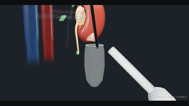
Schematic scene preview: Living-donor nephrectomy
Preparing the 3D anatomy…

UrOops3D · UroRef · © Nity G

Click me next Section 2 of 8 Anatomy The structures this operation works between, and the one it must not cross.

The structures in the scene

Living-donor nephrectomy: how the structures relateRight side down, the left kidney from the front, colon and spleen reflected. Left renal vein: behind and above; staple clear of the aorta Renal artery (danger). Left renal vein: enters from behind; clipped before rolling Lumbar vein (danger). Left renal vein: lift as a block; inside it the ureter dies The golden triangle (danger). Left renal vein: its vein enters from above; divided first The adrenal. Left renal vein: reflected medially; torn by traction Spleen, pancreatic tail and colon (danger). Left renal vein: made before the vessels are divided The extraction incision.Left renal vein!Renal artery!Lumbar vein!The golden triangleThe adrenal!Spleen, pancreatictail and colonThe extractionincisionbehind and above;staple clear of theaortaenters from behind;clipped beforerollinglift as a block;inside it theureter diesits vein entersfrom above; dividedfirstreflected medially;torn by tractionmade before thevessels are dividedTargetDangerLandmarkContext
Right side down, the left kidney from the front, colon and spleen reflected. Schematic, not to scale; relationships only.
  • The donor lies on the right side and the camera looks at the left kidney from the front with the colon and spleen reflected medially off Gerota.
  • The gonadal vein rises from the psoas to the underside of the renal vein, the ureter lies lateral to it, and the tissue between them and the lower pole is the golden triangle.
  • The renal vein crosses in front of the aorta to the cava with the adrenal vein entering from above and the lumbar vein from behind; the artery sits behind and above it.
  • ›The left renal vein - long, in front of the aorta to the cava, joined by the adrenal vein above, the gonadal below and a lumbar vein behind; the reason the left is preferred.
  • ›The renal artery - behind and above the vein; freed to the aorta without stripping, and stapled first, as long as it can be taken.
  • ›The golden triangle - bounded by the gonadal vein, the lower pole and the ureter; the ureteric blood supply lives in it and it is lifted as a block.
  • ›The lumbar vein - enters the back of the left renal vein near the gonadal; short, thin and tethered, clipped on both sides.
  • ›The adrenal - peeled off the upper pole after its vein is divided; left in the donor on its own supply.
  • ›The spleen, pancreatic tail and colon - reflected medially as a unit with the mesocolon; injured by traction more than by instruments.
  • ›The extraction incision - a Pfannenstiel or lower midline made before the vessels are divided; the bag is in before the stapler fires.
Click me next Section 3 of 8 Steps The operation in order, with the landmark that guards each move.

Operative steps

  1. Who: the donor and the work-up A competent adult who volunteers freely, assessed on a pathway that is independent of the recipient team. The Human Tissue Authority independent assessor confirms consent without pressure or payment. Blood group and tissue typing with a crossmatch against the recipient, or entry into the paired exchange scheme if incompatible. Measured GFR by isotope clearance, corrected for age, above the threshold in the BTS and Renal Association guidance; blood pressure, urinalysis for protein and blood, HbA1c, BMI, and a cardiorespiratory assessment. Anything that would predict kidney disease later in the donor stops the process. The donor hears the risks plainly: death in about one in three thousand, major complications in a few per cent, a permanent loss of about a third of GFR, and lifelong follow-up.
  2. Which kidney: the left, and the imaging CT angiography with delayed urographic phase maps the arteries, the veins and the collecting systems on both sides. The left kidney is preferred because the left renal vein is long, running in front of the aorta to the cava, and a long vein makes the recipient anastomosis safe. The right is taken when the left has multiple arteries or a lesion, or when the right is the smaller or the poorer kidney by split function, because the rule is that the donor keeps the better kidney. The number of arteries is counted, early branching noted, and a retro-aortic or circumaortic left vein identified before the first port, because that anatomy changes how the vein is taken. The imaging is read by the surgeon, not just the report.
  3. Position and ports General anaesthesia, a urethral catheter, thromboprophylaxis with stockings and calf compression, and a litre or more of crystalloid running before the dissection starts, because a well-filled donor keeps the kidney perfused through the pneumoperitoneum. Lateral decubitus at about sixty degrees with the table gently broken, the kidney side up, all pressure points padded, the arm on a rest. Pneumoperitoneum at 12 to 15 millimetres of mercury, a camera port at or beside the umbilicus, two or three working ports along the lateral rectus border and subcostally, and a hand port through a Pfannenstiel incision if the hand-assisted technique is used, which also serves as the extraction site. The robot docks from the back in the robotic variant.
  4. Reflecting the colon and the plane on Gerota The white line of Toldt is incised from the splenic flexure to the pelvic brim and the descending colon reflected medially, taking the splenocolic and splenorenal ligaments so the spleen and the tail of the pancreas fall away with the colon. The plane is between the mesocolon in front and Gerota's fascia behind, avascular when it is the right one; bleeding means the dissection has strayed into the mesocolon. Gerota is left intact over the kidney at this stage. The psoas is found at the lower pole and followed medially to the gonadal vein and the ureter, which lie on it. On the right the duodenum is kocherised off the cava.
    where it bites: the splenic capsule and the tail of the pancreas on the left; the duodenum on the right. Traction, not the instrument, is what tears them.
  5. The gonadal vein, the ureter and the golden triangle The gonadal vein is followed up to the renal vein on the left, or to the cava on the right, and it is the guide to the hilum. The ureter lies lateral to it on the psoas. The golden triangle is the tissue bounded by the gonadal vein medially, the lower pole of the kidney above and the ureter laterally: the ureteric blood supply runs in it, and it is lifted as a block with the ureter, never dissected inside. The ureter and gonadal vein with their fat are elevated together off the psoas down to the pelvic brim, where the ureter will be divided at the end, as long as it can be made. The gonadal vein is clipped and divided where it joins the renal vein.
    where it bites: a ureter stripped of its adventitia or dissected out of the golden triangle is a ureter that necroses in the recipient and leaks at ten days.
  6. The adrenal and lumbar veins With the ureter and gonadal vein lifted, the renal vein is exposed on its anterior surface. On the left the adrenal vein enters its upper border, sometimes with the inferior phrenic joining it, and is clipped and divided so the adrenal can be left behind on its own supply. One or more lumbar veins enter the posterior surface of the left renal vein close to the gonadal vein, running down behind it to the lumbar plexus of veins. They are found by rolling the renal vein gently forward, clipped on both sides and divided. This frees the vein to its full length to the cava. The adrenal is peeled off the upper pole and the upper pole freed from the diaphragm and the spleen.
    where it bites: the lumbar vein behind the left renal vein is short, thin-walled and tethered; torn, it retracts into the psoas and bleeds where it cannot be seen.
  7. The renal artery and vein to the aorta and the cava The renal artery lies behind and above the vein. It is found by following the vein medially and dissecting the tissue between, or by approaching from below along the psoas, and freed circumferentially back to the aorta on the left, or to its origin behind the cava on the right, without stripping the adventitia, and lymphatics clipped rather than cut. A second artery seen on the CT is found and freed in the same way. The kidney is then mobilised completely: posterior attachments off the psoas and quadratus, the upper pole free, and the ureter free to the pelvic brim, so it hangs by the artery, the vein and the ureter alone. Mannitol and, in most units, frusemide are given and the diuresis confirmed.
  8. Stapling the vessels and the extraction The extraction incision is made before the vessels are divided, a Pfannenstiel or a small lower midline with its peritoneum intact, or the hand port already in place, and the bag introduced. The ureter is clipped at the pelvic brim and divided above the clip. The artery is divided first with a vascular stapler as close to the aorta as it can safely be placed, then the vein with a second stapler flush with the cava; the artery goes first so the kidney does not become congested. Polymer clips alone are not used on the donor renal artery. The kidney is placed in the bag and delivered through the incision by hand without twisting the pedicle. Warm ischaemia starts when the artery is stapled and stops when cold perfusion begins.
    where it bites: the staple line must lie on healthy vessel with the full length taken; a stapler fired across the aorta or a stump too short to close is the donor's emergency.
  9. The kidney on ice, and the donor after On the bench the kidney goes into a bowl of ice slush and the artery is cannulated and flushed with cold preservation solution at 4 degrees, hyperosmolar citrate or UW or HTK, until the venous effluent is clear, within a warm time that should be under five minutes. The staple lines are trimmed, the anatomy checked and the kidney packed on ice for the recipient theatre. In the donor the staple lines are inspected under reduced pressure, the extraction wound closed in layers, and the ports closed. The donor mobilises the same day and goes home at two to four days. Creatinine rises by about a third and settles; the donor is reviewed at six weeks and then yearly for life with blood pressure, eGFR and urinalysis, on a register.
Click me next Section 4 of 8 Danger zones Where this operation bites, and why it bites there.
Critical relationship

Danger zones

The spatial relationships the animation is built to make automatic. Before you read each one, ask yourself: at this point in the operation, what is about to go wrong, and what is my first move? The save for each sits in the Complications tab.

  • RiskThe lumbar vein behind the renal vein - torn on rolling the vein forward, it retracts and bleeds from the psoas; it is found, clipped both sides and divided before the vein is freed.
  • RiskThe renal artery at the aorta - a stapler placed on the aorta itself, or a short stump that will not close, is a vascular emergency in a healthy person.
  • RiskThe golden triangle and the ureteric adventitia - dissected inside, the ureter loses its blood supply and necroses in the recipient.
  • RiskThe spleen, pancreas, duodenum and pleura - the organs beside the upper pole on each side, torn by retraction and diaphragm dissection.
Practise it Take me into the scene Opens the animation at a danger moment and asks you to predict the mechanism and your first move before it reveals.
Click me next Section 5 of 8 Complications The save for each one: the first move, the escalation trigger, the op-note line.

Complications and immediate saves

Trigger each of these live in the Animation tab, then read the save: the first move, the escalation trigger, and the line that goes in the op note.

Major vascular bleeding

  • a torn lumbar vein, a slipped clip on the gonadal or adrenal vein, or a stapler misfire on the renal artery or vein: the field red in a healthy donor who has no reason to be harmed.
The save
  1. Pressure on the point with a swab or the suction, raise the pneumoperitoneum briefly, and tell the anaesthetist and the scrub team in one sentence. first move
  2. A vein: hold pressure two minutes, then a clip or a suture under vision; the lumbar vein is followed into the psoas and clipped, not coagulated blind.
  3. A stapler that has misfired or a stump that bleeds: grasp the stump with an atraumatic grasper and suture it, or place a second stapler if there is length.
  4. Bleeding not controlled in minutes, or arterial bleeding at the aorta: open through the extraction incision or a midline at once, with vascular help called. escalation trigger
  5. Op note: the vessel, the control, the blood loss and transfusion, whether conversion occurred, and the donor told plainly on the ward. document

Injury to the spleen, pancreas, bowel or pleura

  • a splenic capsular tear from retraction on the left, a pancreatic tail injury in the wrong plane, a duodenal or colonic injury from a hot instrument, or the pleura opened behind the upper pole.
The save
  1. Stop retracting; look at the injury with the pressure lowered and decide whether it is bleeding, leaking or neither. first move
  2. A splenic capsular tear: haemostatic agent and pressure; a deep tear is discussed with general surgery, and splenectomy is the last resort.
  3. A bowel injury seen: repair in two layers laparoscopically or open; a pancreatic injury: a drain and the pancreas team told.
  4. An injury not recognised until the ward, with peritonitis, high drain amylase or a pleural effusion: CT and a return to theatre with the right team. escalation trigger
  5. Op note: the organ, the mechanism, the repair and the drain plan; the recipient team told if the graft is affected. document

Damage to the graft

  • a short artery, a torn vein, an intimal flap from traction, a stripped ureter or a capsular tear: the donor is fine and the recipient pays.
The save
  1. Tell the recipient team before the kidney leaves the room; the bench is where a short or damaged vessel is assessed and repaired. first move
  2. A short artery is extended with the recipient's internal iliac or a vein patch; a short right vein is lengthened with a caval cuff only in an open right donor, or with gonadal vein on the bench.
  3. A stripped or short ureter is used with the recipient's native ureter as a ureteroureterostomy rather than to the bladder.
  4. A vessel that cannot be reconstructed or a kidney that will not flush: the transplant consultant decides whether the graft is usable. escalation trigger
  5. Op note: warm ischaemia time, the vessel lengths, the number of arteries, any injury and the bench repair, on the donor and the graft form. document

Conversion to open

  • bleeding that will not stop, anatomy that does not match the CT, dense adhesions, or a graft in danger: the decision to open is a good one when it is made early.
The save
  1. Say the word aloud and open through the extraction incision or a midline while the assistant holds pressure; the instruments for open are already in the room. first move
  2. Once open, control the pedicle by hand, then the bleeding point under vision; the kidney is completed as an open donor nephrectomy.
  3. The donor was consented for conversion at the outset, with the rate quoted from the unit's own figures.
  4. A donor who is unstable after control, or a kidney whose viability is in doubt: blood, critical care and the recipient theatre told immediately. escalation trigger
  5. Op note: the reason for conversion, the timing, the findings, and the incision, for the donor register. document

The donor after: pain, hernia, thrombosis and the single kidney

  • shoulder-tip and wound pain, chronic pain or numbness at the Pfannenstiel, an incisional hernia, venous thromboembolism, and the long-term cost of a single kidney: a lower GFR, a higher blood pressure, and a small lifetime risk of kidney failure.
The save
  1. Thromboprophylaxis until fully mobile, early mobilisation, regular analgesia and a wound checked before discharge. first move
  2. Creatinine at discharge and six weeks; a rise of about a third that settles is expected, a rise that continues is investigated.
  3. Blood pressure, eGFR and urinalysis yearly for life on the living donor register, with lifestyle advice and treatment of hypertension.
  4. New proteinuria, a falling eGFR or resistant hypertension: nephrology review; a donor is never lost to follow-up. escalation trigger
  5. Note: the risks quoted at consent, the discharge creatinine, the follow-up plan and the register entry. document
Click me next Section 6 of 8 Troubleshooting When the case turns: the problem, then the next move.

Intra-operative troubleshooting

When the case turns: the problem, then the next move.

  • ActionThe colon will not fall away and the plane bleeds - the dissection is in the mesocolon; come back to the white line and find the avascular plane on Gerota, then take the splenic attachments high so the spleen goes with the colon.
  • ActionThe ureter cannot be found - find the gonadal vein on the psoas and look lateral to it at the pelvic brim, where the ureter crosses the iliac vessels; lift the gonadal vein and the ureter comes with it.
  • ActionA second artery appears that was not on the CT - free it fully to the aorta as with the first; tell the recipient team, and take it with its own staple line or with the main artery on a common cuff if they arise together.
  • ActionThe renal vein is retro-aortic or circumaortic - the retro-aortic vein is short and posterior; the dissection stays close to the kidney, the stapler goes on as much length as is safe, and the recipient is warned that the vein is short.
  • ActionThe kidney goes dusky after the artery is freed - spasm from handling: warm saline on the vessel, papaverine if the unit uses it, wait, check the filling and pressure; do not staple a kidney that has not pinked up unless the spasm will not resolve.
  • ActionThe stapler fails to fire or jams on the vein - do not pull it; leave it closed, place a second stapler proximally if there is length, or open through the extraction incision and control the stump with a clamp.
Click me next Section 7 of 8 FRCS viva Say it out loud, then mark yourself against the model answer.

FRCS viva questions

The examiner stems this scene prepares you for. Try to answer out loud first, then reveal, and learn the discriminator, the detail that turns a pass into a strong pass.

QHow is a living kidney donor assessed, and what are they told?reveal answer
  • I would describe a pathway that is separate from the recipient's and designed to protect the donor.
  • Donation must be voluntary, informed and without payment, confirmed by an independent assessor under the Human Tissue Act.
  • Medically I would confirm the absence of anything that predicts future kidney disease: a measured isotope GFR above the age-adjusted threshold in the BTS and Renal Association living donor guidance, no proteinuria or haematuria, blood pressure controlled without organ damage, no diabetes, and a BMI and cardiorespiratory fitness safe for anaesthesia.
  • Immunologically, blood group and HLA typing with a crossmatch against the recipient, or entry into the paired exchange scheme if incompatible.
  • Anatomically, CT angiography with a urographic phase to count arteries and veins on each side and to choose the side, keeping the better kidney with the donor.
  • Psychosocially, a formal assessment and a period to withdraw.
  • I would tell them the perioperative mortality is about one in three thousand, that major complications occur in a few per cent, that they will lose about a third of their GFR permanently with a small increase in the lifetime risk of kidney failure and hypertension, and that they will be followed for life.
Strong pass

Say both

  • The independent assessor and the separation of donor and recipient pathways as the safeguard
  • The mortality quoted as a number with the lifelong follow-up that follows from the single kidney
Examiner note
The independent assessor and the separation of donor and recipient pathways as the safeguard, and the mortality quoted as a number with the lifelong follow-up that follows from the single kidney.
QWhy is the left kidney preferred, and when would you take the right?reveal answer
  • The left renal vein is long, passing in front of the aorta to reach the cava, so the graft comes with a vein that can be sewn to the external iliac vein without tension, whereas the right renal vein is short and enters the cava directly, giving a stump that may be too short for a safe anastomosis, and one that cannot be lengthened laparoscopically with a caval cuff.
  • The left is therefore the default.
  • I would take the right when the CT shows the left with multiple arteries or early branching that would make reconstruction complex, when the right kidney is the smaller or the poorer kidney by split function, since the donor must keep the better kidney, or when there is a lesion such as a cyst or stone on the right that would be better removed.
  • In a woman who may become pregnant some units also prefer to take the right because right hydronephrosis of pregnancy is commoner.
  • A retro-aortic or circumaortic left vein is not a contraindication but changes the dissection and shortens the vein, and the recipient surgeon is told.
Strong pass

The rule that the donor keeps the better kidney stated as the principle that overrides the preference for the left.

QDescribe the steps of a laparoscopic left donor nephrectomy and the danger points.reveal answer
  • With the donor in the right lateral position and filled with crystalloid, I would establish a pneumoperitoneum and reflect the descending colon medially on the plane between the mesocolon and Gerota's fascia, taking the splenic attachments so the spleen and pancreatic tail fall away.
  • I would find the gonadal vein on the psoas and follow it to the renal vein, lifting the ureter and the gonadal vein with the tissue of the golden triangle as a block so the ureteric blood supply is preserved.
  • I would clip the gonadal vein at the renal vein, the adrenal vein at the upper border, and the lumbar vein entering the posterior surface, which is the vessel that bleeds if torn.
  • I would free the artery to the aorta behind the vein without stripping it, mobilise the kidney completely, give mannitol, make the Pfannenstiel extraction incision and introduce the bag before dividing anything.
  • I would clip and divide the ureter at the pelvic brim, staple the artery first close to the aorta and then the vein flush with the cava, not using polymer clips alone on the artery, bag the kidney and extract it by hand, and flush it with cold preservation solution within a warm ischaemia time under about five minutes.
Strong pass

Say all three

  • The golden triangle named and left alone
  • The lumbar vein found before the vein is freed
  • The extraction incision and bag prepared before the stapler fires
Examiner note
The golden triangle named and left alone, the lumbar vein found before the vein is freed, and the extraction incision and bag prepared before the stapler fires.
QWhat are the complications of living-donor nephrectomy, for the donor and for the graft?reveal answer
  • For the donor I would separate the immediate from the lifelong.
  • Immediately: major vascular bleeding from the lumbar, gonadal or adrenal veins or a staple line failure on the renal vessels, which is the reason a healthy donor can die, at about one in three thousand;
  • injury to the spleen, pancreas, colon, duodenum or pleura, which may need conversion to open, in a few per cent;
  • and the general risks of a laparoscopic operation, port site bleeding, wound infection, ileus, venous thromboembolism, chest infection and cardiac events.
  • Later: chronic wound pain, numbness and incisional hernia at the extraction site.
  • Lifelong: an immediate fall in GFR of about a third with partial compensation to about seventy per cent of baseline, a modest rise in blood pressure and proteinuria over decades, and a small absolute increase in end-stage kidney disease compared with equally healthy non-donors, which is why the follow-up is yearly for life.
  • For the graft: a short or injured artery or vein, an intimal flap from traction, a ureter devascularised by dissection in the golden triangle leading to necrosis and leak in the recipient, capsular tears, and a prolonged warm ischaemia time causing delayed graft function.
Strong pass

Say both

  • Separating the donor's risks from the graft's
  • Naming ureteric devascularisation as the injury that the donor never notices and the recipient pays for
Examiner note
Separating the donor's risks from the graft's and naming ureteric devascularisation as the injury that the donor never notices and the recipient pays for.
Click me next Section 8 of 8 Evidence The guidance each of these answers is built on.

Evidence and guidance

Two minutes

Close the lap: recall it before you leave.

Say it aloud, then reveal. Reading is not remembering.

  1. 01 The relationship Say the critical spatial relationship out loud, then reveal. reveal
    Memory frame

    The donor lies on the right side and the camera looks at the left kidney from the front with the colon and spleen reflected medially off Gerota.

    Why?
    The gonadal vein rises from the psoas to the underside of the renal vein, the ureter lies lateral to it, and the tissue between them and the lower pole is the golden triangle. The renal vein crosses in front of the aorta to the cava with the adrenal vein entering from above and the lumbar vein from behind; the artery sits behind and above it.
  2. 02 The first move Major vascular bleeding. Your first move? reveal
    First move

    Pressure on the point with a swab or the suction, raise the pneumoperitoneum briefly, and tell the anaesthetist and the scrub team in one sentence.

  3. 03 The escalation trigger Major vascular bleeding. What makes you call for help? reveal
    Escalate when

    Bleeding not controlled in minutes, or arterial bleeding at the aorta: open through the extraction incision or a midline at once, with vascular help called.

  4. 04 The discriminator What turns a pass into a strong pass? reveal
    Strong pass

    Say both

    • The independent assessor and the separation of donor and recipient pathways as the safeguard
    • The mortality quoted as a number with the lifelong follow-up that follows from the single kidney
    Examiner note
    The independent assessor and the separation of donor and recipient pathways as the safeguard, and the mortality quoted as a number with the lifelong follow-up that follows from the single kidney.
  5. 05 The pitfall Where does this operation bite first? reveal
    Pitfall

    The lumbar vein behind the renal vein

    Why?
    torn on rolling the vein forward, it retracts and bleeds from the psoas; it is found, clipped both sides and divided before the vein is freed.

Revision practice with self-marked answers, not an assessment of clinical competence. Kept in this browser only; it does not sync between devices.