
Living-donor nephrectomy
Removal of a kidney from a healthy donor with maximal donor safety and graft vessel/ureter quality as coequal operative objectives.
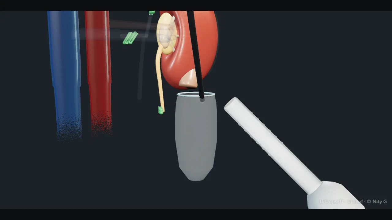 Interactive 3D Interactive laparoscopic donor nephrectomy Open the 3D animation
Teaching catalogue only. This coverage record is not a patient pathway or operative instruction.
Typical place in practice
A fully assessed voluntary living donor accepted through an independent medical, surgical and psychosocial governance pathway.
Alternatives and neighbouring procedures
Deceased-donor transplantation, another suitable living donor, paired exchange, dialysis or conservative kidney care for the intended recipient.
Same procedure family, explicit technique branches
These are variations of this canonical operation. Complication modules below identify whether they apply to all branches or only to named approaches.
Transperitoneal laparoscopic donor nephrectomy
Provides broad anatomical access and rapid graft extraction.
Hand-assisted laparoscopic donor nephrectomy
Allows tactile assistance and manual graft retrieval.
Robotic donor nephrectomy
Uses articulated minimally invasive instrumentation with a planned rapid retrieval route.
Open donor nephrectomy
A selected route for donor anatomy, prior surgery or programme practice.
Right or multiple-vessel donor nephrectomy
Balances donor benefit with short vein or accessory-vessel challenges for the graft.
High-level sequence
Coverage outline only; not a step-by-step technical guide.
- Confirm donor autonomy, fitness, anatomy and side selection
- Mobilise kidney while preserving organ and donor structures
- Prepare maximal safe arterial, venous and ureteric length
- Coordinate timed vascular division, extraction and graft perfusion
- Monitor donor renal function, wound recovery and long-term health
Operative notes
Concise operative notes covering the key steps, landmarks and watch points, in the order you encounter them.
Laparoscopic setup rules
What it does: safe ports and position, any lap case.
- Hasson first port: open cut-down. Safe. (Veress needle: ~4% problems.)
- Side lying. Break the table under the 12th rib.
- Axillary roll. Pillow between legs. Gel pads.
- Boots and warmer on.
- Ports under vision, camera inside.
- 2026 update: Antibiotics: 1 dose, local policy, allergy aware. The old fixed gent + cefuroxime recipe is gone.
From the UroRef Operative Quick Guide. Open the note page →
Every failure mode is its own module.
Timing and variation coverage are recorded for each module.
Major vascular injury or haemorrhage
Healthy donors can sustain life-threatening injury to renal vessels, aorta, vena cava or branches.
Applies to: All variantsBowel, pancreatic, splenic, hepatic or pleural injury
Upper abdominal dissection and access can injure adjacent organs.
Applies to: All variantsConversion to open surgery
Bleeding, anatomy or graft-safety concern may necessitate rapid open exposure.
Applies to: Transperitoneal laparoscopic donor nephrectomy · Hand-assisted laparoscopic donor nephrectomy · Robotic donor nephrectomyVenous thromboembolism, cardiopulmonary event or death
Even a carefully selected healthy donor faces rare serious perioperative harms.
Applies to: All variantsWound pain, hernia, neuropathy or chronic symptoms
Extraction and flank/port wounds can have durable effects.
Applies to: All variantsReduced kidney function or later hypertension
Nephrectomy permanently reduces nephron mass and requires long-term donor follow-up.
Applies to: All variantsGraft vessel, ureter or parenchymal injury
Excess traction, short division or thermal injury can compromise the donated organ.
Applies to: All variantsSources for this module
Living-donor nephrectomy, recipient surgery and transplant complications.
Accessed 2026-08-03 · opens source British Association of Urological Surgeons Patient information leaflets by procedureUK patient-facing procedure, alternatives, recovery and complication framing.
Accessed 2026-08-03 · opens source