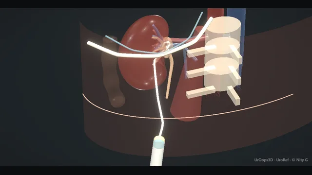
Percutaneous nephrostomy: decompression of the obstructed kidney
A hydronephrotic, obstructed system decompressed through a posterior calyx: ultrasound-guided puncture, urine for culture, wire, tract, pigtail. The neighbours decide the danger: retrorenal colon laterally, pleura above the 12th rib, segmental vessels inside the kidney.
Core tierFRCS endourology, emergency drainageInteractive 3D
Prone, the needle line from skin to posterior calyx. Schematic, not to scale; relationships only.
The needle line runs skin to posterior calyx with three neighbours to clear: the pleura above the 12th rib, the colon on the lateral line, and the segmental arteries running along the calyceal infundibula inside the kidney.
›Posterior calyx - the target: through parenchyma, along the infundibular axis, away from the hilar vessels.
›Dilated pelvis + calyces - the system you are decompressing; efflux of urine is the confirmation you are inside it.
›Retrorenal colon - thin patient or previous surgery can swing it behind the kidney and across the line.
›Pleural reflection - rises above the 12th rib; a supracostal line enters the chest.
Ultrasound surveyThe side is confirmed, the clotting and platelets are acceptable or corrected, antibiotics are given or already running, and the patient is positioned prone; the flank is surveyed with ultrasound before the skin is prepped with intent. The scan confirms the dilated system and a posterior calyx is chosen below the 12th rib, on a line that clears colon laterally and pleura above. The same images show whether liver or spleen dips into the corridor, the depth of parenchyma the needle will cross, and the axis of the infundibulum. The line is decided here, on the scan: a corridor negotiated after the needle is inserted is already a compromised one. where it bites: the line is chosen on the scan, not with the needle.
Calyceal puncture + aspirationThe needle advances along the anaesthetised line through the parenchyma and into the chosen calyx, entering through the papilla along the infundibular axis. Urine is aspirated and sent for culture before anything else follows, because that sample is one of the reasons this route was chosen. Efflux at the needle hub confirms the tip sits in the collecting system, and the needle is steadied for the wire. The system is decompressed deliberately, not emptied: aspirating an infected pelvis dry before the wire is in risks collapsing the very space the next pass must find. where it bites: aspirating the pelvis dry before wire placement risks losing the system.
Wire (Seldinger)A guidewire is passed through the needle and advanced under screening until it coils in the pelvis with spare length inside the system. The coil is the security: it holds the wire in the kidney when the needle comes out and through every exchange that follows. The wire is controlled at the skin by one person throughout, and every pass over it happens with the wire visible at both ends. A wire advanced against resistance perforates the collecting system, and a wire left unattended is a wire that will perforate.
Tract + pigtail drainThe tract is dilated over the wire in graded steps, staying exactly on the line the needle made, and the pigtail is railroaded into the pelvis over the same wire. Screening shows the pigtail curl before the stiffener is released, because the coil is the only thing holding the drain in place. Every movement is small, deliberate and on the image, and nothing is advanced against resistance. Dilation off the needle axis, or over a wire with no intra-renal purchase, displaces the kidney or tears the parenchyma. where it bites: dilating off-axis or over a wire with no intra-renal purchase displaces the kidney or tears the parenchyma.
Secure + checkThe drain is aspirated to confirm free flow, a final sample is sent, and the drain is secured to skin at its entry site without tension or kinking. Position is confirmed on a film or CT before the patient leaves the table, because a pigtail that looks right at the skin can sit outside the system. Output and its colour are documented, and the ward plan names what the drain is for and who acts on it. The operation was decompression, not stone clearance, and it is finished when the kidney is draining, not when the dressing is on.
The spatial relationships the animation is built to make automatic. Before you read each one, ask yourself: at this point in the operation, what is about to go wrong, and what is my first move? The save for each sits in the Complications tab.
RiskInfundibular axis - a puncture across the infundibulum, not along it, is the classic segmental artery injury.
RiskRetrorenal colon - seen on the pre-procedure CT or scan; if it sits behind the kidney, choose the line with it in mind.
RiskSupracostal line - above the 12th rib the pleura is on the path; hydrothorax and haemothorax live here.
When the case turns: the problem, then the next move.
ActionNo efflux from a correctly placed-looking needle - re-image; do not advance deeper hoping for urine - the calyx may be septated or the tip intraparenchymal.
ActionWire will not coil in the pelvis - rotate and withdraw slightly; a wire wedged in an infundibulum perforates if pushed.
ActionKidney pushed away by the needle - stop, withdraw to the capsule and re-advance with a firmer, more perpendicular line.
ActionBloody efflux obscuring the system - aspirate gently, leave the needle for tamponade, re-image before any further puncture.
The examiner stems this scene prepares you for. Try to answer out loud first, then reveal, and learn the discriminator, the detail that turns a pass into a strong pass.
QA septic patient with an obstructed kidney: stent or nephrostomy, and why?reveal answer
The emergency is decompression of an infected obstructed system, and both routes achieve it.
My choice depends on what is available out of hours and on the anatomy.
A percutaneous nephrostomy gives reliable, large-bore drainage, a sample for culture directly from the system, and a route for later stone treatment; it needs imaging equipment and a prone patient.
A retrograde JJ stent avoids a flank puncture and its bleeding and bowel risk, but the obstructed system may be impossible to cannulate, drainage around the stent is narrower, and it usually needs a general anaesthetic in a septic patient.
If I cannot clear the obstruction from below within minutes, or the patient cannot tolerate a prolonged attempt, I stop and go percutaneous.
Either way, antibiotics and resuscitation run in parallel; drainage is the definitive sepsis source control.
Strong pass
Say all three
Framing both as source control
Choosing by what the obstruction and the patient tolerate
Not by operator preference
Examiner note
Framing both as source control, and choosing by what the obstruction and the patient tolerate, not by operator preference.
QWhere exactly do the renal vessels run relative to your puncture, and why does the axis matter?reveal answer
The segmental and interlobar arteries run along the calyceal infundibula and then dive alongside the papillae as arcuate vessels at the corticomedullary junction.
A puncture through a papilla, along the infundibular axis, passes between the segmental branches, which is why the papilla is the avascular plane of entry.
A puncture that crosses an infundibulum, or enters the system through the renal pelvis directly, transects those branches and bleeds, sometimes into a pseudoaneurysm that presents days later.
The same axis logic is why a posterior calyx is chosen: posterior parenchyma is thicker, the avascular line is longest, and the tract lies behind the hilar vessels.
Strong pass
Say both
Naming the papilla as the avascular entry point and the infundibular axis as the bleeding risk
Not just "avoid the hilum"
Examiner note
Naming the papilla as the avascular entry point and the infundibular axis as the bleeding risk, not just "avoid the hilum".
QCloudy urine under pressure on aspiration - what is your sequence?reveal answer
That is an infected obstructed system and the pressure tells me handling matters.
I would aspirate enough to decompress and send the sample for culture, but not aggressively empty the system, because sudden decompression of an infected kidney can drive bacteraemia.
I would complete the nephrostomy with the lowest number of exchanges over the wire, avoid forceful flushing, and sample bloods and cultures around the procedure.
Antibiotics already running is the ideal; if not, they start immediately.
I would watch for post-decompression sepsis - the patient can transiently worsen after source control - with early warning scoring and escalation to critical care if needed.
Strong pass
Say both
Controlled rather than complete decompression of the infected system
Anticipating post-drainage sepsis
Examiner note
Controlled rather than complete decompression of the infected system, and anticipating post-drainage sepsis.
QYour nephrostomy drain has stopped draining on day 2. What do you do?reveal answer
I would not flush it reflexively.
First, check the patient: fever, flank pain or rising inflammatory markers say the system is obstructed again or infected.
Then check the tube for kinking or dislodgement at the skin, and the collection bag and tubing for obstruction.
If the drain is simply blocked or malpositioned, imaging - a nephrostogram or CT - tells me whether the pigtail is still in the pelvis; I would reposition or exchange it over a wire in imaging rather than blind-flush.
If the kidney is decompressed - for instance the obstruction has been bypassed or stented - reduced output may be expected, and the imaging confirms that too.
The decision is imaging-led, and persistent undrained obstruction goes back to theatre or interventional radiology the same day.
Strong pass
Say all three
Patient before tube
Imaging before flushing
Same-day re-intervention for a blocked infected system
Examiner note
Patient before tube, imaging before flushing, and same-day re-intervention for a blocked infected system.
Say it aloud, then reveal. Reading is not remembering.
01The relationshipSay the critical spatial relationship out loud, then reveal.reveal
Memory frame
The needle line runs skin to posterior calyx with three neighbours to clear
Why?
The needle line runs skin to posterior calyx with three neighbours to clear: the pleura above the 12th rib, the colon on the lateral line, and the segmental arteries running along the calyceal infundibula inside the kidney.
02The first moveColonic transfixion. Your first move?reveal
First move
Recognise it: faeculent efflux, gas from the needle, or urine draining per rectum; a CT nephrostogram settles the anatomy.
03The escalation triggerColonic transfixion. What makes you call for help?reveal
Escalate when
Peritonism, intraperitoneal injury or deterioration → general surgical review; may need formal repair or diversion.
04The discriminatorWhat turns a pass into a strong pass?reveal
Strong pass
Say all three
Framing both as source control
Choosing by what the obstruction and the patient tolerate
Not by operator preference
Examiner note
Framing both as source control, and choosing by what the obstruction and the patient tolerate, not by operator preference.
05The pitfallWhere does this operation bite first?reveal
Pitfall
Infundibular axis
Why?
a puncture across the infundibulum, not along it, is the classic segmental artery injury.
Revision practice with self-marked answers, not an assessment of clinical competence. Kept in
this browser only; it does not sync between devices.