
Percutaneous nephrostomy insertion and exchange
Image-guided percutaneous access to the renal collecting system for external drainage, contrast assessment or later antegrade intervention.
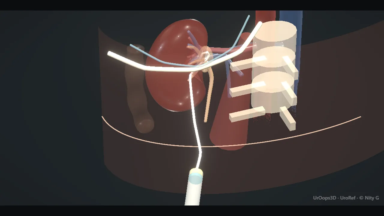 Interactive 3D Interactive operative model Open the 3D animation
Teaching catalogue only. This coverage record is not a patient pathway or operative instruction.
Typical place in practice
Urgent or planned drainage of an obstructed upper tract, urinary diversion after injury or leak, and creation or maintenance of antegrade access.
Alternatives and neighbouring procedures
Retrograde ureteric stenting, definitive treatment, conservative management or surgical drainage according to anatomy and urgency.
Same procedure family, explicit technique branches
These are variations of this canonical operation. Complication modules below identify whether they apply to all branches or only to named approaches.
Ultrasound-guided nephrostomy
Ultrasound identifies the collecting system and adjacent organs during initial puncture.
Fluoroscopic nephrostomy
Contrast and fluoroscopy guide calyceal puncture, wire passage and drain placement.
Combined ultrasound and fluoroscopy
Ultrasound guides safe entry and fluoroscopy confirms wire and catheter position.
Nephrostogram and tube exchange
An existing tract is assessed and the tube replaced while access is preserved.
High-level sequence
Coverage outline only; not a step-by-step technical guide.
- Confirm side, indication, coagulation context and cross-sectional anatomy
- Plan a calyceal path that avoids pleura, bowel and major vessels
- Puncture and confirm collecting-system entry
- Secure wire access and place a freely draining catheter
- Confirm position, secure the tube and document an exchange or removal pathway
Operative notes
Concise operative notes covering the key steps, landmarks and watch points, in the order you encounter them.
Nephrostomy / kidney access
What it does: drain the kidney through the back, over a wire.
- Prone. (Or supine for ECIRS; see the PCNL operative note.)
- Posterior axillary line. Midway, crest to 12th rib.
- Through parenchyma into a POSTERIOR calyx. (Calyx = a cup of the kidney's collecting system.) Fewer vessels. Stable track.
- 16-18G needle under ultrasound. Urine back into a syringe: send it for culture. No urine: re-image, do not push.
- Wire in, coiled in the pelvis on screening. Needle out. Dilate over the wire.
- Pigtail 8-10 Fr over the wire. Curl in the pelvis. Wire out. Aspirate to check. Stitch to skin.
- Watch: Bleeding (rarely embolisation).
- Watch: Pneumothorax above the 12th rib.
- Watch: Colon, spleen, liver injury. Sepsis.
From the UroRef Operative Quick Guide. Open the note page →
Every failure mode is its own module.
Timing and variation coverage are recorded for each module.
Haemorrhage or vascular injury
The tract may injure renal parenchymal or segmental vessels, producing haematuria, perirenal bleeding or a later vascular lesion.
Applies to: Ultrasound-guided nephrostomy · Fluoroscopic nephrostomy · Combined ultrasound and fluoroscopy · Nephrostogram and tube exchangePleural injury, hydrothorax or pneumothorax
A high intercostal tract may traverse pleura or diaphragm and cause respiratory compromise or fluid in the chest.
Applies to: Ultrasound-guided nephrostomy · Fluoroscopic nephrostomy · Combined ultrasound and fluoroscopyVisceral injury
Colon and solid organs may lie in the proposed tract, particularly with unusual anatomy or previous surgery.
Applies to: Ultrasound-guided nephrostomy · Fluoroscopic nephrostomy · Combined ultrasound and fluoroscopyUrinary infection or sepsis
Puncture, contrast injection or impaired drainage in an infected obstructed system can precipitate systemic illness.
Applies to: Ultrasound-guided nephrostomy · Fluoroscopic nephrostomy · Combined ultrasound and fluoroscopy · Nephrostogram and tube exchangeUrine extravasation or perinephric collection
A displaced or poorly draining tube can leak urine outside the collecting system.
Applies to: Ultrasound-guided nephrostomy · Fluoroscopic nephrostomy · Combined ultrasound and fluoroscopy · Nephrostogram and tube exchangeTube displacement, blockage or disconnection
Loss of secure external drainage may recreate obstruction, leak or infection and can result in tract loss.
Applies to: Ultrasound-guided nephrostomy · Fluoroscopic nephrostomy · Combined ultrasound and fluoroscopy · Nephrostogram and tube exchangeFailure to gain or retain access
A non-dilated system, difficult anatomy or wire displacement can make safe tube placement impossible.
Applies to: Ultrasound-guided nephrostomy · Fluoroscopic nephrostomy · Combined ultrasound and fluoroscopy · Nephrostogram and tube exchangeSources for this module
SWL, ureteroscopy, RIRS, PCNL, bladder stones and decompression options.
Accessed 2026-08-03 · opens source European Association of Urology EAU Guidelines on Urological InfectionsInfection prevention, recognition and procedure-associated infection context.
Accessed 2026-08-03 · opens source British Association of Urological Surgeons Patient information leaflets by procedureUK patient-facing procedure, alternatives, recovery and complication framing.
Accessed 2026-08-03 · opens source