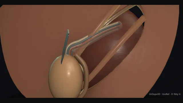
A groin-crease orchidopexy taught along the line of the inguinal canal: the testis parks at the external ring, its vessels and vas run obliquely through the canal wrapped in a patent processus vaginalis, and the operation wins the distance from the external ring to the bottom of the scrotum without tension.
The pouch at the end holds the testis by fitting it, not by squeezing it.
Core tierFRCS paediatric urology, genital surgeryInteractive 3D
Inguinal canal to scrotum, one line from internal ring to scrotal floor. Schematic, not to scale; relationships only.
The canal and the scrotum are two ends of one line.
The undescended testis sits at the upper end, at or just below the external ring, its cord running obliquely back through the canal to the internal ring where the vessels and vas diverge: the vessels up the retroperitoneum, the vas down into the pelvis.
The operation converts this oblique course into a direct vertical drop into the scrotal floor, and every manoeuvre, sac ligation at the neck, retroperitoneal release through the ring, pouch seating at the bottom, buys a little more of that vertical distance.
The sub-dartos pouch is the anchor at the lower end, and its neck is where the cord finally stops moving.
›Inguinal canal + rings - the oblique course from the internal ring superolaterally to the external ring over the pubic tubercle; the axis along which every mobilising move is made.
›Testicular vessels - the single arterial supply of the testis, running the length of the cord; injured by stripping, kinking or over-enthusiastic diathermy.
›Vas deferens - firm and pale, adherent within the wall of the processus vaginalis; the structure the sac dissection endangers.
›Patent processus vaginalis - the peritoneal sac wrapping the cord; separated and ligated at its neck, which simultaneously repairs the associated hernia.
›Sub-dartos pouch - the pocket in the dartos layer that holds the testis in the scrotum; its neck is the balance between escape and strangulation.
Palpable vs impalpableExamination, repeated under anaesthesia on the day, sets the pathway. A palpable testis, sitting in the canal or at the external ring, is approached through a groin crease incision so the whole length of the canal is available for mobilisation. An impalpable testis goes to laparoscopy first, because its position must be known before any incision is made. A scrotal incision alone is reserved for the low, mobile testis confirmed in the scrotum under anaesthesia: planning scrotal-only for a testis that then cannot be reached abandons the cord length the canal approach provides. where it bites: planning a scrotal-only approach for a testis that then cannot be reached abandons the cord length the canal approach provides.
Groin crease incisionA skin-crease incision is made over the external ring, along the line of the canal, and deepened through the subcutaneous fat and Scarpa's fascia to the external oblique aponeurosis. The aponeurosis is opened onto the ring and along its fibres, which puts the whole canal under direct vision. The incision sits over the ring and not over the testis: the testis is mobilised up to the incision along the canal axis, and working along that axis is what buys the length needed later. where it bites: the incision is placed over the ring, not over the testis: the testis is mobilised to the incision along the canal axis.
Deliver the testisThe testis is delivered into the wound with the cord intact, and the coverings over the cord are opened along their fibres. Three structures are then identified and named before anything is divided: the testicular vessels, the vas deferens, and the patent processus vaginalis lying between them. Nothing is cut until all three are positive, because the sac hides the vas within its wall, and a structure that has not been found is a structure that can be injured. where it bites: nothing is divided until all three structures are positively identified; the sac hides the vas in its wall.
Separate + ligate the sacThe patent processus vaginalis is separated from the cord by gentle sweeping from the internal ring down to the testis, keeping the plane on the sac and the field dry enough to see. The vas sits adherent within the sac wall, so no clip or ligature closes until the vas is accounted for, and the sac is handled cold and blunt rather than hot and sharp. The sac is then transfixed at its neck with an absorbable ligature and divided: ligation of the sac is also the repair of the associated hernia, done at the same moment. where it bites: the vas is adherent within the sac wall and is the structure at risk in this dissection: sweeping rather than sharp clearance, cold rather than hot division.
Mobilise to gain lengthThe cord is freed proximally, back to the internal ring and then through it into the retroperitoneal fat, where the vessels hold their true length. Lateral cremasteric bands are divided and the testis is checked against the scrotal floor: it must reach without tension, helped where needed by formal retroperitoneal release. Length comes from release, never from pulling, because a stretched testicular vessel behaves like a ligated one, and the atrophy declares itself weeks later in clinic rather than on the table. where it bites: length comes from release, never from pulling: a stretched vessel is a ligated vessel, and the atrophy declares weeks later.
Sub-dartos pouchA pouch is developed in the dartos of the scrotum, through the same groin incision or a small scrotal one, sized to admit the testis comfortably. The testis is drawn down into it along a straight path, with the cord confirmed untwisted and untethered before seating. The pouch neck is then closed around the cord, calibrated around the cord rather than the testis: a neck too tight strangulates the blood supply and a neck too loose lets the testis re-ascend, so the pouch holds by fit and not by compression. where it bites: a neck too tight strangulates the cord, a neck too loose lets the testis re-ascend: the pouch holds by fit, not by compression.
Closure + reviewThe wounds are closed in layers and local anaesthetic infiltrated for the first hours. Before discharge a review plan is made with clinic appointments at which position and size are recorded against the normal side, because re-ascent and atrophy both declare themselves after discharge and both are caught by examination rather than by symptoms. Discharging without that appointment loses the window in which a climbing testis can be re-parked while the scrotum still has capacity. where it bites: discharge without a review appointment loses the window to catch a climbing testis while the scrotum still has capacity.
The spatial relationships the animation is built to make automatic. Before you read each one, ask yourself: at this point in the operation, what is about to go wrong, and what is my first move? The save for each sits in the Complications tab.
RiskTesticular vessels along the cord - a single supply with no collateral reserve in the child; tension, twist or diathermy here equals atrophy.
RiskVas in the sac wall - hidden in exactly the plane the sac dissection uses; identified before division, always.
RiskThe neck of the pouch - too tight and the cord is strangulated, too loose and the testis re-ascends; calibrated around the cord, not the testis.
Trigger each of these live in the Animation tab, then read the save: the first move, the escalation trigger, and the line that goes in the op note.
Testicular atrophy
the testicular vessels are stripped, kinked, twisted or divided during mobilisation; the testis shrinks over the following weeks and the damage is permanent.
The save
Prevention on the table: handle the cord as a bundle, avoid skeletonising the vessels, and confirm the cord lies untwisted and untethered before closing.first move
At review, compare size and consistency with the normal side and record it; a firm small testis is the finding that matters.
No acute intervention rescues a devascularised testis: the injury happened at mobilisation and declares itself late.
Counsel the family honestly at each review about what the size difference means, and correct any torsion of the cord found on examination.
A painful swollen testis early after surgery → urgent review to exclude torsion of the mobilised cord.escalation trigger
Op note + clinic notes: condition of the vessels at surgery, tension on the cord, and measured size at each review.document
Re-ascent of the testis
the testis migrates back out of the pouch towards the ring: a loose pouch neck, residual cord tension, or a pouch made too shallow.
The save
Detect it at scheduled review by palpating the testis position against the scrotal floor, not by the scar.first move
Distinguish a retractile testis, which can be brought down and stays, from a re-ascended testis, which cannot be parked in the scrotum.
A genuinely high testis is re-operated electively while the scrotum retains capacity; delay makes the second mobilisation harder.
At re-operation the cord is re-mobilised and the pouch refashioned or the testis fixed afresh in the dartos.
Progressive climb, a testis becoming impalpable, or a second recurrence → specialist paediatric urology referral.escalation trigger
Note: position at each review against the last, and the timing agreed for re-operation.document
Vas injury
the vas is caught in a ligature or clip on the sac, or divided during dissection, usually unrecognised at the time.
The save
Prevention: identify the vas in the sac wall before any ligature is placed, and keep the dissection close to the sac, cold and sharp-free.first move
Suspect it when the sac tears low, when a pale cord-like structure appears in the field, or when a clip has closed on more than sac.
If division is recognised on the table, tag both ends and document the site: anastomosis in a child is a specialised, staged decision.
If found late, baseline documentation of the injury is the action; fertility implications are discussed at an appropriate age.
Recognised bilateral vas injury → immediate specialist referral; this is a fertility-critical event.escalation trigger
Op note: exact finding, whether ends were tagged, and the referral made. An unrecorded injury cannot be counselled for.document
When the case turns: the problem, then the next move.
ActionTestis will not reach the scrotum - return to the internal ring and mobilise retroperitoneally along the vessels; if it still will not reach, stage the repair or consider a vessel-sparing or Fowler-Stephens strategy rather than force it.
ActionSac tears during separation - re-grasp the sac wall cleanly and continue the sweep; a ragged sac is ligated higher where it is clean, and the area is inspected for the vas before any clip.
ActionTestis impalpable on the table after palpable clinic examination - gentle re-examination under anaesthesia, then groin exploration; if absent, proceed to laparoscopy to find or exclude an intra-abdominal or vanishing testis.
ActionBleeding from the sac edge or dartos - precise diathermy away from the cord structures, and pressure first; blind clipping near the cord is how vas and vessels are lost.
The examiner stems this scene prepares you for. Try to answer out loud first, then reveal, and learn the discriminator, the detail that turns a pass into a strong pass.
QWhy is an undescended testis brought down in infancy rather than later?reveal answer
Three arguments drive the timing.
First, histology: the germ cells of the undescended testis begin to deteriorate within the first years of life, and earlier placement in the cooler scrotal environment protects the fertility potential that remains.
Second, torsion: an undescended testis is more mobile and more prone to torsion than a scrotal testis anchored in the dartos, and orchidopexy fixes it.
Third, examination: a testis in the scrotum can be examined throughout life, which matters because a history of undescended testis carries a higher risk of testicular cancer, and a palpable testis is what makes early detection possible.
Surgery is therefore scheduled in infancy, once the child is fit and the testis is sizeable enough to operate on with safety, and after the period in which spontaneous descent can still occur has passed.
Hormonal treatment is not routinely used, reflecting modest and non-durable response.
Counselling includes the fertility picture: paternity rates after unilateral orchidopexy approach normal, while bilateral disease carries a materially higher risk of impaired fertility, which is exactly why the bilateral testis is operated on without delay.
Strong pass
Structuring the answer around the three consequences, germ-cell loss, torsion risk and lifelong examinability, rather than quoting a timing threshold alone.
QHow do you manage an impalpable testis?reveal answer
The impalpable testis has three possible explanations: it is intra-abdominal, it has vanished, or rarely it is truly absent, and the operation that sorts between them is laparoscopy.
Examination under anaesthesia comes first, because a testis impalpable in clinic occasionally becomes palpable once the child is relaxed.
If it remains impalpable, laparoscopy is performed.
Intra-abdominal findings fall into a pattern: healthy vessels and vas entering the internal ring suggest a testis in the groin or a vanishing testis lower down;
vessels and vas heading cranially lead to the intra-abdominal testis.
A low intra-abdominal testis with adequate vessel length is mobilised laparoscopically into the scrotum in one stage.
A high testis with a short vascular pedicle is the indication for a staged Fowler-Stephens approach: the testicular vessels are divided at the first operation so the testis revascularises from its secondary supply along the vas and the cremasteric collateral bed, and it is brought down at a second procedure some months later.
Vanishing testis, represented by blind-ending vessels, needs no further surgery, and the nubbin in the scrotum is left or removed depending on findings and preference.
Strong pass
Say both
Describing the laparoscopic decision tree at the internal ring
The principle that Fowler-Stephens works by deliberately dividing the primary supply to promote collateral inflow
Examiner note
Describing the laparoscopic decision tree at the internal ring, and the principle that Fowler-Stephens works by deliberately dividing the primary supply to promote collateral inflow.
QThe testis will not reach the scrotum despite full mobilisation. What are your options?reveal answer
This is a cord length problem, and the options are all ways of buying length or of protecting the blood supply while length is created.
First, check the mechanical releases have genuinely been done: division of the lateral bands of cremaster, full retroperitonealisation of the vessels through the internal ring, and sometimes the Prentiss manoeuvre, in which the cord is transposed medially through a window at the internal ring, straightening its oblique course into the scrotum and shortening the effective path.
If the vessels remain short, the decision is between a staged Fowler-Stephens strategy and, in selected low testes, a one-stage vessel division with immediate orchidopexy, accepting reliance on the vasal and cremasteric collaterals.
A testis that reaches the scrotum only under tension must not be fixed there under that tension: the vessels will not tolerate it and the result is a scrotal atrophied testis, which is worse than a delayed good result.
A staged plan is also reasonable for the surgeon as well as the child: an orchidopexy parked at the mid-scrotum with a plan for a second stage is a recognised strategy.
Finally, in the adolescent presenting late with a unilateral intra-abdominal testis, orchidectomy may be the better operation, since the fertility contribution is limited and examinability dominates.
Strong pass
Say both
Naming the Prentiss manoeuvre and the tension rule
Framing late adolescent intra-abdominal disease as an orchidectomy decision rather than an automatic fixation
Examiner note
Naming the Prentiss manoeuvre and the tension rule, and framing late adolescent intra-abdominal disease as an orchidectomy decision rather than an automatic fixation.
QWhat do you tell the parents about outcomes after orchidopexy?reveal answer
The honest framing is that the operation achieves a scrotal position in the great majority, and that the testicular outcomes to discuss are atrophy, re-ascent and the long-term issues of fertility and cancer risk.
Atrophy is the most feared complication: the testis lives on a single artery, and if that supply is compromised during mobilisation the testis shrinks over months, which is why size is checked in clinic against the normal side.
Re-ascent means the testis climbs back towards the groin, usually from a loose pouch or residual tension, and needs a second procedure while the scrotum still has capacity.
I would explain the review schedule explicitly, because both of these are found at follow-up, not on the day.
On fertility, unilateral disease generally carries a good outlook for paternity, while bilateral undescended testes carry a materially higher risk of impaired fertility, which is why bilateral cases are prioritised.
On cancer risk, a history of undescended testis raises the lifetime risk of testicular cancer, and orchidopexy does not erase that risk, which is precisely why achieving an examinable scrotal position matters: the family should be taught testicular self-examination in adolescence.
Torsion risk falls once the testis is fixed.
Strong pass
Say both
Saying that orchidopexy does not cancel the cancer risk but converts it into a risk that can be watched by examination
Pairing each complication with where it is detected
Examiner note
Saying that orchidopexy does not cancel the cancer risk but converts it into a risk that can be watched by examination, and pairing each complication with where it is detected.
Say it aloud, then reveal. Reading is not remembering.
01The relationshipSay the critical spatial relationship out loud, then reveal.reveal
Memory frame
The canal and the scrotum are two ends of one line.
Why?
The undescended testis sits at the upper end, at or just below the external ring, its cord running obliquely back through the canal to the internal ring where the vessels and vas diverge: the vessels up the retroperitoneum, the vas down into the pelvis. The operation converts this oblique course into a direct vertical drop into the scrotal floor, and every manoeuvre, sac ligation at the neck, retroperitoneal release through the ring, pouch seating at the bottom, buys a little more of that vertical distance. The sub-dartos pouch is the anchor at the lower end, and its neck is where the cord finally stops moving.
02The first moveTesticular atrophy. Your first move?reveal
First move
Prevention on the table: handle the cord as a bundle, avoid skeletonising the vessels, and confirm the cord lies untwisted and untethered before closing.
03The escalation triggerTesticular atrophy. What makes you call for help?reveal
Escalate when
A painful swollen testis early after surgery → urgent review to exclude torsion of the mobilised cord.
04The discriminatorWhat turns a pass into a strong pass?reveal
Strong pass
Structuring the answer around the three consequences, germ-cell loss, torsion risk and lifelong examinability, rather than quoting a timing threshold alone.
05The pitfallWhere does this operation bite first?reveal
Pitfall
Testicular vessels along the cord
Why?
a single supply with no collateral reserve in the child; tension, twist or diathermy here equals atrophy.
Revision practice with self-marked answers, not an assessment of clinical competence. Kept in
this browser only; it does not sync between devices.