
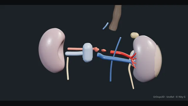
UrOops3D, danger-zone atlas
Operative animation, Organ retrieval

Deceased-donor kidney and multiorgan retrieval

The donor after brain death or after the heart has stopped, the abdominal team at a distant hospital in the night, the aorta cannulated and the organs cooled in place, and two kidneys leaving in ice with their vessels and ureters intact.

This page is the two donor pathways, the consent and the clock, the cannulation and the cross-clamp, the en bloc retrieval, the bench, and the documentation that lets a recipient centre trust what arrives.

Advanced tier FRCS transplantation, organ donation and retrieval Interactive 3D

Operative visualisation

Schematic scene preview: Deceased-donor kidney retrieval
Preparing the 3D anatomy…

UrOops3D · UroRef · © Nity G

Click me next Section 2 of 8 Anatomy The structures this operation works between, and the one it must not cross.

The structures in the scene

Organ retrieval: how the structures relateOpen abdomen, xiphisternum to pubis, the aorta and cava exposed. Kidneys either side of the aorta and cava: tip sits below the renal arteries Perfusion cannula at the infrarenal aorta (danger). Kidneys either side of the aorta and cava: cross-clamp here starts the cold ischaemia clock Supracoeliac aorta. Kidneys either side of the aorta and cava: an accessory cut makes a kidney hard to implant Renal arteries and their accessories (danger). Kidneys either side of the aorta and cava: stripped clean, the transplant ureter necroses Ureters and the golden triangle (danger). Kidneys either side of the aorta and cava: shared vessels and planes with the other teams Liver, pancreas and their vessels (danger). Kidneys either side of the aorta and cava: vented for the effluent; a cuff for the right kidney Inferior vena cava.Kidneys either side ofthe aorta and cava!Perfusion cannula atthe infrarenal aortaSupracoeliac aorta!Renal arteries andtheir accessories!Ureters and thegolden triangle!Liver, pancreas andtheir vesselsInferior vena cavatip sits below therenal arteriescross-clamp herestarts the coldischaemia clockan accessory cutmakes a kidney hardto implantstripped clean, thetransplant ureternecrosesshared vessels andplanes with theother teamsvented for theeffluent; a cufffor the right…TargetDangerLandmark
Open abdomen, xiphisternum to pubis, the aorta and cava exposed. Schematic, not to scale; relationships only.
  • The field is the open abdomen from xiphisternum to pubis with the right colon and small bowel swept up and to the left, so that the aorta and the cava lie exposed from the diaphragm to the bifurcation with a kidney on each side of them.
  • The cannula enters the aorta just above the bifurcation and the cross-clamp sits at the supracoeliac aorta; the ureters run down from the lower poles over the psoas toward the bladder.
  • When the block lifts, the kidneys leave together on a segment of aorta and cava with the lumbar vessels divided behind them.
  • ›The infrarenal aorta at the bifurcation - encircled with two ties, the inferior mesenteric artery ligated, the cannula placed here with its tip below the renal arteries.
  • ›The supracoeliac aorta - reached through the crus or the chest; the cross-clamp goes here and the cold ischaemia clock starts with it.
  • ›The inferior vena cava - vented into the chest or a bag so the perfusion has somewhere to go; divided to leave a cuff with the right kidney.
  • ›The renal arteries and their accessories - seen from inside the opened aorta on the bench; a lower pole accessory supplies the ureter.
  • ›The ureters and the golden triangle - divided low near the bladder with the tissue between lower pole, ureter and gonadal vein kept intact.
  • ›The liver, pancreas and their vessels - shared planes with the other teams; the coeliac axis and superior mesenteric artery go with them and never at the expense of a renal artery.
  • ›The perfusion cannula and the ice - gravity-fed cold fluid through the aorta and slush around every organ; the effluent watched at the cava until it runs clear.
Click me next Section 3 of 8 Steps The operation in order, with the landmark that guards each move.

Operative steps

  1. Who: the donor after brain death or circulatory death Two donors. The donor after brain death, DBD, has had brainstem death confirmed by two doctors on two sets of tests to the Academy of Medical Royal Colleges code of practice; the heart beats, the ventilator runs, and the retrieval is a controlled operation with a warm dissection before the circulation is stopped. The donor after circulatory death, DCD, is the patient in intensive care in whom treatment is to be withdrawn: death is confirmed after five minutes of absent circulation, and only then does the team begin, against a clock, because the organs have been warm and unperfused since the blood pressure fell. Kidneys tolerate that warm period better than the liver or the pancreas, which is why the kidneys are so often the DCD organs.
  2. The team, the consent and the clock The specialist nurse for organ donation has taken consent from the family and checked the register, under deemed consent in England, Wales and Scotland where it applies, with the family still able to object. The National Organ Retrieval Service abdominal team, a lead surgeon, an assistant, a scrub practitioner and a perfusionist, travels with its own instruments, cannulae and preservation fluid; a cardiothoracic team joins for a DBD heart or lungs. Before the knife: a briefing naming the donor, the organs consented, the blood group, the virology, and for DCD the point at which the team stands down, usually if death has not occurred within about three hours of withdrawal, and the functional warm ischaemia limits for each organ.
    where it bites: in DCD the clock runs from the fall in blood pressure, not from the knife; the team is scrubbed and the trolley open before treatment is withdrawn.
  3. The incision and the exploration A long midline incision from the xiphisternum to the pubis, extended as a cruciate incision or joined to a sternotomy when the chest is being taken, gives the exposure. In the DBD donor the abdomen is explored first: the liver, the pancreas and the bowel are inspected and palpated for tumour, the kidneys felt through the peritoneum, and any finding that might change allocation is telephoned to the duty office at once. In DCD the exploration waits: the aorta is cannulated within minutes and the inspection is done under cold perfusion. The right colon and the small bowel are then mobilised medially on the root of the mesentery, the Cattell-Braasch manoeuvre, exposing the aorta, the cava, both kidneys and the ureters in one field.
  4. The aorta and the cava: dissection and cannulation The infrarenal aorta is dissected just above the bifurcation and encircled with two ties, the inferior mesenteric artery ligated, and the cava beside it exposed for venting. In the DBD donor the supracoeliac aorta is also encircled through the crus of the diaphragm, or the descending thoracic aorta reached through the chest, so that a cross-clamp can go on quickly later. Heparin, about 300 units per kilogram, is given by the anaesthetist and allowed to circulate. The aortic cannula is placed through a transverse aortotomy between the ties, secured, and connected to the perfusion line, air free. In DCD the whole sequence, from skin to aortic cannula, is done in the first few minutes without any other dissection.
    where it bites: a cannula passed up into a renal artery or the superior mesenteric perfuses one organ and starves the rest; it sits in the aorta with the tip below the renal arteries.
  5. Cross-clamp, cold perfusion and ice The supracoeliac or thoracic aorta is clamped, the cava is vented into the chest or through a wide cannula into a bag, and the cold preservation fluid runs into the aorta under gravity or pressure: University of Wisconsin or histidine-tryptophan-ketoglutarate solution for a multiorgan retrieval, hypertonic citrate for a kidney-only retrieval, several litres, until the effluent from the cava runs clear and the kidneys are pale and cold. Sterile ice slush is packed around every organ as the perfusion runs. The cold ischaemia clock starts at the cross-clamp. In the DBD donor the ventilator is stopped at this point; in DCD the heart has already stopped and this moment ends the functional warm ischaemia.
    where it bites: a kidney that stays pink and warm while the others blanch has an unperfused artery, accessory or main; it is looked for now and the cannula or the clamp adjusted.
  6. The kidneys en bloc or separately The liver and the pancreas leave first, on planes agreed with the team leader so that the coeliac axis, the superior mesenteric artery and the portal vein go with their organs and the renal arteries are never encroached on. The kidneys are then taken en bloc: the ureters are divided low near the bladder with all the tissue between the lower pole, the ureter and the gonadal vein preserved, the aorta and the cava are divided below the cannula and above the renal vessels, and the block of both kidneys, Gerota's fascia intact, is lifted off the posterior abdominal wall by dividing the lumbar vessels behind it. In a small child both kidneys are kept on the aorta and cava for en bloc implantation.
    where it bites: the tissue between lower pole, ureter and gonadal vein carries the ureteric blood supply; a ureter stripped clean here necroses in the recipient.
  7. The bench and the packing On the bench in a bowl of ice the block is separated: the aorta is opened along its posterior wall between the lumbar orifices to show both renal artery ostia and any accessory, then divided anteriorly so that each kidney keeps a patch of aorta; the cava is left with the right kidney to lengthen its short vein. Each kidney is inspected for tumour, cysts, scars and injury, the vessels for atheroma and intimal tears, the ureter for length and its tissue. Any doubt is described exactly. Spleen and nodes go for tissue typing, and a biopsy is taken if the donor was marginal. Each kidney is triple-bagged in cold fluid, packed in ice in a labelled box with its side, and the box sealed.
  8. Documentation and the recipient centres The kidney is only as good as the paper that travels with it. The retrieval form records the donor identity and blood group, the times of withdrawal, asystole, knife, cross-clamp and cold perfusion, the fluid and its volume, the anatomy of each kidney including every artery and vein and any injury, the ureter length, the biopsy, and the surgeon's judgement of quality. The duty office allocates through the national kidney offering scheme and the recipient centre is telephoned directly about anything unusual. The donor is closed with a running suture and treated with the same respect as any patient, and the family is told the operation is complete. The ischaemia times, warm and cold, decide how long the recipient centre can safely wait.
Click me next Section 4 of 8 Danger zones Where this operation bites, and why it bites there.
Critical relationship

Danger zones

The spatial relationships the animation is built to make automatic. Before you read each one, ask yourself: at this point in the operation, what is about to go wrong, and what is my first move? The save for each sits in the Complications tab.

  • RiskThe renal arteries and accessories - an accessory artery cut in the dissection, or a main artery divided short of its patch, makes a kidney hard or impossible to implant.
  • RiskThe ureteric blood supply - the tissue between the lower pole, the ureter and the gonadal vein; stripped clean, the transplant ureter necroses weeks later.
  • RiskThe perfusion - a malpositioned cannula, an air lock, an unvented cava or a late clamp leaves a kidney warm and unperfused.
  • RiskThe other teams' organs - the liver and the pancreas share vessels and planes with the kidneys; a division made without agreement damages a graft that is not yours.
Practise it Take me into the scene Opens the animation at a danger moment and asks you to predict the mechanism and your first move before it reveals.
Click me next Section 5 of 8 Complications The save for each one: the first move, the escalation trigger, the op-note line.

Complications and immediate saves

Trigger each of these live in the Animation tab, then read the save: the first move, the escalation trigger, and the line that goes in the op note.

Renal artery or vein injury and the unusable graft

  • a renal artery divided short, an accessory cut in the dissection, an intimal tear from the cannula or a vein torn from the cava: a kidney that arrives with vessels the recipient surgeon cannot join.
The save
  1. Open the aorta posteriorly on the bench and count every ostium before any vessel is divided; look for an accessory at each pole. first move
  2. Keep a patch of aorta with every artery and the cava with the right vein; a short vein is lengthened with cava on the bench.
  3. An injured vessel is described precisely, marked with a suture and repaired on the bench if it can be, with 6-0 or 7-0 Prolene.
  4. A kidney with a torn main artery or a devascularised pole: telephone the recipient centre before it travels so that the decision to use, repair or decline is theirs with the facts. escalation trigger
  5. Retrieval form: every artery and vein with its length and patch, any injury, any repair, and a diagram. document

Ureteric devascularisation

  • a ureter stripped of its periureteric tissue or divided too short: fine at the bench, necrotic and leaking in the recipient at two to three weeks.
The save
  1. Divide the ureter low, near the bladder, and lift it with all the fat between the lower pole, the gonadal vein and the ureter attached. first move
  2. Never skeletonise the ureter to see it; the gonadal vein is taken with it and the lower pole accessory artery is preserved.
  3. A ureter that is short or looks pale is described as such, so the recipient surgeon plans a short anastomosis or a pyelo-ureterostomy to the native ureter.
  4. A ureter injured or divided at the pelvis: the kidney is still usable; the recipient centre is told and plans a native ureter reconstruction from the start. escalation trigger
  5. Retrieval form: the length of the ureter, the state of its tissue and any lower pole vessel that runs to it. document

Poor perfusion and prolonged ischaemia

  • a kidney that stayed pink while the others blanched, a cannula in the wrong vessel, an air lock, or a DCD agonal phase that ran long: delayed graft function or a kidney that never works.
The save
  1. Watch both kidneys blanch as the perfusion starts and the caval effluent run clear; if one stays pink, check the cannula position and the clamp now. first move
  2. Vent the cava widely and keep the ice slush around every organ; a kidney at the bottom of a warm abdomen is not cold.
  3. Flush again on the bench through the artery until the venous effluent is clear; a kidney that will not flush is described, not assumed.
  4. Functional warm ischaemia beyond the agreed limit in DCD, or a perfusion that failed: the duty office and the recipient centre are told the times before allocation proceeds. escalation trigger
  5. Retrieval form: the times of withdrawal, of the blood pressure falling, of asystole, knife, cannulation and cross-clamp, and the perfusion quality of each kidney. document

Injury to another team's organ

  • the hepatic artery, the superior mesenteric artery or the pancreas divided or torn while the kidneys are freed, or a renal artery taken with the liver: one graft saved at the cost of another.
The save
  1. Agree the planes and the sequence at the briefing and again at the bench: who takes which vessel, and who divides the aorta and where. first move
  2. The abdominal team leader divides the shared vessels under vision, with both surgeons looking, never by feel.
  3. An injury to the other organ is told to its team at once, and repaired on the bench together if it can be.
  4. A replaced or accessory hepatic artery arising from the superior mesenteric, or a renal artery within the liver team's cuff: stop and reconcile the anatomy before either organ is divided. escalation trigger
  5. Retrieval form: the vessels taken with each organ and any injury, with the organ that has the vessel named so the recipient teams can reconstruct. document

Unrecognised pathology, identification and packaging error

  • a tumour in the kidney or elsewhere that was not seen, a kidney labelled with the wrong side, blood group or donor number, or a box that arrives warm or unsealed.
The save
  1. Inspect and palpate every organ and the bowel in the DBD donor before the cross-clamp, and both kidneys out of their fat on the bench. first move
  2. Any lesion is biopsied or sent for frozen section if the hospital can do it, and the duty office is told before allocation.
  3. Each kidney is bagged three times, labelled with side and donor number by two people reading aloud, packed in ice with the paperwork and blood samples in the box.
  4. A mass, a suspicious node or an unexpected finding at the bench: the duty office and every recipient centre with an organ from this donor are informed, and allocation paused. escalation trigger
  5. Retrieval form: the exploration findings, any lesion and its biopsy, and the label check countersigned. document
Click me next Section 6 of 8 Troubleshooting When the case turns: the problem, then the next move.

Intra-operative troubleshooting

When the case turns: the problem, then the next move.

  • ActionThe aorta is atheromatous and will not take a cannula - cannulate higher on a softer segment or through a common iliac artery; a cannula forced through plaque sends emboli into every organ.
  • ActionThe perfusion will not run - check the clamp is on, the line is unkinked and free of air, the cannula tip is in the aorta and the cava is vented; the effluent must have somewhere to go.
  • ActionOne kidney stays pink - the artery is not being perfused; move the cannula, look for an accessory arising above the clamp or a plaque at the ostium, and flush that kidney separately on the bench.
  • ActionThe DCD donor has not died within the agreed time - stand down as briefed, tell the family through the specialist nurse, and record it; the organs are not taken.
  • ActionA tumour is found in the bowel or the liver - stop, biopsy, ring the duty office; allocation pauses for every organ until the histology or the decision is known.
  • ActionTwo arteries on one kidney and a third on the other - keep every artery on a single patch of aorta if they are close, separate patches if not, and draw it; the recipient surgeon joins them on the bench.
Click me next Section 7 of 8 FRCS viva Say it out loud, then mark yourself against the model answer.

FRCS viva questions

The examiner stems this scene prepares you for. Try to answer out loud first, then reveal, and learn the discriminator, the detail that turns a pass into a strong pass.

QWhat is the difference between donation after brain death and donation after circulatory death, and why does it matter to the kidney?reveal answer
  • A donor after brain death has had death confirmed by brainstem testing, two doctors performing two sets of tests to the Academy of Medical Royal Colleges code, while the heart continues to beat on the ventilator.
  • Retrieval is a controlled operation: the organs are perfused and warm until the moment of cross-clamp, so I would have time to explore the abdomen, dissect the aorta and the cava and prepare every organ before the cold perfusion starts, and the warm ischaemia is essentially nil.
  • A donor after circulatory death is a patient in whom treatment is withdrawn, usually a controlled Maastricht category three;
  • death is confirmed after five minutes of absent circulation, and only then can the team begin, so the organs have been hypoperfused from the point the blood pressure fell.
  • The super-rapid technique cannulates the aorta within minutes and the dissection is done cold.
  • For the kidney the consequence is functional warm ischaemia, which raises the rate of delayed graft function, though long-term outcomes are broadly similar to DBD, and it sets the stand-down time, about three hours from withdrawal, and the acceptable agonal limits.
  • Cold ischaemia should be shorter after DCD than after DBD for the same reason.
Strong pass

Say both

  • Functional warm ischaemia defined from the fall in blood pressure rather than from asystole
  • Delayed graft function named as its consequence
Examiner note
Functional warm ischaemia defined from the fall in blood pressure rather than from asystole, and delayed graft function named as its consequence.
QDescribe the retrieval of the kidneys from a deceased donor.reveal answer
  • After the briefing that confirms consent, blood group, virology and the organ plan, I would open the abdomen from xiphisternum to pubis and, in a DBD donor, explore for tumour.
  • I would mobilise the right colon and small bowel medially to expose the aorta and the cava.
  • I would encircle the infrarenal aorta above the bifurcation with two ties, ligate the inferior mesenteric artery, and expose the supracoeliac aorta through the crus.
  • After systemic heparin I would cannulate the aorta between the ties with the tip below the renal arteries.
  • I would cross-clamp the supracoeliac aorta, vent the cava, start the cold preservation fluid and pack ice around the organs, watching both kidneys blanch and the effluent clear.
  • After the liver and pancreas teams have taken their organs on agreed planes, I would divide the ureters low with their periureteric tissue intact, divide the aorta and cava below the cannula and above the renal vessels, and lift both kidneys en bloc off the posterior wall by dividing the lumbar vessels.
  • On the bench I would open the aorta posteriorly, identify every ostium, separate the kidneys each with an aortic patch and the cava with the right, inspect, flush, bag and label them, and complete the retrieval form with the times and the anatomy.
Strong pass

Say both

  • The cannula tip below the renal arteries with the cava vented
  • The en bloc lift with the aorta opened posteriorly to count the ostia before separation
Examiner note
The cannula tip below the renal arteries with the cava vented, and the en bloc lift with the aorta opened posteriorly to count the ostia before separation.
QHow is the ureter protected at retrieval, and why?reveal answer
  • The transplant ureter has no collateral supply once it is in the recipient: it depends entirely on the vessels that reach it from the renal artery, running in the periureteric tissue from the renal pelvis and lower pole, sometimes from a lower pole accessory artery.
  • The native ureter in situ also draws from the aorta, the iliac vessels and the bladder, and all of those are lost at division.
  • So at retrieval I would divide the ureter as low as possible near the bladder to give length, and lift it with all the tissue between the lower pole of the kidney, the gonadal vein and the ureter, the so-called golden triangle, attached and undisturbed;
  • I would never skeletonise the ureter to see it, and I would take the gonadal vein with it rather than dissect it off.
  • I would preserve every lower pole artery, since one may be the ureteric supply, and I would describe the length and colour of the ureter on the retrieval form.
  • A ureter stripped at retrieval looks normal on the bench and at implantation, and declares itself at two to three weeks as necrosis with a urine leak at the anastomosis, which is the commonest urological complication of transplantation and the one the retrieval surgeon owns.
Strong pass

Say all three

  • The transplant ureter's single source of supply explained
  • The golden triangle named
  • The delayed presentation of the necrotic ureter linked back to the retrieval
Examiner note
The transplant ureter's single source of supply explained, the golden triangle named, and the delayed presentation of the necrotic ureter linked back to the retrieval.
QWhat are cold and warm ischaemia, and what are the limits for a kidney?reveal answer
  • Warm ischaemia is the time the organ spends without perfusion at body temperature.
  • In a DBD donor it is essentially zero, since perfusion continues until the cross-clamp and the cold flush starts at the same moment.
  • In DCD it is the functional warm ischaemia time, measured from the point at which the systolic blood pressure falls below about 50 millimetres of mercury after withdrawal to the start of cold perfusion, and the acceptable limit is organ specific: the liver and pancreas tolerate about thirty minutes, the kidney considerably longer, with most UK protocols accepting kidneys within about two hours of withdrawal and standing down at about three;
  • I would present these figures as approximate.
  • Cold ischaemia runs from the cross-clamp and cold perfusion to reperfusion in the recipient, and the kidney tolerates it well because metabolism at four degrees is a fraction of normal, but the risk of delayed graft function rises with every hour;
  • the aim is under about eighteen hours for a DCD kidney and under twenty-four for a DBD kidney.
  • There is also a second warm ischaemia at implantation, from the kidney leaving the ice to reperfusion, which the recipient surgeon keeps to a minimum.
  • The retrieval form records every one of these times, because the recipient centre plans around them.
Strong pass

Say all three

  • Functional warm ischaemia defined by the pressure threshold
  • The organ-specific tolerance stated
  • The second warm ischaemia at implantation named
Examiner note
Functional warm ischaemia defined by the pressure threshold, the organ-specific tolerance stated, and the second warm ischaemia at implantation named.
Click me next Section 8 of 8 Evidence The guidance each of these answers is built on.

Evidence and guidance

Two minutes

Close the lap: recall it before you leave.

Say it aloud, then reveal. Reading is not remembering.

  1. 01 The relationship Say the critical spatial relationship out loud, then reveal. reveal
    Memory frame

    The field is the open abdomen from xiphisternum to pubis with the right colon and small bowel swept up and to the left, so that the aorta and the cava lie exposed from the diaphragm to the bifurcation with a kidney on each side of them.

    Why?
    The cannula enters the aorta just above the bifurcation and the cross-clamp sits at the supracoeliac aorta; the ureters run down from the lower poles over the psoas toward the bladder. When the block lifts, the kidneys leave together on a segment of aorta and cava with the lumbar vessels divided behind them.
  2. 02 The first move Renal artery or vein injury and the unusable graft. Your first move? reveal
    First move

    Open the aorta posteriorly on the bench and count every ostium before any vessel is divided; look for an accessory at each pole.

  3. 03 The escalation trigger Renal artery or vein injury and the unusable graft. What makes you call for help? reveal
    Escalate when

    A kidney with a torn main artery or a devascularised pole

    More detail
    A kidney with a torn main artery or a devascularised pole: telephone the recipient centre before it travels so that the decision to use, repair or decline is theirs with the facts.
  4. 04 The discriminator What turns a pass into a strong pass? reveal
    Strong pass

    Say both

    • Functional warm ischaemia defined from the fall in blood pressure rather than from asystole
    • Delayed graft function named as its consequence
    Examiner note
    Functional warm ischaemia defined from the fall in blood pressure rather than from asystole, and delayed graft function named as its consequence.
  5. 05 The pitfall Where does this operation bite first? reveal
    Pitfall

    The renal arteries and accessories

    Why?
    an accessory artery cut in the dissection, or a main artery divided short of its patch, makes a kidney hard or impossible to implant.

Revision practice with self-marked answers, not an assessment of clinical competence. Kept in this browser only; it does not sync between devices.