
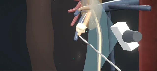
UrOops3D, danger-zone atlas

Schematic model.

Operative animation, Paediatric stone procedures

Paediatric urinary stone procedures

A child with a stone is worked up with ultrasound first and a metabolic screen from the start, and the route is chosen by the size and site of the stone: shock waves for most renal stones, ureteroscopy for the lower ureter, mini-PCNL for the larger or lower-pole stone.

The mini-PCNL is shown in full: prone, an ultrasound-guided puncture of a posterior lower calyx, one dilation to a small sheath, the miniature nephroscope and the laser, the fragments washed out and a small nephrostomy.

The lessons are a small ureter, a growing kidney, the radiation and anaesthetics a child accumulates, and a stone that will come back unless its cause is found.

Advanced tier FRCS paediatric urology; stones and endourology Interactive 3D

Operative visualisation

Schematic scene preview: Paediatric stone procedures
Preparing the 3D anatomy…

UrOops3D · UroRef · © Nity G

How to use this module. Play through the ten steps, or use Back and Next. Watch the route chosen, the needle find the calyx on ultrasound, one dilation, the laser and the washout, then ureteroscopy for a lower-ureteric stone. The triggers show bleeding, sepsis, a ureteric injury and residual fragments.

Click me next Section 2 of 8 Anatomy The structures this operation works between, and the one it must not cross.

The structures in the scene

Paediatric stone procedures: how the structures relateThe stone in the lower pole, the access to it and what lies around it. Stone in the posterior lower calyx: one small tract; the fragments leave by the sheath Single small tract and sheath. Stone in the posterior lower calyx: crossed by the tract; each tract costs kidney Developing renal parenchyma (danger). Stone in the posterior lower calyx: above the access line; the upper limit Twelfth rib and pleura (danger). Stone in the posterior lower calyx: lateral to the access line Descending colon (danger). Stone in the posterior lower calyx: route of fragments; torn by a scope too large Small ureter (danger). Stone in the posterior lower calyx: finds the calyx and guides the needle, no radiation Ultrasound probe on the flank.Stone in the posteriorlower calyxSingle small tractand sheath!Developing renalparenchyma!Twelfth rib andpleura!Descending colon!Small ureterUltrasound probe onthe flankone small tract;the fragments leaveby the sheathcrossed by thetract; each tractcosts kidneyabove the accessline; the upperlimitlateral to theaccess lineroute of fragments;torn by a scope toolargefinds the calyx andguides the needle,no radiationTargetDangerLandmarkContext
The stone in the lower pole, the access to it and what lies around it. Schematic, not to scale; relationships only.
  • The child prone, the probe on the left flank.
  • The access line runs forward and inward from the posterior flank, below the twelfth rib and the pleural reflection and medial to the colon, through the papilla into a posterior lower calyx, the stone just beyond it.
  • The ureter runs down medially to a lower-ureteric stone.
  • ›Renal stone - in the lower-pole calyx; on ultrasound a bright echo with an acoustic shadow behind it.
  • ›Posterior lower calyx - the target of the puncture: its axis points back toward the flank skin.
  • ›Renal papilla and parenchyma - crossed by the needle and the single tract; in a child still growing.
  • ›Renal pelvis - where the wire is coiled and where contrast or blood is seen.
  • ›Twelfth rib and pleura - above the access line; the reflection marks the upper limit.
  • ›Descending colon - lateral to the kidney and the access line.
  • ›Ureter - small in a child; the route of the catheter, the ureteroscope and the fragments.
  • ›Ultrasound probe and imaging plane - on the flank; the needle is kept in its plane.
  • ›Small sheath and miniature nephroscope - the single small tract and the scope that passes down it.
  • ›Ureteric stone - in the lower ureter, the second path, dusted by ureteroscopy.
Click me next Section 3 of 8 Steps The operation in order, with the landmark that guards each move.

Operative steps

  1. The child and the work-up Loin pain, haematuria or a urinary infection in a child is investigated with ultrasound first, with a plain film; low-dose non-contrast CT is kept for when they leave doubt. The urine is cultured, and the blood and urine tests of the metabolic screen are sent from the start.
    where it bites: a CT for every child adds radiation the ultrasound would have spared.
  2. The route by size and site The route is the least morbid one likely to clear the stone, chosen by its size and site as the guideline sets out: shock waves for most renal stones, ureteroscopy for the lower ureter, and mini-PCNL for the larger stone and for the lower pole, where shock-wave fragments clear poorly.
    where it bites: each extra session is another anaesthetic in a young child; the route that clears in one is often the kinder one.
  3. Anaesthetic, catheter, prone A general anaesthetic is given with the urine known to be sterile and an antibiotic at induction. A ureteric catheter is placed cystoscopically up to the pelvis, a urethral catheter beside it, and the child is turned prone on chest rolls, padded and kept warm. Contrast fills the collecting system.
    where it bites: a small child loses heat quickly under drapes and irrigation; warming and shielding are set up before the first puncture.
  4. Ultrasound-guided puncture The probe on the flank finds a posterior lower calyx and lines up the path to it. The needle is advanced in the plane of the beam through the papilla into the calyx, its tip watched on the screen. The stylet is withdrawn and urine at the hub confirms the position.
    where it bites: the pleura and the twelfth rib lie above the line and the colon lateral to it; pus at the hub stops the operation.
  5. One dilation to a small sheath A wire is passed through the needle and coiled in the pelvis, and the needle is removed. A small skin incision is made along the wire, and the tract is dilated once, in a single pass, to the small sheath, whose mouth is seated in the calyx under vision and screening.
    where it bites: the parenchyma of a growing kidney is torn by every dilation; one small tract tears less of it than a full-size one.
  6. The miniature nephroscope and the laser The miniature nephroscope goes down the sheath and is kept in line with it, never levered against the parenchyma. The laser fibre is passed through its channel and the stone is broken into pieces small enough to leave through the sheath, with the irrigation kept at a low pressure.
    where it bites: levering the scope tears the infundibulum, and high irrigation pressure drives infected fluid into the blood.
  7. Washing the fragments out The scope is drawn back into the sheath so that the outflow carries the fragments out, one after another; a larger piece is caught with a grasper. Every calyx is inspected, with screening where it helps, and the stone is sent for analysis.
    where it bites: a fragment left in a calyx is the nucleus of the next stone.
  8. Nephrostomy or tubeless A small nephrostomy is advanced over the wire into the calyx, the sheath is removed over it and the ureteric catheter comes out. After an uncomplicated clearance a selected child is left tubeless, with or without a stent. The child is turned back and woken.
    where it bites: bleeding or a large residual burden is a reason to leave the nephrostomy in.
  9. The second path: ureteroscopy For a stone in the lower ureter, cystoscopy and a wire passed beyond the stone come first. The smallest semirigid ureteroscope goes up beside the wire with no access sheath, the stone is dusted with the laser, the ureter is inspected on the way out, and a stent is left only when it is needed.
    where it bites: a small ureter torn by a scope too large for it; a scope that will not pass is not forced.
  10. Analysis, work-up and follow-up Every stone is analysed and every child has a full metabolic work-up: timed urine for calcium, oxalate, citrate, uric acid and cystine, and blood chemistry. Plenty to drink, less salt, normal dietary calcium and treatment of the cause follow, with ultrasound surveillance.
    where it bites: without the work-up the cause is missed and the stone returns.
Click me next Section 4 of 8 Danger zones Where this operation bites, and why it bites there.
Critical relationship

Danger zones

The spatial relationships the animation is built to make automatic. Before you read each one, ask yourself: at this point in the operation, what is about to go wrong, and what is my first move? The save for each sits in the Complications tab.

  • RiskSmall urethra and ureter - a scope or sheath too large tears or strictures them; the instrument is chosen to fit, and never forced.
  • RiskDeveloping renal parenchyma - every tract and every session costs kidney; one small tract, and no more sessions than needed.
  • RiskPleura and adjacent organs during percutaneous access - the pleura and rib above the line, the colon lateral to it; the needle is kept in view on ultrasound.
  • RiskCumulative radiation and anaesthetic exposure - ultrasound for diagnosis and puncture, pulsed and collimated screening, and a route that clears in as few sessions as possible.
Practise it Take me into the scene Opens the animation at a danger moment and asks you to predict the mechanism and your first move before it reveals.
Click me next Section 5 of 8 Complications The save for each one: the first move, the escalation trigger, the op-note line.

Complications and immediate saves

Trigger each of these live in the Animation tab, then read the save: the first move, the escalation trigger, and the line that goes in the op note.

Bleeding from the tract

  • a vessel in the tract or the infundibulum, torn by the puncture, the dilation or a levered scope; the view fills with blood that wells up the sheath.
The save
  1. Stop the laser, lower the irrigation and push the sheath in to tamponade the tract. first move
  2. Tell the anaesthetist; check the haemoglobin and set transfusion against the child's weight and blood volume.
  3. Place a small nephrostomy and clamp it, and abandon the clearance for another day.
  4. Bleeding that continues through the tube or after it is unclamped: angiography and embolisation. escalation trigger
  5. Record the tract, the point of bleeding, the measured loss, any transfusion and the plan. document

Sepsis from an infected stone

  • cloudy urine or pus at the hub on the puncture, or a fever after the operation: an infected stone, with infected urine behind it.
The save
  1. Pus at the hub: stop. No dilation and no laser. first move
  2. Drain the kidney with a small nephrostomy down the needle track, and send the pus for culture.
  3. Sepsis care at once: cultures, antibiotics and fluids by weight, with the paediatric team. escalation trigger
  4. Antibiotics adjusted to the culture, and the stone treated at a later sitting when the child is well and the urine sterile.
  5. Record the finding at the hub, the drainage, the cultures and the plan to return. document

Ureteric injury from a scope too large

  • a ureteroscope too large for a small ureter pushed on: the wall stretched and torn, irrigant outside it, and a stricture later.
The save
  1. Stop and do not force the scope; keep the wire in place. first move
  2. A retrograde study on the wire to see whether the wall is torn and how far.
  3. A stent across the injury for some weeks; return when the ureter has dilated passively, with a smaller scope.
  4. An avulsion or a long tear needs a formal repair by a paediatric urologist. escalation trigger
  5. Record the scope, the site and extent of the injury, the stent and the follow-up imaging for stricture. document

Residual fragments and recurrence

  • fragments left in a calyx at follow-up, and one growing into a new stone: residual stone becomes recurrent stone unless the cause is found.
The save
  1. Confirm the fragments and their size on ultrasound, and look again at the stone analysis. first move
  2. Complete the metabolic work-up if it has not been done, and treat what it finds.
  3. Plenty to drink, less salt and normal dietary calcium, with the dietitian and the child's family.
  4. Fragments that grow, obstruct or cause infection: treat them again by the route their size and site call for. escalation trigger
  5. Record the fragments, the work-up results, the prevention plan and the interval of ultrasound follow-up. document
Click me next Section 6 of 8 Troubleshooting When the case turns: the problem, then the next move.

Intra-operative troubleshooting

When the case turns: the problem, then the next move.

  • ActionThe calyx is not seen well enough on ultrasound - more contrast or fluid through the ureteric catheter to distend the system; screening for the last part of the puncture if needed.
  • ActionUrine does not come from the hub - the tip is short or through the calyx: adjust under ultrasound before any wire or dilator is passed.
  • ActionFragments will not wash out through the sheath - break them smaller, draw the scope back into the sheath, and catch the last ones with a grasper.
  • ActionThe ureteroscope will not pass the orifice - do not force it: a stent and a return in a few weeks, when the ureter has dilated passively.
  • ActionA stone that cannot be seen on fluoroscopy - a uric acid stone may be radiolucent: ultrasound guidance, and urine alkalinisation to be considered as treatment.
  • ActionA small infant with a tiny stone on ultrasound - observation: the guideline advises watching infant microlithiasis unless symptoms appear or the stone grows.
Click me next Section 7 of 8 FRCS viva Say it out loud, then mark yourself against the model answer.

FRCS viva questions

The examiner stems this scene prepares you for. Try to answer out loud first, then reveal, and learn the discriminator, the detail that turns a pass into a strong pass.

QA child presents with a renal stone. How would you investigate them?reveal answer
  • I would start with ultrasound and a plain film, which the EAU paediatric guideline recommends as the primary imaging, and keep low-dose non-contrast CT for when the diagnosis is doubtful.
  • I would culture the urine.
  • Every child with a stone should have a complete metabolic evaluation: a family and dietary history, stone analysis, and serum electrolytes, creatinine, calcium, phosphate, uric acid and alkaline phosphatase, with parathyroid hormone if the calcium is high.
  • Then timed urine for calcium, oxalate, citrate, uric acid, magnesium and phosphate, with cystine if suspected.
  • The guideline names hypocitraturia as the commonest abnormality, followed by hypercalciuria; cystinuria accounts for 2 to 6% of stones in children.
Strong pass

Say all three

  • Insisting on a metabolic work-up for every child
  • Not only for recurrence
  • Naming hypocitraturia and hypercalciuria
Examiner note
Insisting on a metabolic work-up for every child, not only for recurrence, and naming hypocitraturia and hypercalciuria.
QHow do you choose between shock wave lithotripsy, ureteroscopy and PCNL in a child?reveal answer
  • I would choose by size and site using the guideline table.
  • For a renal pelvic stone under 1 cm, shock waves; between 1 and 2 cm, shock waves, PCNL or retrograde intrarenal surgery; over 2 cm, PCNL.
  • For a lower-pole stone over 1 cm, PCNL, because shock-wave clearance there is only 50 to 62%.
  • Shock waves work less well as stones grow: about 90% stone-free under 1 cm, 80% at 1 to 2 cm and 60% over 2 cm.
  • For the upper ureter, shock waves; for the lower ureter, ureteroscopy.
  • In the trial of Ahmed and colleagues, in 128 children with 1 to 2 cm renal stones, mini-PCNL was stone-free in 93.8% against 41.3% after shock waves.
Strong pass

Say both

  • Quoting the guideline thresholds by site
  • The lower-pole clearance figure that pushes the choice to PCNL
Examiner note
Quoting the guideline thresholds by site, and the lower-pole clearance figure that pushes the choice to PCNL.
QWhat is different about ureteroscopy and PCNL in a child?reveal answer
  • I would use the smallest instruments that will do the job.
  • For ureteroscopy that is a small semirigid scope for the distal ureter and a flexible scope for the kidney, often without an access sheath.
  • Mokhless and colleagues treated 60 preschool children with 1 to 2 cm renal stones by retrograde surgery with hydrodilatation alone and no sheath, 86.6% stone-free after one session against 70% with shock waves.
  • If the scope will not pass I would stent and return.
  • For PCNL the guideline lists mini, ultra-mini and micro techniques, single-session stone-free rates of 86.9 to 98.5%, transfusion in under 10% and infection in under 15%, and says supine is as good as prone.
Strong pass

Say both

  • Knowing that a ureter that will not take the scope is stented and revisited
  • Never dilated by force
Examiner note
Knowing that a ureter that will not take the scope is stented and revisited, never dilated by force.
QWhat anaesthetic and radiation considerations apply to a child with stones?reveal answer
  • I would plan for the fewest anaesthetics.
  • The guideline notes that children under 10 need a general or dissociative anaesthetic even for shock waves, so a route that clears in one session can be kinder than repeated lithotripsy;
  • in the Ahmed trial recurrence at two years was 6.1% after mini-PCNL against 15.8% after shock waves.
  • For radiation I would use ultrasound for diagnosis and follow-up and for the puncture, keep CT low-dose and for doubt only, and use pulsed, collimated fluoroscopy;
  • the guideline credits ultrasound guidance and digital fluoroscopy with lowering the dose.
  • I would also keep the child warm, because a small child cools quickly.
Strong pass

Counting anaesthetics and radiation over the child's whole stone career, not one procedure.

QHow do you prevent recurrence after the stone has been cleared?reveal answer
  • I would base it on the stone analysis and the metabolic work-up.
  • For every child: plenty of fluid, moderate salt restriction and normal dietary calcium, since the guideline warns that restricting calcium increases stone risk.
  • Then I would treat the cause: thiazide for hypercalciuria, potassium citrate for hypocitraturia, alkalinisation with citrate for uric acid stones and allopurinol if that fails, and alkalinisation above pH 7 for cystinuria with a thiol agent if needed.
  • The guideline puts recurrence at 35 to 50%, so I would follow with ultrasound to catch residual fragments and regrowth early.
Strong pass

Say both

  • Not restricting calcium
  • Matching the drug to the abnormality the work-up found
Examiner note
Not restricting calcium, and matching the drug to the abnormality the work-up found.
Click me next Section 8 of 8 Evidence The guidance each of these answers is built on.

Evidence and guidance

Operative Dictations in Urologic Surgery
Armenakas NA, Fracchia JA, Golan R. Percutaneous nephrolithotomy (chapter 75) and Ureteroscopy for stones (chapter 79). Wiley, 2019
Adult chapters; the operative order follows them. Chapter 75: a urethral catheter, prone on chest rolls and padded, fluoroscopy in the field, the tract dilated over the wire and the sheath advanced, the stone fragmented and every fragment removed and sent for analysis. Then a check of every calyx, and a nephrostomy or stent at the surgeon's discretion; its variations include sonographic access. Chapter 79: a wire into the renal pelvis first, the scope to the stone under vision, fragmenting or dusting with the holmium laser, inspection of the ureter for residual stone or trauma, and a stent at the surgeon's discretion. The adult sheath and scope sizes are kept off the page; the miniaturisation, the single small tract and the paediatric choices are general practice from the EAU guideline and the papers below.
Open guideline
EAU Guidelines on Paediatric Urology, 2026
Chapter 18, urinary stone disease: causes, diagnosis, management and Table 4, read 19 October 2026
Hypocitraturia the commonest metabolic abnormality, then hypercalciuria; cystinuria 2 to 6% of stones; recurrence 35 to 50%. Ultrasound and plain film as primary imaging, low-dose non-contrast CT when doubtful; a complete metabolic evaluation for every child (all strong). Table 4: renal pelvis under 1 cm shock waves, 1 to 2 cm shock waves, PCNL or retrograde surgery, over 2 cm PCNL; lower pole over 1 cm PCNL; upper ureter shock waves; lower ureter ureteroscopy.
Open guideline
EAU Guidelines on Paediatric Urology, 2026
Chapter 18, sections on shock waves, PCNL, ureteroscopy, radiation and prevention, read 19 October 2026
Shock waves: general or dissociative anaesthesia under 10 years; stone-free about 90% under 1 cm, 80% at 1 to 2 cm, 60% over 2 cm; lower calyx 50 to 62%. PCNL single-session stone-free 86.9 to 98.5%, transfusion under 10%, infection under 15%; mini, ultra-mini and micro techniques; supine as good as prone. Ultrasound guidance and digital fluoroscopy lower the dose. Prevention: fluids, moderate salt restriction, normal calcium intake; thiazide, citrate, allopurinol and alkalinisation above pH 7 for cystinuria by cause. Infant microlithiasis observed.
Open guideline
Archivio Italiano di Urologia e Andrologia
Ahmed MNA, Selmy GI, Deif HA, et al. Extracorporeal shock wave lithotripsy versus percutaneous nephrolithotomy, complication rate and recurrence rate in management of pediatric renal stone, a prospective randomized trial. Arch Ital Urol Androl 2025;97:14286
Abstract read 19 October 2026. Randomised trial, 128 children with a solitary renal stone of 1 to 2 cm. Stone-free at eight weeks 93.8% after mini-PCNL against 41.3% after shock waves; complications 26.2% and 20.6%, not significantly different; recurrence at two years 6.1% against 15.8%.
Open guideline
Journal of Urology
Mokhless IA, Abdeldaeim HM, Saad A, Zahran AR. Retrograde intrarenal surgery monotherapy versus shock wave lithotripsy for stones 10 to 20 mm in preschool children: a prospective, randomized study. J Urol 2014;191:1496-1499
Abstract read 19 October 2026. Randomised trial, 60 preschool children. Retrograde surgery begun with a small semirigid scope and completed with a flexible one, no access sheath, hydrodilatation only. Stone-free after one session 86.6% against 70% with shock waves; 96.6% and 93.3% at three months; no major complication in either group.
Open guideline

Curriculum. Supports learning relevant to the ISCP Urology Curriculum syllabus topics Paediatric urology and Stones and endourology. Not a workplace-based assessment and not evidence of competence.

Two minutes

Close the lap: recall it before you leave.

Say it aloud, then reveal. Reading is not remembering.

  1. 01 The relationship Say the critical spatial relationship out loud, then reveal. reveal
    Memory frame

    The child prone, the probe on the left flank.

    Why?
    The access line runs forward and inward from the posterior flank, below the twelfth rib and the pleural reflection and medial to the colon, through the papilla into a posterior lower calyx, the stone just beyond it. The ureter runs down medially to a lower-ureteric stone.
  2. 02 The first move Bleeding from the tract. Your first move? reveal
    First move

    Stop the laser, lower the irrigation and push the sheath in to tamponade the tract.

  3. 03 The escalation trigger Bleeding from the tract. What makes you call for help? reveal
    Escalate when

    Bleeding that continues through the tube or after it is unclamped: angiography and embolisation.

  4. 04 The discriminator What turns a pass into a strong pass? reveal
    Strong pass

    Say all three

    • Insisting on a metabolic work-up for every child
    • Not only for recurrence
    • Naming hypocitraturia and hypercalciuria
    Examiner note
    Insisting on a metabolic work-up for every child, not only for recurrence, and naming hypocitraturia and hypercalciuria.
  5. 05 The pitfall Where does this operation bite first? reveal
    Pitfall

    Small urethra and ureter

    Why?
    a scope or sheath too large tears or strictures them; the instrument is chosen to fit, and never forced.

Revision practice with self-marked answers, not an assessment of clinical competence. Kept in this browser only; it does not sync between devices.