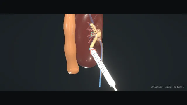
Left kidney, prone. Schematic model; sizes owner-supplied.
Operative animation, PCNL
Percutaneous nephrolithotomy
From CT planning to puncture, tract dilation, stone clearance and drainage. The whole case is decided by the line you choose from skin to calyx - and what that line passes on the way.
Advanced tierFRCS endourology, complex stoneInteractive 3D
How to use this module. Play through the eleven steps, or use Back and Next. The Screen panel shows a schematic image intensifier view; the working wire is white and the safety wire teal. Open Cross-section to relate the path to the axial view. Try the error states to see what a bad line looks like.
Prone, the posterior kidney and what surrounds the tract. Schematic, not to scale; relationships only.
PCNL is one line in three dimensions: skin to calyx.
Posterior calyx, through the papilla, below the pleura, clear of the colon.
Miss the line and the complications are the pleura, the colon and the bleed.
›Posterior calyces - the safe target; posterior calyces line up with a posterolateral tract.
›Papilla & infundibulum - puncture through the papillary tip along the avascular line of Brodel, the watershed between the anterior and posterior segmental supply; infundibular puncture risks the interlobar vessels and major bleeding.
›Pleura & 11th/12th ribs - supracostal access gains the upper pole but crosses the pleural reflection.
›Lumbodorsal fascia - the layer that resists: the needle gives through it, and it is opened before the dilators.
›Retrorenal colon - a posterolateral colon (more common when supine/with a mobile colon) lies in the path of a lateral tract.
›Staghorn stone - the branched target filling the collecting system; clearance is the endpoint.
Before the dayUrine is cultured and any infection treated before the date, and antibiotic prophylaxis is given by local policy: a single dose where there are no risk factors, a course beforehand where there are (hydronephrosis, tube drainage, a complex stone burden, diabetes or recurrent infection).
Retrograde set-upIn lithotomy a rigid (17 to 22 Fr) or flexible cystoscope passes a 0.035 inch PTFE or hybrid wire up the ureter, and a ureteric catheter follows it. A 5 to 6 Fr open-ended catheter is standard; a 7 Fr occlusion balloon catheter, its balloon inflated at the PUJ, stops fragments running down the ureter and distends the system. The catheter is taped to a 14 to 16 Fr Foley. Contrast diluted to about 30 to 50 per cent fills the pelvis without masking the stone; methylene blue, if added, makes the efflux obvious at puncture. Sizes and dilutions on this page are owner-supplied typical values.
CT planningThe CT is read before the patient is positioned, and the posterior calyx that best lines up with the stone bulk is chosen. A lower-pole calyx serves a simple stone; a staghorn usually needs an upper-pole, often supracostal, tract. The skin-to-calyx line is then traced slice by slice, noting everything it crosses on the way: skin, fat, the lumbodorsal fascia and muscle before the kidney. The line must clear the pleura above and the colon laterally; where it does not, the calyx or the position changes now, at the console rather than in the flank. One well-aligned tract is the plan, because extra tracts and sheath torque are where the bleeding lives. The patient is then turned prone, every pressure point padded. where it bites: the line must clear the pleura above and the colon laterally before a needle moves.
Calyceal punctureAn 18G needle, 15 to 20 cm, diamond or bevel tip (or a 21 or 22G Chiba first in a two-step technique), goes in under the C-arm. Bull's-eye: the arm is tilted until the hub and the tip overlap as one dot, and the needle is advanced down the beam; triangulation: 0 degrees for the mediolateral line, 20 to 30 degrees oblique for depth. Ultrasound is an alternative: a curvilinear 3.5 to 5 MHz probe with a needle guide and colour Doppler to keep off the interlobar vessels. The needle gives twice, at the lumbodorsal fascia and at the renal capsule, and its tip enters the calyx through the papilla, along the calyceal axis and the avascular line of Brodel. where it bites: puncturing the infundibulum or pelvis directly tears the interlobar vessels beside the calyx; the papilla is the avascular entry, the line of Brodel, and everything beside it bleeds.
Confirm accessThe stylet comes out and urine, contrast or blue drips from the hub. That efflux confirms the tip is inside the collecting system, and nothing is dilated until that confirmation exists.
Two wiresA 0.035 inch hydrophilic angled-tip wire goes through the needle, and a 5 Fr angled catheter (Kumpe or Cobra) steers it through the infundibulum into the pelvis and down the ureter. It is exchanged for a very stiff working wire (Amplatz Super Stiff or Lunderquist) so the dilators track without buckling. An 8/10 Fr coaxial dilator over it lets a second, safety wire go down alongside, and the 10 Fr outer is then removed; the safety wire is clipped to the drape. Both wires lie down the ureter to the bladder, through and through if the retrograde route grabs one, or coiled in the pelvis or a distant calyx. In the animation the working wire is white and the safety wire teal, and each is labelled. where it bites: dilating over a single wire leaves no way back in if it is lost.
Tract dilationA 1 to 1.5 cm skin incision is made along the wire with a No. 11 blade, and the lumbodorsal fascia is opened with a fascial incising needle or scissors, because it is the hardest layer to dilate. An 8 Fr Teflon guide catheter stiffens the track. Amplatz dilators follow over it in 2 Fr steps, 10, 12, 14 and on to 28 or 30 Fr, each with a gentle rotating push along the wire axis and each advanced only until its tip reaches the calyx on screening. The skin-level depth mark is noted on the first dilator and every later one goes to the same mark. Alken metal dilators, balloon dilation, one-shot dilation and mini-PCNL tracts are the alternatives. The Dilation chip in the animation plays the balloon alternative: a balloon dilator over the working wire, its tip at the calyx, inflated at the tract until the waist at the fascia gives and the balloon is 30 Fr along its length with the sheath loaded on it; it is deflated and withdrawn once the sheath is seated. One technique or the other, never both. where it bites: a dilator driven past the calyx perforates the pelvis; a kinked wire sends it off track.
The sheathThe Amplatz sheath (30 Fr; 24 to 34 Fr are available) is loaded on the last dilator and slid into the calyx, its position confirmed on the image; dilator and guide come out, leaving the sheath and both wires. The sheath is then both the working channel for the nephroscope and the tamponade for bleeding, which is why it stays in the kidney for the whole case.
Stone clearanceThe nephroscope passes down the sheath and the stone is fragmented, the pieces leaving through the sheath with irrigation. The sheath is angulated to reach neighbouring calyces rather than forced, because torque on a seated sheath tears parenchyma. Flexible nephroscopy at the end checks the calyces the rigid scope cannot reach before anything is called clear. The endpoint is a system that is clear on inspection; fragments left out of reach are booked for a staged second look rather than the tract being over-run.
DrainageAt the end of clearance the choice is a nephrostomy tube, an antegrade JJ stent, or a tubeless exit in selected uncomplicated cases. The decision follows the field: bleeding, residual fragments or a perforated system earn a tube, because the tube drains, tamponades and marks the tract for a second look. A nephrostogram through the tube confirms position and documents drainage before the patient leaves the table. Whatever drains the kidney, the drain plan is written into the op note with its follow-up imaging, because a drain without a plan becomes a drain without an exit.
The spatial relationships the animation is built to make automatic. Before you read each one, ask yourself: at this point in the operation, what is about to go wrong, and what is my first move? The save for each sits in the Complications tab.
RiskSupracostal tract - above the 12th rib the pleura is at risk: hydrothorax or pneumothorax.
RiskMedial / infundibular puncture - tears the interlobar vessels; the route to bleeding that may need embolisation.
RiskLateral tract + retrorenal colon - colonic transfixion; usually extraperitoneal and manageable if recognised.
The examiner stems this scene prepares you for. Try to answer out loud first, then reveal, and learn the discriminator, the detail that turns a pass into a strong pass.
QHow do you minimise the risk of bleeding when obtaining percutaneous access?reveal answer
Bleeding is largely determined by where I puncture, so my access is planned.
I aim for a posterior calyx and puncture through the papilla - the relatively avascular plane - rather than the infundibulum or directly into the renal pelvis, which risk the segmental and interlobar vessels.
I choose the calyx that lines up with the stone bulk to avoid needing multiple tracts or excessive torque on the sheath, which tears parenchyma.
I use image guidance (fluoroscopic bull's-eye or triangulation, or ultrasound) to keep the needle central in the calyx, dilate over a wire in a controlled fashion, and seat the Amplatz sheath, which also tamponades the tract.
I avoid over-angulation and respect a single, well-chosen tract.
Strong pass
Bleeding is an access-planning problem
Papilla→Puncture through it, not the infundibulum or pelvis
Why→The papilla is avascular
Calyx→The one that lines up with the stone
Examiner note
Puncture through the papilla (avascular) not the infundibulum/pelvis, and choosing the calyx that lines up with the stone - framing bleeding as an access-planning problem.
QWhen would you choose supracostal access and what is the trade-off?reveal answer
I would choose supracostal access - above the 12th, occasionally the 11th rib - when I need the upper pole or better alignment with the stone bulk, for example a large upper-pole or staghorn stone where an upper-pole tract clears more in a single access.
The trade-off is the pleura: the tract crosses the pleural reflection, so the risk of hydrothorax, pneumothorax and intrathoracic complications rises.
I mitigate it by puncturing at the end of expiration, staying as low as the anatomy allows, and having a low threshold to image the chest and place a drain if there is a significant collection.
So it is a calculated exchange of better stone access for a defined thoracic risk I plan for.
Strong pass
Say both
Justifying supracostal access by stone position (upper-pole / staghorn)
Naming the specific pleural mitigations (end-expiration puncture, low threshold to drain)
Examiner note
Justifying supracostal access by stone position (upper-pole / staghorn) and naming the specific pleural mitigations (end-expiration puncture, low threshold to drain).
QYou suspect a colonic injury post-PCNL. How do you confirm and manage it?reveal answer
I would confirm it with imaging: a nephrostogram showing contrast or the tube in the colon, or CT.
Management depends on the situation.
If it is recognised, the colon is retroperitoneal, and the patient is well without peritonism, most are managed conservatively: I withdraw the nephrostomy from the kidney back into the colon so it acts as a colostomy, place a separate ureteric stent to divert urine away from the injury, give antibiotics, keep the two tubes apart, and re-image before removing anything.
If there is an intraperitoneal injury, peritonitis, or sepsis, this needs laparotomy and surgical repair with general-surgical involvement.
Strong pass
Say both
Splitting management cleanly into the well
Extraperitoneal patient (conservative: nephrostomy-as-colostomy + diverting stent) versus the unwell / intraperitoneal one (laparotomy)
Examiner note
Splitting management cleanly into the well, extraperitoneal patient (conservative: nephrostomy-as-colostomy + diverting stent) versus the unwell / intraperitoneal one (laparotomy).
QDescribe how you would manage significant delayed bleeding after PCNL.reveal answer
Delayed bleeding, classically a few days to a couple of weeks post-op, often signals a pseudoaneurysm or arteriovenous fistula.
I would manage it as an emergency: resuscitate with ABC, large-bore access, fluids and cross-matched blood, and transfuse as needed.
If there is a nephrostomy I would clamp it to tamponade the tract.
The definitive step is CT angiography and selective angioembolisation by interventional radiology, which is both diagnostic and therapeutic and preserves renal tissue.
I would avoid open exploration, which in this setting usually ends in nephrectomy, reserving it for the exsanguinating patient who cannot be stabilised for embolisation.
Strong pass
Say all three
Recognising the pseudoaneurysm / AV fistula picture
Going to angioembolisation as nephron-sparing - explicitly avoiding open surgery
Which means nephrectomy
Examiner note
Recognising the pseudoaneurysm / AV fistula picture and going to angioembolisation as nephron-sparing - explicitly avoiding open surgery, which means nephrectomy.
Say it aloud, then reveal. Reading is not remembering.
01The relationshipSay the critical spatial relationship out loud, then reveal.reveal
Memory frame
One line: skin to calyx
Posterior calyx
Through the papilla
Below the pleura
Clear of the colon
Miss the line and the complications are the pleura, the colon and the bleed.
Why?
PCNL is one line in three dimensions: skin to calyx. Posterior calyx, through the papilla, below the pleura, clear of the colon. Miss the line and the complications are the pleura, the colon and the bleed.
02The first movePleural injury / hydrothorax. Your first move?reveal
First move
Recognise it
Desaturation
High airway pressures
Reduced air entry
Fluid above the diaphragm on screening
More detail
Recognise it: desaturation, high airway pressures, reduced air entry, or fluid above the diaphragm on screening.
03The escalation triggerPleural injury / hydrothorax. What makes you call for help?reveal
Escalate when
Significant haemo-, hydro- or pneumothorax, or tension
Intercostal chest drain
Senior and respiratory input
More detail
A significant haemo/hydro/pneumothorax, or tension physiology → intercostal chest drain and senior / respiratory input.
04The discriminatorWhat turns a pass into a strong pass?reveal
Strong pass
Bleeding is an access-planning problem
Papilla→Puncture through it, not the infundibulum or pelvis
Why→The papilla is avascular
Calyx→The one that lines up with the stone
Examiner note
Puncture through the papilla (avascular) not the infundibulum/pelvis, and choosing the calyx that lines up with the stone - framing bleeding as an access-planning problem.
05The pitfallWhere does this operation bite first?reveal
Pitfall
Above the 12th rib, the pleura is at risk
Supracostal tract: hydrothorax or pneumothorax.
Why?
Supracostal tract: above the 12th rib the pleura is at risk: hydrothorax or pneumothorax.
Revision practice with self-marked answers, not an assessment of clinical competence. Kept in
this browser only; it does not sync between devices.