UrOops3D, danger-zone atlas

Schematic model.

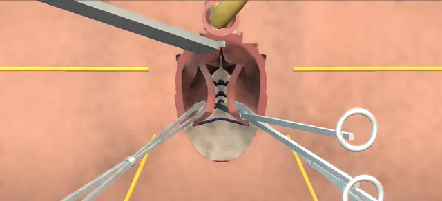
Operative animation, Rectocele repair

Rectocele repair: posterior colporrhaphy

A native-tissue repair of a symptomatic rectocele through the vagina.

A triangle of posterior vaginal epithelium is removed, the epithelium is dissected off the fascia that covers the rectum, and the rectovaginal fascia is plicated in the midline from the apex down to the fourchette so that it holds the rectum back.

Where the perineal body is attenuated, a perineorrhaphy is added. The lesson is the rectal wall, which lies directly under the scissors and the needle, and a vagina that must not be closed too tight.

Advanced tier FRCS female and reconstructive urology, prolapse Interactive 3D

Operative visualisation

Schematic scene preview: Rectocele repair
Preparing the 3D anatomy…

UrOops3D · UroRef · © Nity G

How to use this module. Play through the eight steps, or use Back and Next. Watch the triangle leave in its clamp, the flaps rise off the fascia and each suture gather it as it is tied; the profile views show the rectal wall behind. The triggers show a rectal injury, a stitch through the rectum and a tight repair.

Click me next Section 2 of 8 Anatomy The structures this operation works between, and the one it must not cross.

The structures in the scene

Rectocele repair: how the structures relateThe rectovaginal fascia from the vagina, and what lies on each side of it. Rectovaginal fascia: plicated in the midline: lies under it; a triangle is excised and dissected off Posterior vaginal epithelium. Rectovaginal fascia: plicated in the midline: lies on it; shallow bites keep it out Anterior rectal wall: the bulge (danger). Rectovaginal fascia: plicated in the midline: lateral to it; left out of the bites Levator ani, each side (danger). Rectovaginal fascia: plicated in the midline: plicated from the apex down to it Posterior fourchette. Rectovaginal fascia: plicated in the midline: ends on it; rebuilt only if attenuated Perineal body and its muscles. Rectovaginal fascia: plicated in the midline: behind the perineal body; the examining finger Anal canal (danger).Rectovaginal fascia:plicated in the midlinePosterior vaginalepithelium!Anterior rectalwall: the bulge!Levator ani, eachsidePosterior fourchettePerineal body andits muscles!Anal canallies under it; atriangle is excisedand dissected offlies on it; shallowbites keep it outlateral to it; leftout of the bitesplicated from theapex down to itends on it; rebuiltonly if attenuatedbehind the perinealbody; the examiningfingerTargetDangerLandmarkContext
The rectovaginal fascia from the vagina, and what lies on each side of it. Schematic, not to scale; relationships only.
  • Seen from the foot of the table in lithotomy, the urethra is at the top and the anus at the bottom, and the posterior vaginal wall is the floor of the field.
  • Under its epithelium lies the perirectal fascia and then the anterior rectal wall, which is the bulge.
  • The rectovaginal fascia is found by dissecting lateral to the bulge and is brought across the midline from the apex down to the fourchette.
  • The levator ani lies lateral to it on each side.
  • The perineal body lies below it, between the fourchette and the anal canal.
  • ›Posterior vaginal wall - bulges forward over the rectocele; a triangle of its epithelium is removed and its edges are brought together and closed.
  • ›Perirectal fascia - the layer directly on the rectum beneath the epithelium; the plane of the first dissection.
  • ›Rectovaginal fascia - exposed lateral to the bulge and plicated across the midline; the layer that holds the repair.
  • ›Anterior rectal wall - the content of the bulge; kept out of the dissection and out of every bite.
  • ›Levator ani - lateral to the fascia on each side; left out of the plication sutures.
  • ›Posterior fourchette - the base of the triangle and the lower end of the plication.
  • ›Perineal body - between the fourchette and the anal canal; rebuilt by the perineorrhaphy when it is attenuated.
  • ›Bulbocavernosus and transverse perineal muscles - meet in the perineal body; approximated across the perineal body in a perineorrhaphy.
  • ›Urethra and bladder - anterior; the catheter keeps the bladder empty throughout.
  • ›Anal canal - behind the perineal body; the route of the examining finger.
Click me next Section 3 of 8 Steps The operation in order, with the landmark that guards each move.

Operative steps

  1. The bulge A woman with a symptomatic rectocele. Antibiotics are given and she is placed in dorsal lithotomy on padded stirrups; the genitalia, the perineum and the lower abdomen are prepared. A urethral catheter is passed and the bladder is emptied. A Deaver retractor is placed on the anterior vaginal wall and a self-retaining retractor is set to hold the exposure. The posterior vaginal wall bulges forward, and behind it lies the anterior wall of the rectum.
    where it bites: the bulge is rectum; every layer opened from here on lies on its anterior wall.
  2. The triangle The posterior vaginal epithelium is infiltrated with local anaesthetic and adrenaline, which eases the dissection and helps haemostasis. Two Allis clamps are placed at the posterior fourchette, on the lateral margins of the vaginal wall, and a third in the midline at the proximal border of the rectocele. With the clamps as a guide, a triangle of vaginal epithelium is incised between them, its base distal at the fourchette.
    where it bites: the incision is through epithelium only; the fascia and the rectum are the next two layers.
  3. Dissection With Metzenbaum scissors the epithelium is dissected off the underlying perirectal fascia, away from the rectum. During this dissection a finger can be placed in the rectum to show how close the anterior rectal wall is. With downward traction on the rectum, the dissection is extended lateral to the rectocele until the rectovaginal fascia is exposed on each side.
    where it bites: the anterior rectal wall is directly under the plane; rectal injury is a listed complication of this operation.
  4. Plication sutures Interrupted absorbable sutures are placed to plicate the rectovaginal fascia, starting at the apex and moving distally to the posterior fourchette. The bites are shallow, to avoid incorporating the rectum in the repair, and care is taken to leave out the levator ani. Every suture is placed before any is tied.
    where it bites: a deep bite takes the rectal wall; a wide one takes the levator, and plicating the levator may increase dyspareunia.
  5. Tie Once all the sutures are in place they are tied individually, in succession. The two sides of the fascia meet over the rectum and the bulge reduces.
    where it bites: the repair narrows the vagina as it is tied; the calibre is judged before anything is closed over it.
  6. Perineorrhaphy Done only if it is indicated, as here for an attenuated perineal body. A further triangle of perineal and vaginal skin is excised at the posterior commissure, which makes the posterior defect a diamond and exposes the perineal body. The bulbocavernosus and transverse perineal muscles are brought together at the midline with vertical absorbable sutures.
    where it bites: the anal canal lies directly behind the perineal body.
  7. Calibre The calibre of the vagina is checked to avoid excessive narrowing. In a sexually active woman it should admit at least two fingers. A repair that is too tight is corrected at this point, while the sutures can still be reached.
    where it bites: excessive narrowing is an avoidable cause of dyspareunia after this repair.
  8. Closure and rectal examination The edges of the vaginal epithelium are trimmed as needed and the midline incision is closed with a running absorbable suture; after a perineorrhaphy the perineal skin is closed in the same way. Haemostasis is secured with careful diathermy. At the end a finger is placed in the rectum to confirm an intact rectal wall, with no bleeding and no palpable suture. The retractors are removed, the wound is irrigated and the catheter is connected to a drainage bag.
    where it bites: only this examination finds a suture through the rectal wall before she leaves theatre.
Click me next Section 4 of 8 Danger zones Where this operation bites, and why it bites there.
Critical relationship

Danger zones

The spatial relationships the animation is built to make automatic. Before you read each one, ask yourself: at this point in the operation, what is about to go wrong, and what is my first move? The save for each sits in the Complications tab.

  • RiskAnterior rectal wall under the scissors - the next layer beneath the perirectal fascia; a finger in the rectum can show how close it is.
  • RiskAnterior rectal wall under the needle - shallow bites keep it out of the repair; the rectal examination at the end checks for a palpable suture.
  • RiskLevator ani - lateral to the fascia; left out of the bites, because plicating it may increase dyspareunia.
  • RiskVaginal calibre - checked before closure; at least two fingers in a sexually active woman.
  • RiskPerineal body and anal canal - a perineorrhaphy works directly in front of the anal canal.
Practise it Take me into the scene Opens the animation at a danger moment and asks you to predict the mechanism and your first move before it reveals.
Click me next Section 5 of 8 Complications The save for each one: the first move, the escalation trigger, the op-note line.

Complications and immediate saves

Trigger each of these live in the Animation tab, then read the save: the first move, the escalation trigger, and the line that goes in the op note.

Rectal injury in the dissection

  • the scissors pass through the perirectal fascia and open the anterior rectal wall; rectal injury is a listed complication of this operation.
The save
  1. Stop dissecting. With the examining finger still in place, find the edges of the hole and how far it runs. first move
  2. Irrigate the field, and change contaminated instruments and gloves before going on.
  3. Call a colorectal colleague to theatre: the repair of the rectal wall and what else the bowel needs are decided with them now. escalation trigger
  4. Whether the prolapse repair then goes ahead over the closed rectum is decided together.
  5. Op note: where the hole was, its size, the layers closed and by whom; the patient is told afterwards. document

Stitch through the rectal wall

  • a plication bite taken too deep carries the suture through the rectal wall; the rectal examination at the end of the operation is how it is found.
The save
  1. A suture is felt in the rectal lumen: identify from the vaginal side which one it is. first move
  2. Cut that suture out and withdraw it. Place it again with a shallow bite, guided by the rectal finger.
  3. Examine the rectum again: the wall is intact and no suture is felt.
  4. A tear in the wall, bleeding from the rectum or any doubt about its integrity is a rectal injury: colorectal help in theatre. escalation trigger
  5. Op note: the finding, what was removed and replaced, and the result of the repeat examination. document

Vagina closed too tight

  • the plication or the perineorrhaphy has taken in too much and the calibre check fails; dyspareunia is among the known complications of the operation, and the calibre check exists to prevent this cause of it.
The save
  1. Stop before the epithelium is closed and find which sutures make the ring. first move
  2. Take those sutures down and place them again with less tissue in each bite, then check the calibre again.
  3. Trim less epithelium than first planned, so that the closure itself does not narrow the canal.
  4. A calibre that cannot be restored without giving up the repair is a decision for the senior surgeon in theatre. escalation trigger
  5. Op note: the calibre accepted at the end, in fingers, and anything taken down to reach it. document
Click me next Section 6 of 8 Troubleshooting When the case turns: the problem, then the next move.

Intra-operative troubleshooting

When the case turns: the problem, then the next move.

  • ActionThe plane under the epithelium is hard to find - the infiltration is what opens it: the epithelium is lifted off the fascia before the scissors go in. A finger in the rectum shows where the rectal wall is.
  • ActionThe perineal body is of normal thickness - no perineorrhaphy. The second triangle and the muscle sutures are omitted, and the vaginal incision is closed after the calibre check.
  • ActionA high-grade prolapse, and the fascia alone looks inadequate - the levator muscles may be plicated in the midline. The operative text warns that this may increase the likelihood of dyspareunia, so it is a choice made with that in mind.
  • ActionWhether to reinforce the repair - the operative text lists a graft or mesh as a variation. NICE recommends posterior vaginal repair without mesh, and its guidance on transvaginal mesh for anterior or posterior wall prolapse restricts that procedure to research.
  • ActionStress urinary incontinence as well - a midurethral sling can be done at the same sitting; it is a separate decision with its own consent.
  • ActionOozing from the dissected surfaces at the end - the vagina may be packed with a rolled gauze after the operation to help haemostasis.
Click me next Section 7 of 8 FRCS viva Say it out loud, then mark yourself against the model answer.

FRCS viva questions

The examiner stems this scene prepares you for. Try to answer out loud first, then reveal, and learn the discriminator, the detail that turns a pass into a strong pass.

QA woman has a symptomatic rectocele. Pelvic floor training and a pessary have not helped. What do you offer, and what do you tell her?reveal answer
  • I would first make sure the bulge is what troubles her, and I would ask closely about her bowels and about intercourse, because the operation can change both.
  • NICE asks that the options are discussed again, including no treatment and continued non-surgical management, and that surgery is offered to women whose symptoms have not improved with non-surgical treatment or who have declined it.
  • For posterior vaginal wall prolapse it recommends posterior vaginal repair without mesh.
  • I would explain that it is done through the vagina: the fascia between the vagina and the rectum is plicated across the rectum to hold it back, and the perineal body is rebuilt if it is thin.
  • I would give her the complications the operative text lists: bleeding, infection, rectal injury, painful intercourse for her or her partner, constipation, pain on defecation and recurrence.
  • On function I would be careful with numbers.
  • In the retrospective series of Kahn and Stanton from one London unit, at a mean of 42.5 months, prolapse symptoms fell from 64 to 31 per cent, but constipation rose from 22 to 33 per cent and sexual dysfunction from 18 to 27 per cent;
  • most of those women had other vaginal or bladder neck surgery as well.
  • In the randomised trial of Paraiso and colleagues, symptoms, quality of life and sexual function improved in every group, and the rate of dyspareunia had not changed significantly at one year.
  • NICE asks for a review six months after surgery.
Strong pass

Say both

  • Asking about bowel and sexual function before promising anything
  • Knowing that the operation the guideline recommends is a repair without mesh
Examiner note
Asking about bowel and sexual function before promising anything, and knowing that the operation the guideline recommends is a repair without mesh.
QTalk me through a posterior colporrhaphy.reveal answer
  • Antibiotics, then dorsal lithotomy on padded stirrups.
  • I pass a urethral catheter and empty the bladder, place a Deaver retractor on the anterior wall and set a self-retaining retractor.
  • I infiltrate the posterior wall with local anaesthetic and adrenaline.
  • I place two Allis clamps at the posterior fourchette on the lateral margins of the vaginal wall and a third on the midline at the proximal border of the rectocele, and with those as a guide I incise a triangle of epithelium with its base at the fourchette.
  • With Metzenbaum scissors I dissect the epithelium off the perirectal fascia, away from the rectum; if I am in any doubt about the plane I examine the rectum as I go.
  • With downward traction on the rectum I carry the dissection lateral to the rectocele until I can see the rectovaginal fascia on both sides.
  • I then place interrupted absorbable sutures to plicate that fascia, starting at the apex and working down to the fourchette, with shallow bites so that I do not take the rectum, and I leave the levator ani out.
  • When they are all in I tie them one at a time.
  • If the perineal body is attenuated I excise a further triangle at the posterior commissure and bring the bulbocavernosus and transverse perineal muscles together with vertical sutures.
  • I check that the vagina takes at least two fingers, trim the epithelium and close the incision with a running suture.
  • I finish with a finger in the rectum to confirm an intact wall, no bleeding and no palpable suture.
Strong pass

Say both

  • Placing every suture before tying any
  • Ending with the calibre check and the rectal examination
Examiner note
Placing every suture before tying any, and ending with the calibre check and the rectal examination.
QWhy is the levator left out of the repair, and why is no mesh used?reveal answer
  • The operative text allows levator plication for a high-grade prolapse but warns that it may increase the likelihood of dyspareunia, so I would not include the levator in a standard repair.
  • The trial evidence does not settle it: the Cochrane review of surgery for posterior compartment prolapse found a single trial of 52 women comparing levator plication with midline fascial plication and could draw no conclusion.
  • On technique more generally, the randomised trial of Paraiso and colleagues found similar anatomical and functional results after posterior colporrhaphy and site-specific repair, with anatomical failure at one year in 4 of 28 and 6 of 27 women, and more failures, 12 of 26, when a porcine graft was added.
  • On mesh, the same Cochrane review concluded that the evidence does not support any mesh or graft at the time of posterior vaginal repair, and it reported a mesh exposure rate of 7 per cent in the synthetic arm of the one trial that used it.
  • NICE recommends posterior vaginal repair without mesh, and its guidance on transvaginal mesh for anterior or posterior wall prolapse says the procedure should only be used in the context of research.
  • The operative text lists graft or mesh reinforcement as a variation; I would not offer it outside research.
Strong pass

Say both

  • Separating what the operative text permits from what the guideline recommends
  • Knowing that these questions rest on small trials
Examiner note
Separating what the operative text permits from what the guideline recommends, and knowing that these questions rest on small trials.
QDuring the dissection you see your glove through a hole in the rectum. What now? And what is the rectal examination at the end for?reveal answer
  • I would stop dissecting.
  • With my finger still in the rectum I would define the edges of the hole and how far it extends, irrigate the field, and ask a colorectal colleague to come to theatre: the repair of the rectal wall and the decision on what else the bowel needs are made with them, at once.
  • Whether the prolapse repair then goes ahead over the closed rectum is a joint decision.
  • I would record the site, the size, the layers closed and who closed them, and I would tell the patient afterwards.
  • The examination at the end has a different purpose.
  • The plication bites are taken without sight of the rectal lumen, so I finish every repair with a rectal examination to confirm that the wall is intact and that there is no bleeding and no palpable suture.
  • A suture I can feel has gone through the wall: I would cut it out, place it again with a shallower bite and examine once more.
Strong pass

Say both

  • Treating a palpable suture as a finding to act on in theatre
  • Calling the colorectal surgeon first for an open rectum
Examiner note
Treating a palpable suture as a finding to act on in theatre, and calling the colorectal surgeon first for an open rectum.
QHer main complaint is difficulty emptying the bowel. Will the repair cure it, and would you repair it through the vagina or the anus?reveal answer
  • I would not promise that.
  • Kahn and Stanton concluded from their series that the presence of the anatomical defect does not imply dysfunction, and that posterior colporrhaphy does not necessarily correct bowel and sexual dysfunction and may contribute to it;
  • they advised close questioning about bowel habit and selective bowel investigation before surgery.
  • So I would take a bowel history, treat constipation, and involve a colorectal colleague where the symptoms are out of proportion to the bulge.
  • On the route, the Cochrane review found that recurrent posterior wall prolapse was probably more likely after transanal than after transvaginal repair, with a risk ratio of 4.12 from two trials of 87 women, and that awareness of prolapse was probably more common after the transanal approach;
  • its data on adverse effects were scanty.
  • I would repair it through the vagina.
Strong pass

Say both

  • Not equating a rectocele with obstructed defecation
  • Knowing which route the randomised evidence favours
Examiner note
Not equating a rectocele with obstructed defecation, and knowing which route the randomised evidence favours.
Click me next Section 8 of 8 Evidence The guidance each of these answers is built on.

Evidence and guidance

Operative Dictations in Urologic Surgery
Armenakas NA, Fracchia JA, Golan R. Rectocele repair. Chapter 95. Wiley, 2019
The indication, the six essential steps and their order, the template dictation followed here, the variations (a finger in the rectum during the dissection, levator plication and its risk of dyspareunia, graft or mesh reinforcement, a concurrent midurethral sling, a vaginal pack) and the complication list: bleeding, infection, rectal injury, dyspareunia, constipation, defecatory pain, recurrent rectocele. The chapter makes the perineorrhaphy conditional; it is shown here for an attenuated perineal body. Its numbered steps put the calibre check before closure and ask for at least two fingers in a sexually active woman; its dictation records the check after closure, with three fingers. The page follows the numbered steps. The chapter lists complications without their management: the first, escalate and document steps, the troubleshooting line on finding the plane, and the danger lines on the anal canal, on narrowing as the sutures are tied and on the palpable suture are general surgical practice. The scene is schematic and at the scale of its field: instruments and fingers are drawn small, the rectum is drawn pressed downward by closed forceps although the chapter does not say what applies the traction, and the closure is shown without any narrowing of the canal.
Open guideline
NICE
Urinary incontinence and pelvic organ prolapse in women: management (NG123), published April 2019, updated June 2019: recommendations 1.7.1, 1.7.5, 1.7.6, 1.8.1, 1.8.2, 1.8.23 and 1.8.24, read 13 October 2026
Discuss no treatment, non-surgical and surgical options; consider supervised pelvic floor muscle training and a vaginal pessary; offer surgery to women whose symptoms have not improved with or who have declined non-surgical treatment; the discussion to cover changes in urinary, bowel and sexual function and the risk of recurrent prolapse; offer posterior vaginal repair without mesh to women with a posterior vaginal wall prolapse; offer a review 6 months after surgery.
Open guideline
NICE
Transvaginal mesh repair of anterior or posterior vaginal wall prolapse (HealthTech guidance HTG456, formerly IPG599), December 2017: recommendation 1.1, read 13 October 2026
Current evidence on safety shows serious but well-recognised safety concerns, and evidence of long-term efficacy is inadequate in quality and quantity; the procedure should only be used in the context of research.
Open guideline
Cochrane Database of Systematic Reviews
Mowat A, Maher D, Baessler K, Christmann-Schmid C, Haya N, Maher C. Surgery for women with posterior compartment prolapse. Cochrane Database Syst Rev 2018;3:CD012975
Abstract read 13 October 2026. Ten randomised trials, 1,099 women; evidence quality very low to moderate. Recurrent posterior wall prolapse probably more likely after transanal than transvaginal repair (risk ratio 4.12, 95% CI 1.56 to 10.88; two trials, 87 women), and awareness of prolapse probably more common. Single trials of site-specific repair against midline fascial plication (74 women) and of levator plication against midline fascial plication (52 women): insufficient evidence for any conclusion. Mesh exposure 7% in the synthetic group of the one trial of a synthetic graft. The authors conclude that the evidence does not support any mesh or graft at posterior vaginal repair.
Open guideline
British Journal of Obstetrics and Gynaecology
Kahn MA, Stanton SL. Posterior colporrhaphy: its effects on bowel and sexual function. Br J Obstet Gynaecol 1997;104:82-86
Abstract read 13 October 2026. Retrospective series from one London unit: 231 women and 244 posterior colporrhaphies, 171 interviewed and 140 examined at a mean of 42.5 months; 209 had prior or concurrent vaginal or bladder neck surgery. Prolapse symptoms fell (64% to 31%); constipation (22% to 33%), incomplete emptying (27% to 38%), faecal incontinence (4% to 11%) and sexual dysfunction (18% to 27%) rose. The vaginal defect was corrected in 76%. The authors conclude that the anatomical defect does not imply dysfunction. The abstract does not describe the technique of repair.
Open guideline
American Journal of Obstetrics and Gynecology
Paraiso MFR, Barber MD, Muir TW, Walters MD. Rectocele repair: a randomized trial of three surgical techniques including graft augmentation. Am J Obstet Gynecol 2006;195:1762-1771
Abstract read 13 October 2026. 106 women with stage II or greater posterior wall prolapse randomised to posterior colporrhaphy, site-specific repair, or site-specific repair with a porcine graft. Anatomical failure at one year: 4 of 28 (14%), 6 of 27 (22%) and 12 of 26 (46%), P = 0.02. Symptoms, quality of life and sexual function improved in all groups; no significant change in the rate of dyspareunia at one year.
Open guideline

Curriculum. Supports learning relevant to the ISCP Urology Curriculum special interest module Female, functional and reconstructive urology. Not a workplace-based assessment and not evidence of competence.

Two minutes

Close the lap: recall it before you leave.

Say it aloud, then reveal. Reading is not remembering.

  1. 01 The relationship Say the critical spatial relationship out loud, then reveal. reveal
    Memory frame

    Seen from the foot of the table in lithotomy, the urethra is at the top and the anus at the bottom, and the posterior vaginal wall is the floor of the field.

    Why?
    Under its epithelium lies the perirectal fascia and then the anterior rectal wall, which is the bulge. The rectovaginal fascia is found by dissecting lateral to the bulge and is brought across the midline from the apex down to the fourchette. The levator ani lies lateral to it on each side. The perineal body lies below it, between the fourchette and the anal canal.
  2. 02 The first move Rectal injury in the dissection. Your first move? reveal
    First move

    Stop dissecting.

    More detail
    With the examining finger still in place, find the edges of the hole and how far it runs.
  3. 03 The escalation trigger Rectal injury in the dissection. What makes you call for help? reveal
    Escalate when

    Call a colorectal colleague to theatre: the repair of the rectal wall and what else the bowel needs are decided with them now.

  4. 04 The discriminator What turns a pass into a strong pass? reveal
    Strong pass

    Say both

    • Asking about bowel and sexual function before promising anything
    • Knowing that the operation the guideline recommends is a repair without mesh
    Examiner note
    Asking about bowel and sexual function before promising anything, and knowing that the operation the guideline recommends is a repair without mesh.
  5. 05 The pitfall Where does this operation bite first? reveal
    Pitfall

    Anterior rectal wall under the scissors

    Why?
    the next layer beneath the perirectal fascia; a finger in the rectum can show how close it is.

Revision practice with self-marked answers, not an assessment of clinical competence. Kept in this browser only; it does not sync between devices.