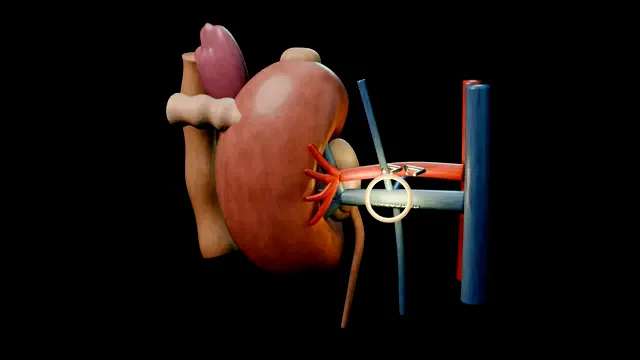
The hilum is the case: artery first, vein second, early control before any traction. Around the kidney sit the spleen, the pancreatic tail and the colon, and the renal vein tear is the complication that defines the operation.
Advanced tierFRCS uro-oncology, renal surgeryInteractive 3D
Imaging + positioningBefore access, the case is mapped rather than discovered: confirm side, measure the 6-7 cm lower-pole tumour, count the renal arteries, trace the renal vein and its adrenal, gonadal and lumbar tributaries, and exclude venous thrombus. The patient is secured in 45-60 degrees of lateral decubitus with the table broken to open the space between costal margin and iliac crest. Pressure points are protected, the dependent leg is flexed and the kidney rest is used only where the team can verify that it is not creating pressure injury. The displayed port map and viewing cone then show how the 30 degree laparoscope will approach the hilum. Spatial target: Map the synthetic 6-7 cm lower-pole tumour, count one main artery or two in the accessory variant, and rehearse 45-60 degrees of lateral positioning. Trace: Scenario-authored feature IDs F13, F17 and F21. Status: Scene targets; no claim that EAU or BAUS validates these numeric animation details.
Ports + colon reflectionAccess starts with a safe initial entry and the remaining ports are placed under direct vision in positions that let the whole kidney be worked from upper pole to pelvis. The colon is reflected medially along the white line of Toldt, sweeping the mesentery off Gerota’s fascia with blunt dissection and the diathermy kept on the mesenteric side. The check that the plane is right is the ureter on psoas and, on the left, the gonadal vein running cranially towards the renal vein; on the right, orientation instead returns to the short renal vein beside the cava. The whole reflection stays outside Gerota’s fascia, because entering it early shreds the plane into perinephric fat and every later step is harder for it. Spatial target: Establish a displayed 12-15 mmHg pneumoperitoneum and keep all four ports under vision and triangulated on the hilum. Trace: Scenario-authored feature IDs F22 and F29. Status: Technique rehearsal; the pressure and port pattern are not attributed to EAU or BAUS.
Ureter to the hilumThe ureter is identified where the colon reflection exposed it on psoas and followed upward toward the lower pole. It is slung, two clips are placed on the patient side, and it is divided immediately on the specimen side of them: two visible clips in total. The specimen-side end is raised with adjacent tissue as a controlled handle so lower-pole lift presents the hilum without pulling directly on a vein. On the left, the gonadal vein leads cranially into the renal vein; on the right, the short renal vein and exposed cava replace that tributary map and require a separate approach. Dissecting blindly into fat to find the hilum without controlled traction is the error, because the instruments then go where the vein lives. Spatial target: Use two visible ureteric clips in total, both on the patient-side stump, then divide immediately specimen-side to them. Trace: Scenario-authored feature IDs F6 and F23. Status: Control geometry; the explicit total matches the two visible clip meshes. where it bites: blind traction to "find" the hilum is the mechanism of the vein tear.
Artery firstThe artery is found posterior and slightly superior to the vein, reached by lifting the lower pole and working behind the vein rather than over it. The synthetic pulse animation is an orientation cue, not a diagnostic threshold. In this technique rehearsal, a visible working length is developed before three patient-side and two specimen-side clips are placed and the vessel is divided between fixed control points. Both controlled stumps remain in view. Arterial control first visibly pales the synthetic kidney and decompresses the venous side. Only after main-artery control is complete does the sequence perform the accessory-artery sweep, because a missed branch leaves a live stump bleeding behind a field that looks finished. Spatial target: Develop a displayed 1.5-2 cm artery window and reconcile five clips: three patient-side and two specimen-side, before division. Trace: Scenario-authored feature ID F24. Status: Technique target; not presented as an EAU or BAUS clip-count standard. where it bites: a missed accessory artery leaves a bleeding retroperitoneal stump.
Renal vein dividedWith the artery secure and the accessory-artery sweep complete, the vein is freed around its full circumference back to the great vessel so a 45 mm vascular stapler can be applied without touching anything behind. The translucent jaw path dwells first on the adrenal tributary above and then the lumbar tributary behind; both must sit outside the bite, while the gonadal vein remains on the patient-side stump. The vein is divided between paired staple rows under direct vision, with no traction while the blade crosses. A short vein, level 0 thrombus or an early adrenal take-off shortens the safe working length and must already be known from imaging. Spatial target: Use the displayed 45 mm vascular load only after arterial control, with both jaws and every named tributary clear. Trace: Scenario-authored feature ID F25. Status: Device choreography; not presented as an EAU or BAUS stapler prescription.
Upper pole + adrenal planeAfter hilar division, the kidney remains supported by its upper-pole, lateral and posterior attachments and can be drawn away from the splenic hood under direct control. For this lower-pole tumour, an amber dotted adrenal-sparing plane is rehearsed and the adrenal settles back onto its retained fat pad. EAU RCC 2026 supports adrenal preservation when there is no clinical evidence of adrenal invasion. The assistant holds the splenic hood while the splenorenal and remaining lateral attachments are divided. On the right, liver retraction and duodenal mobilisation replace the splenic and pancreatic relationships. The error is a loaded retractor or grasper swept high and lateral in one careless move. Spatial target: Rehearse an adrenal-sparing plane with two or three synthetic crossing vessels when imaging shows no clinical adrenal invasion. Trace: Scenario-authored feature ID F26 for the vessel count and plane; EAU RCC 2026 disease-management guidance for the broad adrenal-preservation indication. Status: EAU supports the broad preservation decision, not the synthetic vessel count or illustrated dissection minutiae. where it bites: the spleen and pancreatic tail are one careless sweep away at the upper pole.
Bag + extractionThe rolled bag enters through the 12 mm port, unfurls, receives the kidney inside Gerota’s fascia and cinches above the intact specimen. Pneumoperitoneum then falls from 12 to 8 mmHg for a seven-point bed check: artery stump, vein staple line, spleen or liver, pancreas or duodenum, colon, ureteric stump and large-port sites. Those sites are inspected under vision while the ports remain in place, before extraction and port withdrawal. The bagged specimen leaves through a protected Pfannenstiel incision without morcellation. The extraction fascia and indicated 10-12 mm port fascia are then closed, the abdominal dome deflates and the final clip and gauze counts reconcile. Spatial target: Reduce the display from 12 to 8 mmHg, inspect all seven bed targets including large-port sites while ports remain in place, and require gauze zero before extraction and closure. Trace: Scenario-authored feature IDs F27, F54 and F55; BAUS laparoscopic radical nephrectomy information is broad patient-information context only. Status: BAUS is not represented as validating the pressure, seven-target sequence and count gate.
The spatial relationships the animation is built to make automatic. Before you read each one, ask yourself: at this point in the operation, what is about to go wrong, and what is my first move? The save for each sits in the Complications tab.
RiskRenal vein / IVC junction - traction on a vein that is not yet arterial-controlled; the tear retracts and bleeds catastrophically.
RiskUpper pole sweep - spleen on the left, liver on the right; retractor injuries happen here.
RiskPancreatic tail - lies across the left renal vein; a leak is diagnosed on day three, not day zero.
When the case turns: the problem, then the next move.
ActionHilum will not come into view - more colon reflection and ureteric traction, not more dissecting into fat.
ActionCannot find the artery - the artery is posterior and superior to the vein; lift the kidney and go behind the vein rather than through it.
ActionStapler will not close on the vein - check for an adrenal or lumbar branch loading the jaw; divide or clip them first.
ActionSpecimen too big for the bag - stop forceful traction and widen the protected extraction incision; do not morcellate the cancer specimen in this teaching case.
The examiner stems this scene prepares you for. Try to answer out loud first, then reveal, and learn the discriminator, the detail that turns a pass into a strong pass.
QWhy is arterial control first the rule in nephrectomy, and what if you cannot achieve it?reveal answer
Dividing the vein with the artery intact engorges the kidney and can tear a short renal vein or the caval junction, and an unrestrained artery back-bleeds.
Artery first decompresses the venous side, shrinks the kidney and gives a controlled field for the vein staple line.
If the artery cannot be safely skeletonised, the alternatives are early vascular control from a posterior approach, a hilar En-bloc stapler load in an emergency field, or conversion to open control.
The principle survives every approach: proximal arterial control before venous division, or accept a field you cannot see through.
Strong pass
The engorged kidney, not just "artery first"
Mechanism→Name the engorged-kidney mechanism
Option→The en-bloc stapler in a catastrophic field
Examiner note
Naming the engorged-kidney mechanism, not just reciting "artery first", and knowing the en-bloc stapler option in a catastrophic field.
QYour patient has a left renal tumour with level 0 thrombus in the renal vein. How does that change the operation?reveal answer
The thrombus changes planning and venous control without changing the fixed scene order.
I map its medial limit on pre-operative imaging, secure arterial inflow first and complete the accessory-artery sweep.
I then expose only the circumferential venous window needed to show the thrombus tip and demonstrably normal vein medial to it; I do not claim complete kidney mobilisation before this venous step.
Using a no-touch approach, the stapler or clamp remains medial to the thrombus with every jaw edge visible before division.
The upper-pole, lateral and posterior attachments are released afterwards.
I warn anaesthesia about embolic and bleeding risk, avoid grasping the vein, and escalate to an appropriately experienced specialist team if safe caval-side control cannot be demonstrated or thrombus extends into the cava.
Strong pass
Say both
Stapling or clamping medial to the thrombus with imaging-confirmed margins
Escalating caval extension to a specialised team
Examiner note
Stapling or clamping medial to the thrombus with imaging-confirmed margins, and escalating caval extension to a specialised team.
QWhen would you offer partial rather than radical nephrectomy?reveal answer
Partial nephrectomy is preferred for T1 tumours when technically feasible, with particular importance in a solitary kidney, bilateral tumours or chronic kidney disease where nephron preservation matters.
Radical nephrectomy remains appropriate when nephron-sparing surgery is not feasible and minimally invasive radical surgery will not compromise oncological, functional or peri-operative outcomes.
The decision is made on CT or MRI anatomy and the patient’s renal reserve.
Anchoring the answer in renal function and tumour anatomy
Naming the CKD and solitary-kidney triggers
Examiner note
Anchoring the answer in renal function and tumour anatomy, and naming the CKD and solitary-kidney triggers.
QHow do you consent a patient for laparoscopic radical nephrectomy?reveal answer
I cover the generic laparoscopic risks: conversion to open, bleeding needing transfusion, visceral injury including bowel, spleen or liver, and port-site hernia.
Specific to nephrectomy: loss of the kidney with dependence on the contralateral kidney, and the possibility of dialysis if that kidney is not normal, so I check it pre-operatively.
I discuss the extraction incision, possible drain, expected recovery and return to activity.
I explain that histology determines the final diagnosis and may alter the onward plan.
I document the discussion including the conversion possibility, and record that contralateral renal function was assessed.
Written information and time for questions complete the consent.
Source trace: BAUS laparoscopic radical removal of kidney patient information.
Strong pass
Say both
Making the contralateral kidney part of the consent conversation
The honest possibility of open conversion
Examiner note
Making the contralateral kidney part of the consent conversation, and the honest possibility of open conversion.
Say it aloud, then reveal. Reading is not remembering.
01The relationshipSay the critical spatial relationship out loud, then reveal.reveal
Memory frame
Artery deep and superior, vein shallow and anterior
Artery→Deep and superior
Vein→Shallow and anterior
After→Upper-pole, lateral and posterior attachments still hold the specimen
Keep the spleen, pancreas and colon out of the instrument's path.
Why?
Before vascular division, the hilum connects the kidney to the midline vessels: artery deep and superior, vein shallow and anterior. After artery and vein control, the specimen remains supported by upper-pole, lateral and posterior attachments until those are released. Every step either delivers the hilum into view safely or keeps a neighbour (spleen, pancreas, colon) out of the instrument’s path.
02The first moveRenal vein / IVC tear. Your first move?reveal
First move
Pressure first, then raise the pneumoperitoneum
Gauze roll or grasper tamponade
Raise the pneumoperitoneum
No blind clips into blood
More detail
Immediate pressure with a gauze roll or grasper tamponade and raise the pneumoperitoneum; do not blind-clip into blood.
03The escalation triggerRenal vein / IVC tear. What makes you call for help?reveal
Escalate when
Unstable, or an IVC tear you cannot control laparoscopically
Immediate open conversion.
More detail
Unstable patient or tear into the IVC that cannot be controlled laparoscopically → immediate open conversion.
04The discriminatorWhat turns a pass into a strong pass?reveal
Strong pass
The engorged kidney, not just "artery first"
Mechanism→Name the engorged-kidney mechanism
Option→The en-bloc stapler in a catastrophic field
Examiner note
Naming the engorged-kidney mechanism, not just reciting "artery first", and knowing the en-bloc stapler option in a catastrophic field.
05The pitfallWhere does this operation bite first?reveal
Pitfall
Traction on a vein before the artery is controlled
The tear retracts and bleeds catastrophically.
Why?
Renal vein / IVC junction: traction on a vein that is not yet arterial-controlled; the tear retracts and bleeds catastrophically.
Revision practice with self-marked answers, not an assessment of clinical competence. Kept in
this browser only; it does not sync between devices.