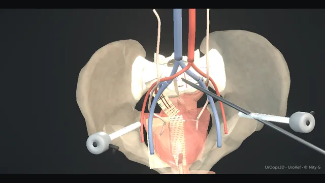
Laparoscopic sacrocolpopexy for vaginal vault prolapse
A Y-shaped mesh runs from the front and back of the vaginal vault up to the anterior longitudinal ligament at the sacral promontory, raising the vault to its anatomical position under direct vision.
The presacral venous plexus on the bone behind the promontory is the catastrophic zone; the ureters crossing the pelvic brim towards the bladder are the silent one; and complete reperitonealisation over the graft is the step that keeps bowel away from the mesh for good.
Advanced tierFRCS female and reconstructive urology, prolapseInteractive 3D
Laparoscopic, the plumb line from vaginal vault to sacral promontory. Schematic, not to scale; relationships only.
The operation is a plumb line from the vault to the sacrum.
The flat Y graft runs from both faces of the vault to the anterior longitudinal ligament at the promontory.
The fixation field is not avascular by default: the left common iliac vein lies nearby, the middle sacral artery and vein may track close to the midline, variable presacral veins lie on the bone, and the right ureter and iliac vessels bound the lateral field.
The final peritoneal closure separates the graft from bowel; vaginal mesh exposure remains a distinct vault complication.
›Sacral promontory + anterior longitudinal ligament - the fixation point; the strongest static anchor in the pelvis, approached through a defined peritoneal window.
›Presacral venous plexus - thin, valveless veins lying on bone beneath the promontory; torn easily, unable to retract, the source of the operation's catastrophic bleed.
›Common iliac vessels - the left vein in particular crosses the promontory approach; identified before the peritoneum is opened, not after it bleeds.
›Middle sacral artery and vein - small midline or near-midline vessels that can cross the fixation corridor on the anterior sacrum.
›Ureters - cross the brim anterior to the iliacs and run lateral to the vault dissection; mobilised medially by the very dissection that risks them.
›Vaginal vault + its two spaces - the target, freed between bladder front and rectum back until fully mobile; the graft limbs meet it on both faces.
Ports + exposureThe patient is placed in lithotomy with steep head-down tilt, ports are positioned to give a working view of the whole pelvic brim, and adhesions between bowel and the promontory or the vault are divided before anything else. Before any dissection the landmarks are named across the table: promontory, common iliac vessels, ureters, rectum, vault. The promontory is confirmed as a defined point on bone by tracing the aortic bifurcation and both common iliacs down into the sacral hollow. The left common iliac vein above and the ureters to either side are located first, because a peritoneal window opened before its neighbours are known is how the bleeding starts. where it bites: starting the peritoneal dissection before the left common iliac vein and the ureter are located: the promontory approach crosses both their territories.
Vault dissectionA vaginal probe or uterine sound pushed up by the assistant gives the apex a visible edge, and the peritoneum is opened transversely over it. The vesicovaginal space is developed in front and the rectovaginal space behind, the bladder advanced anteriorly and the rectum posteriorly with sharp spread, until the vault is fully mobile between the two planes. The dissection stays strictly midline, because drifting lateral draws the ureters medially into the field on their way to the bladder. A correctly developed space shows smooth, avascular fascia that accepts the spread without bleeding, while a plane that oozes or shows muscle fibre is the wrong layer. where it bites: the ureters run lateral and anterior to this plane and are pulled medially by the dissection itself; identified and observed, not assumed.
Sacral promontory dissectionThe peritoneum over the promontory is grasped and lifted, then divided to expose the anterior longitudinal ligament. The fixation corridor is variable, so the relevant structures are identified rather than inferred: the left common iliac vein cephalad and lateral, the right ureter and iliac vessels laterally, and the middle sacral artery and vein close to the midline. Cadaveric measurements place the middle sacral artery and vein, on average, only millimetres from the midsacral promontory. The dissection stays on the ligament with presacral veins respected beneath. where it bites: the middle sacral vessels and variable presacral veins may traverse the intended fixation corridor; expose before placing a suture or tack.
Y-graft fixationThe two mesh limbs are fixed to the anterior and posterior faces of the mobilised vault with permanent suture, taking good bites of fibromuscular tissue but never passing through the vaginal epithelium, which would seed erosion. The body of the graft is then fixed to the anterior longitudinal ligament with suture or tacks, placed along the ligament where it is thick and not into fat or periosteum alone. The graft is brought to length, not winched tight: tension is correct when the vault sits in its anatomical position on its own, without being held. An over-tensioned graft erodes, and a graft fixed into fat pulls out, so each bite is tested before the next is placed. where it bites: over-tensioning the graft and dissecting too lateral at the promontory: the first erodes, the second bleeds.
ReperitonealisationThe peritoneal edges from the vault and sacral windows are closed over the mesh and any potential internal-herniation spaces are closed according to the operative plan. The purpose shown here is to separate mesh from bowel and reduce direct mesh-bowel contact; it must not be taught as the principal prevention for vaginal mesh exposure, which is a separate vault and tissue-healing complication. where it bites: leaving mesh in direct contact with bowel or an unclosed potential hernia space.
Checks + counsellingHaemostasis is confirmed under low insufflation pressure, the vault position is checked per vaginam with the table flattened, and both ureters are watched along their length with peristalsis and efflux seen on each side. Drains are rarely needed and are reserved for the dissection that proved harder than planned. The mesh conversation then continues for years: erosion, exposure and the rationale for the graft are part of the operation and its follow-up, not an appendix to the consent form. The patient leaves knowing what a mesh complication looks like, who to call, and why the follow-up does not stop at the first review. where it bites: discharging without the mesh-specific follow-up and symptom safety-netting: late presentations are silent until they are not.
The spatial relationships the animation is built to make automatic. Before you read each one, ask yourself: at this point in the operation, what is about to go wrong, and what is my first move? The save for each sits in the Complications tab.
RiskPresacral venous plexus - valveless veins on bone beneath the promontory; the catastrophic haemorrhage of this operation, behaving more like a venous lake than a tearable vessel.
RiskUreter at the brim and vault plane - silent when injured, expensive when missed; stays in view throughout, and is checked before the case ends.
RiskVault apex under tension - over-tensioned mesh and dissected-out vault mucosa are the substrate for erosion; the graft sits the vault in place, it does not winch it.
Trigger each of these live in the Animation tab, then read the save: the first move, the escalation trigger, and the line that goes in the op note.
Presacral venous bleeding
the plexus on the sacrum tears at the bone edge during promontory dissection; venous, low pressure, catastrophic volume, with a field that fills silently from a plane suction cannot reach.
The save
Immediate and sustained pressure: a swab, a fingertip or a blunt instrument directly on the bleeding point; the reflex to look first is the reflex that loses the volume.first move
Tell the team: anaesthetist informed, bloods sent, crossmatch activated and escalation declared while pressure is held.
Raise the pneumoperitoneum pressure temporarily as a tamponade aid while the field is prepared.
Definitive local control: tack or thumb pressure, haemostatic agents and bone wax at the bone edge; blind clipping or suturing into the plexus extends the injury.
Ongoing loss, haemodynamic instability or failure of local control → conversion, vascular support and cell salvage; presacral haemorrhage is a team event, not a solo one.escalation trigger
Op note: site and mechanism, methods used in sequence, estimated loss, blood products, and the critical care plan.document
Ureteric injury
the vault or peritoneal dissection wanders lateral and the ureter is caught where it crosses the brim towards the bladder; silent when it happens, expensive when it is missed.
The save
Recognise the intra-operative clues: unexpected clear fluid in the field, a ureter that suddenly stops peristalsing, or a suspicious waist in its course.first move
Inspect the length of both ureters in the dissected field before closing, and use intra-operative optical dye or a stent where doubt remains.
A recognised partial or thermal injury is stented across; a complete division or segmental loss is repaired on the table or by immediate specialist input.
Post-operatively, flank pain, ileus or a leak collection after an uneventful list is a ureter until imaging says otherwise.
Delay in diagnosis, a closed infected system or a devascularised segment → urgent drainage and reconstructive planning.escalation trigger
Op note: ureters seen and how confirmed, any injury with site and management, stent details with a registered removal date.document
Mesh erosion (late)
months to years later mesh presents through the vaginal vault; discharge or bleeding is the complaint, vault examination is the diagnosis.
The save
Confirm with a vault examination and, where the story is unclear, imaging and cystoscopy to exclude bladder or bowel involvement.first move
Assess the exposure: a small asymptomatic exposure can be managed conservatively with review and oestrogen where appropriate.
Symptomatic discharge, bleeding or involvement of adjacent organs → partial or complete mesh excision, by a surgeon who does this work regularly.
Involve bowel or bladder specialists early where the exposure tracks towards either; erosion through to viscera is a combined decision.
Recurrent symptoms, retained infected mesh or fistula → specialist pelvic floor centre for excisional and reconstructive planning.escalation trigger
Notes and letters: findings at examination, conservative or operative plan, and the mesh-specific follow-up schedule.document
When the case turns: the problem, then the next move.
ActionPromontory not clearly identified in a deep pelvis - Confirm the aortic bifurcation and both common iliacs first and work down; the promontory is the first prominent vertebral fixed point between them, not a guess in fat.
ActionPeritoneal plane lost during vault dissection - Return to known structures, bladder or rectum, and re-enter in the correct avascular plane; dissecting in the wrong layer is how bladder and rectum are injured.
ActionVault will not reach the graft without tension - Mobilise further rather than pull harder: complete the rectovaginal release and re-check the vault is fully mobile before accepting any tension.
ActionSuture or anchor at the ligament will not hold - Move slightly caudal on the ligament for better purchase through a stronger bite; shallow bites in fat over the bone hold nothing and endanger the vessels behind.
The examiner stems this scene prepares you for. Try to answer out loud first, then reveal, and learn the discriminator, the detail that turns a pass into a strong pass.
QA patient asks why you are recommending mesh for her vault prolapse when mesh has been in the news. Take me through that consent conversation.reveal answer
The conversation starts by accepting the question is legitimate, because the patient has absorbed a real history and deserves a real answer.
The first layer is what mesh means here: a synthetic graft used to suspend the vault to the sacral promontory in a woman with symptomatic vault prolapse, usually after hysterectomy.
The second layer is honest comparison: non-mesh apical repairs such as sacrospinous fixation or uterosacral ligament suspension, and in selected women colpocleisis, offer different balances of recurrence, vaginal function and operative risk.
Colposuspension is not an apical-prolapse alternative; it treats stress urinary incontinence.
Sacrocolpopexy offers durable anatomical support in exchange for a permanent implant with mesh-specific risks.
Those include vaginal exposure, pain, infection, reoperation and rare bladder or bowel involvement.
The decision belongs to the patient after the alternatives and their trade-offs are made explicit.
Strong pass
Say both
Presenting native tissue repair as a genuine alternative with its own trade of recurrence against implant risk
Naming erosion as a lifelong rather than peri-operative risk
Examiner note
Presenting native tissue repair as a genuine alternative with its own trade of recurrence against implant risk, and naming erosion as a lifelong rather than peri-operative risk.
QWhy the sacral promontory? Explain the anatomy that makes it the anchor for vault support.reveal answer
The promontory is where the spine meets the pelvis, and it is the one fixed, strong, inert structure in a pelvis otherwise made of mobile viscera and distensible walls.
Vault prolapse is a failure of the level one support, the uterosacral and cardinal complex that suspends the apex of the vagina to the pelvic sidewall and sacrum;
once that suspension has failed, and usually after a hysterectomy has removed the organ those ligaments organised around, there is no native structure left with the strength to hold the apex durably.
The promontory offers the anterior longitudinal ligament, a dense fibrous band directly on bone that accepts a secure fixation, and its position restores the natural axis of the vagina, suspended posteriorly and superiorly so that the vault sits over the levator plate and intra-abdominal pressure closes the vagina rather than pushing into it, the same principle that native suspension exploited.
That geometry is why the graft runs the way it does: two limbs on the front and back of the vault because the vagina is a tube supported on both faces, converging to a single body to the ligament, the Y shape that distributes load across the vault rather than pulling on one point.
The price of the address is the neighbourhood: the presacral veins behind the ligament, the left common iliac vein crossing above it, and the ureters running down on either side.
The anatomy that makes the promontory the anchor is also what makes the approach to it the dangerous part of the operation.
Strong pass
Say both
Naming level one support failure as the problem and the anterior longitudinal ligament with the restored vaginal axis as the solution
Rather than answering with an address on a bone
Examiner note
Naming level one support failure as the problem and the anterior longitudinal ligament with the restored vaginal axis as the solution, rather than answering with an address on a bone.
QWhy is presacral venous bleeding so dangerous, and what is your sequence when it happens?reveal answer
The danger is physiological before it is technical.
The presacral plexus drains the sacral venous lakes through thin, valveless walls lying directly on bone.
Torn elsewhere, a vein retracts and thromboses;
torn against bone it can neither retract nor be lifted clear, the bleeding surface is a low-pressure venous lake communicating with a large capacitance bed, and the loss is steady, silent and enormous before the field even looks dramatic.
That physiology dictates the sequence.
First, pressure, immediately and sustained, with a swab, a fingertip or a blunt instrument exactly on the point, because every look before pressure is a look paid for in volume.
Second, the team is told, not merely the assistant: anaesthetist warned, access upgraded, bloods and crossmatch sent, and the escalation declared out loud while pressure is still held.
Third, the tamponade is made to work with the patient: pneumoperitoneum pressure raised temporarily, positions and preparations made while the field stays compressed.
Fourth, definitive local control by the means that fit the anatomy: thumb pressure, tacks or sterile thumbtack devices compressing the lake against bone, haemostatic adjuncts and bone wax at the edge;
what is never done is blind clipping or needling into the plexus, which converts one bleeding point into a field of them.
Fifth, the honest endgame: continuing loss or instability means conversion, vascular input, cell salvage and critical care.
The cases that go wrong are rarely those that bled; they are those where the surgeon looked before pressing.
Strong pass
Explaining the valveless, bone-backed, non-retracting physiology as the reason, and saying that sustained pressure precedes every look at the field.
QHow do you protect and check the ureters during sacrocolpopexy, and what if one is injured?reveal answer
Protection starts before the first cut, with the ureters identified on both sides at the pelvic brim during exposure, because the vault dissection that follows mobilises them medially into harm's way while the operator's attention is on the plane itself.
During the vault dissection the ureter is kept under direct observation rather than assumed away, the peritoneal window opened well medial, and any dissection that drifts lateral, towards the sidewall or the brim, is stopped and re-orientated.
The promontory approach threatens them differently, near the brim crossing, and the same rule applies: see the ureter, divide the peritoneum between the structures, not towards them.
Prophylactic stents are reserved for the re-do field, the irradiated pelvis or the anatomy that cannot be confidently read, and are a crutch for the difficult case rather than a substitute for visualisation.
Checking is a ritual at the end: both ureters inspected along their dissected length, peristalsis observed, and where doubt persists, an optical dye agent or an on-table study settles it before the ports come out.
If an injury is found, the response is graded: a contused or thermally affected ureter is stented across and drained;
a clean partial tear is stented and closed;
a division or segmental loss is repaired immediately with specialist help, reimplantation or reanastomosis as the length allows, and a drain left.
The catastrophe is never the injury itself, it is the unrecognised injury: the patient who leaves theatre with a warm, painless abdomen and returns with a leak, a closed infected kidney and a far worse problem than a stent on the day.
Strong pass
Naming the mechanism, that the vault dissection drags the ureters medially into the plane, and stating that the end-of-case ureteric check is a ritual, not an option.
Say it aloud, then reveal. Reading is not remembering.
01The relationshipSay the critical spatial relationship out loud, then reveal.reveal
Memory frame
The operation is a plumb line from the vault to the sacrum.
Why?
The flat Y graft runs from both faces of the vault to the anterior longitudinal ligament at the promontory. The fixation field is not avascular by default: the left common iliac vein lies nearby, the middle sacral artery and vein may track close to the midline, variable presacral veins lie on the bone, and the right ureter and iliac vessels bound the lateral field. The final peritoneal closure separates the graft from bowel; vaginal mesh exposure remains a distinct vault complication.
02The first movePresacral venous bleeding. Your first move?reveal
First move
Immediate and sustained pressure
More detail
Immediate and sustained pressure: a swab, a fingertip or a blunt instrument directly on the bleeding point; the reflex to look first is the reflex that loses the volume.
03The escalation triggerPresacral venous bleeding. What makes you call for help?reveal
Escalate when
Ongoing loss, haemodynamic instability or failure of local control → conversion, vascular support and cell salvage; presacral haemorrhage is a team event, not a solo one.
04The discriminatorWhat turns a pass into a strong pass?reveal
Strong pass
Say both
Presenting native tissue repair as a genuine alternative with its own trade of recurrence against implant risk
Naming erosion as a lifelong rather than peri-operative risk
Examiner note
Presenting native tissue repair as a genuine alternative with its own trade of recurrence against implant risk, and naming erosion as a lifelong rather than peri-operative risk.
05The pitfallWhere does this operation bite first?reveal
Pitfall
Presacral venous plexus
Why?
valveless veins on bone beneath the promontory; the catastrophic haemorrhage of this operation, behaving more like a venous lake than a tearable vessel.
Revision practice with self-marked answers, not an assessment of clinical competence. Kept in
this browser only; it does not sync between devices.