External sphincterotomy for detrusor sphincter dyssynergia
Indications
Detrusor sphincter dyssynergia in a man with a spinal cord injury, shown on video-urodynamics.
High bladder pressures, autonomic dysreflexia, infections or upper tract dilatation from the obstruction.
A man who will manage a sheath catheter and cannot, or will not, catheterise.
Alternatives
Intermittent catheterisation with botulinum toxin to the detrusor.
Botulinum toxin injected into the sphincter, which wears off.
A urethral stent across the sphincter.
A suprapubic catheter or a urinary diversion.
Risks
Bleeding, from the spongiosum or a venous sinus.
Infection.
Recurrent obstruction and a repeat incision.
Erectile dysfunction; incontinence into the sheath is the intended result.
A permanent endoscopic incision of the external urethral sphincter for a man with a spinal cord injury whose sphincter contracts against his bladder. The bladder then empties by reflex at low pressure into a sheath catheter.
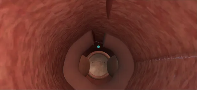
Shown is the resectoscope view: the sphincter ring and the verumontanum, then a cut at twelve o’clock from just distal to the verumontanum through the full thickness of the sphincter into the bulbar urethra, and the ring opening.
The lesson is depth: all of the sphincter, and none of the venous plexus above it or the spongiosum beyond it.
Advanced tierFRCS neuro-urology, spinal cord injury, bladder outletInteractive 3D
How to use this module. Play the seven steps, or use Back and Next. The scope view is the resectoscope; the cutaways are seen from the patient’s left. Watch the cut on the roof deepen until the ring parts and opens. The triggers show bleeding, dysreflexia, a recurrence and the nerves behind erectile dysfunction.
The external sphincter from the resectoscope, and what lies round it. Schematic, not to scale; relationships only.
The resectoscope looks up the urethra from the bulbar segment toward the bladder: twelve o’clock is the top of the picture and the verumontanum sits on the floor ahead.
The sphincter is a muscular ring round the membranous urethra just distal to the verumontanum.
The cut runs along the roof from the level of the verumontanum, through the ring, into the bulbar urethra.
In the cutaway, seen from the patient’s left, the venous plexus lies above the sphincter, the spongiosum surrounds the urethra distal to it, and the cavernous nerves run behind and to each side of it.
›External urethral sphincter - the striated muscle round the membranous urethra, distal to the verumontanum; in dyssynergia it contracts as the bladder does.
›Verumontanum - on the floor of the prostatic urethra; the incision starts just distal to it.
›Twelve o’clock - the roof of the urethra, opposite the verumontanum: the line of the cut.
›Bulbar urethra - distal to the sphincter; the cut runs on into it and stays shallow there.
›Venous plexus - the veins over the front of the sphincter and the apex of the prostate: the depth limit at twelve o’clock.
›Corpus spongiosum - the erectile tissue round the bulbar urethra; entering it bleeds.
›Cavernous nerves - beside the membranous urethra, behind and to each side of it, on their way to the corpora cavernosa.
›Bladder neck - a second possible level of obstruction; incised as well when it obstructs.
›Ureteric orifices - checked at cystoscopy; reflux into them is one reason the outlet is being opened.
The patient and the studyA man with a spinal cord injury and detrusor sphincter dyssynergia. Video-urodynamics shows a high-pressure contraction with the sphincter firing against it and the posterior urethra ballooning above it. He will manage a sheath catheter and does not want, or cannot do, intermittent catheterisation. where it bites: the incision cannot be undone, so the sheath catheter has to be a plan he accepts.
The level of the obstructionUnder anaesthesia, with the blood pressure monitored, the resectoscope is passed under vision. The sphincter is seen as a ring closing the lumen distal to the verumontanum. In the bladder the trabeculated wall, both orifices and the bladder neck are checked, because the neck can be a second level of obstruction. where it bites: a lesion at or above the sixth thoracic level can bring on autonomic dysreflexia during the cystoscopy.
The line at twelve o’clockThe Collings knife is turned to twelve o’clock, the roof of the urethra opposite the verumontanum. The line runs from just distal to the verumontanum, through the sphincter, to the bulbar urethra. Twelve o’clock is where the EAU guideline places the incision with the fewest complications. where it bites: a cut proximal to the verumontanum, or moved off twelve o’clock, reaches tissue that this incision is not meant to open.
The incisionWith cutting current the knife is drawn from the verumontanum toward the meatus, through the ring of the sphincter and on into the bulbar urethra, where the cut stays shallow. The scope returns to the start and the second pass deepens the same line. where it bites: in the bulbar urethra the spongiosum lies just beneath the mucosa; the cut there stays shallow.
Through the full thicknessThe line is deepened until the fibres of the sphincter part and the ring opens at twelve o’clock. The incision goes through the full thickness of the sphincter and no further. Beyond it lie the venous plexus over the sphincter and, distally, the corpus spongiosum. where it bites: too shallow leaves the obstruction; too deep enters the venous plexus or the spongiosum and bleeds.
Haemostasis and drainageThe cut is inspected with the irrigation slowed, and bleeding points are touched with the roller rather than run along the cut. The verumontanum and the bladder neck are seen intact. With the scope out, pressure on the full bladder gives a free stream through the open sphincter. where it bites: heavy coagulation along the cut adds heat close to the nerves to the erectile tissue.
Catheter, sheath and follow-upA catheter is left with its balloon in the bladder and comes out after a day or two. Then the bladder empties by reflex into a sheath catheter and a leg bag. Follow-up is lifelong: video-urodynamics and ultrasound of the upper tracts, because the sphincter can close again. where it bites: a residual or a pressure that rises again is a recurrence until proved otherwise.
The spatial relationships the animation is built to make automatic. Before you read each one, ask yourself: at this point in the operation, what is about to go wrong, and what is my first move? The save for each sits in the Complications tab.
RiskThe external sphincter and the erectile tissue beside it - the cut goes through the whole sphincter and stops; the cavernous nerves lie behind and to each side of the membranous urethra, so the cut stays at twelve o’clock.
RiskThe venous plexus at twelve o’clock - just beyond the full thickness of the sphincter; entering it is where bleeding at this depth comes from.
RiskThe corpus spongiosum in the bulbar urethra - directly under the mucosa distal to the sphincter; the cut there is shallow. Around an implanted stent the same tissue can be eroded.
RiskThe bladder neck and the ureteric orifices - seen at cystoscopy and left intact unless the neck itself obstructs and is incised as a separate decision.
RiskAutonomic dysreflexia - in a lesion at or above the sixth thoracic level, a full bladder or the scope can raise the blood pressure sharply; it is watched throughout.
Trigger each of these live in the Animation tab, then read the save: the first move, the escalation trigger, and the line that goes in the op note.
Bleeding from the spongiosum or a sinus
a cut carried too deep in the bulbar urethra opens the spongiosum, and one too deep over the sphincter opens the venous plexus; the field fills with blood.
Haemorrhage is one of the recognised risks of the operation.
The save
Stop cutting. Slow the irrigation and find the point; coagulate a point that can be seen, with a touch, not a run along the cut.first move
If it will not settle, pass a catheter, put it on traction so that the balloon presses the outlet, and let the bleeding settle.
Once it has, go back in with the resectoscope to look and to finish haemostasis.
Bleeding that continues on traction, or a falling haemoglobin, means senior help and a return to theatre.escalation trigger
Op note: where the bleeding came from, how deep the cut was there, how it was controlled, and the catheter plan.document
Autonomic dysreflexia during the procedure
in a lesion at or above the sixth thoracic level, the full bladder or the scope triggers a surge of blood pressure, often with a slow pulse, headache, sweating and flushing above the lesion.
The EAU guideline defines it as a rise in systolic pressure of more than 20 mmHg from baseline.
The save
Stop: take the instrument out of the urethra and empty the bladder, which removes the trigger.first move
Sit him up, or tilt the head of the table up, and loosen anything tight.
The anaesthetist treats a pressure that stays high: deeper anaesthesia, or a short-acting antihypertensive.
A pressure that does not respond, chest pain, or a fit, is a medical emergency: call for help at once.escalation trigger
Op note: the trigger, the highest pressure, what was given, and the plan to prevent it next time.document
Recurrent obstruction
the sphincter closes again over a healed cut, or the bladder neck obstructs; the residual, the pressures, the infections or the dysreflexia return.
Many men need a repeat incision.
The save
Repeat video-urodynamics to show where the obstruction is and how high the pressure is.first move
A repeat incision on the same twelve o’clock line, through the full thickness again.
If the neck is the level, a bladder neck incision is added.
Upper tract dilatation, or failure after repeat incisions, goes to the neuro-urology team for another bladder management.escalation trigger
Op note: the number of previous incisions, the findings and the level treated.document
Erectile dysfunction
a listed risk of the operation.
The cavernous nerves run beside the membranous urethra, behind and to each side of it, which is why the cut is placed at twelve o’clock, kept to the sphincter, and coagulated sparingly.
The save
Prevention is the save: the twelve o’clock line, the sphincter only, and points touched, not the cut run with current.first move
Consent names it, with what the man’s erections are like before the operation.
Erectile dysfunction afterwards goes to the andrology or spinal injuries team for the usual treatments.escalation trigger
Op note: the clock position of the cut, its extent, and the coagulation used.document
When the case turns: the problem, then the next move.
ActionThe pressure rises as the scope goes in - that is autonomic dysreflexia until shown otherwise. Stop, empty the bladder and sit him up while the anaesthetist treats the pressure; carry on only once it has settled.
ActionThe sphincter cannot be told from the bulbar urethra - find the verumontanum and come back from it: the sphincter is the ring just distal to it.
ActionThe ring does not open after two passes - the cut has not crossed the full thickness. Deepen the same line at twelve o’clock rather than starting a new one beside it.
ActionThe bladder neck looks tight - it may be a second level of obstruction; the EAU guideline describes secondary narrowing of the neck after sphincterotomy and a bladder neck incision for it.
ActionA laser is to be used - Perkash describes a contact laser incision on the same twelve o’clock line from the verumontanum to the bulbous urethra; the line and the depth do not change with the energy.
ActionThe man is not sure he can manage a sheath - do not operate. The incision cannot be undone, and intermittent catheterisation with botulinum toxin to the bladder, a stent, or a suprapubic catheter are the alternatives.
The examiner stems this scene prepares you for. Try to answer out loud first, then reveal, and learn the discriminator, the detail that turns a pass into a strong pass.
QA man with a cervical spinal cord injury has recurrent infections and dysreflexia. Video-urodynamics shows detrusor sphincter dyssynergia. What are his options?reveal answer
The aim is a bladder that stores and empties at low pressure, to protect the upper tracts.
I would first ask how he manages now, what his hand function is, who looks after him and what he wants.
Intermittent catheterisation with botulinum toxin to the detrusor is the usual first choice when it is possible, with an indwelling or suprapubic catheter for some.
If he cannot catheterise and will use a sheath, I would offer to lower the outlet resistance.
The EAU guideline describes external sphincterotomy, a urethral stent and botulinum toxin into the sphincter.
Sphincterotomy is irreversible and limited to men who can use a condom catheter, and the toxin wears off.
I would name the risks: bleeding, a repeat incision, and erectile dysfunction.
Strong pass
Say all three
Choosing by bladder management
Hands and wishes first
Saying that sphincterotomy commits him to a sheath for good
Examiner note
Choosing by bladder management, hands and wishes first, and saying that sphincterotomy commits him to a sheath for good.
QDescribe how you would do the incision.reveal answer
Under anaesthesia, with the blood pressure monitored, I would pass a resectoscope under vision and identify the verumontanum, the sphincter ring and the bladder neck, and inspect the bladder.
I would turn a Collings knife to twelve o’clock and make one incision from just distal to the verumontanum through the sphincter into the bulbar urethra, deepening the same line until the fibres part and the ring opens.
The guideline says the twelve o’clock cold-knife incision has the fewest complications; Perkash describes the same twelve o’clock line from the verumontanum to the bulbous urethra with a contact laser.
I would stay shallow in the bulbar urethra, touch the bleeding points, and leave a catheter for a day or two.
Strong pass
Twelve o’clock, the verumontanum to the bulbar urethra, full thickness of the sphincter and no deeper.
QHow well does it work, and how often does it need repeating?reveal answer
It works for most men but it is often repeated.
The EAU guideline says it resolves dysreflexia, hydronephrosis and recurrent infection and lowers detrusor pressure and the residual, but must be repeated at regular intervals in many patients.
Perkash reported 76 men treated with a contact laser: 69, 92 per cent, voided adequately at a mean of 27 months, and seven needed a repeat within two to five months.
In a French series of 51 men, Lepoittevin and colleagues found reflex voiding kept in 80 per cent at a median of 4.6 years, with a second incision in 39 per cent; dysreflexia fell from 26 men to seven.
These are retrospective single-centre series.
Kim and colleagues found a leak point pressure over 40 cmH2O after the operation went with upper tract damage, so I judge success on the trace, not only on symptoms.
Strong pass
Say both
Quoting a repeat rate alongside the success rate
Judging success by pressure on urodynamics
Examiner note
Quoting a repeat rate alongside the success rate, and judging success by pressure on urodynamics.
QDuring the cystoscopy his blood pressure climbs and his pulse slows. What is happening and what do you do?reveal answer
This is autonomic dysreflexia: an exaggerated sympathetic response to a stimulus below a lesion at or above the sixth thoracic level, here the scope and a full bladder.
The EAU guideline defines it as a rise in systolic pressure of more than 20 mmHg from baseline and warns that it can be life-threatening.
I would stop, take the instrument out and empty the bladder, sit him up or tilt the table head up, and loosen anything tight.
The anaesthetist treats a pressure that stays high.
The review by Krassioukov and colleagues supports upright positioning and removing the trigger by consensus, and a short-acting antihypertensive such as nifedipine if the pressure stays up.
I would start again only once it has settled, and record the event.
Strong pass
Say all three
Removing the trigger first
By emptying the bladder
Before reaching for a drug
Examiner note
Removing the trigger first, by emptying the bladder, before reaching for a drug.
QHow do you follow him up?reveal answer
For life, because the sphincter can close again and the bladder can change.
The EAU guideline recommends assessing the upper tracts at regular intervals in high-risk patients, with ultrasound about every six months, and urodynamics as a baseline and repeated at intervals of no more than one to two years.
Physical examination and urine tests are yearly.
I would ask about dysreflexia, infections and how the sheath stays on, measure the residual, and repeat video-urodynamics if anything changes.
A rising pressure or new dilatation means a recurrence until shown otherwise: a repeat incision, a bladder neck incision, or another way of managing the bladder.
Strong pass
Upper tracts on ultrasound and pressures on urodynamics, for life, with a rising pressure treated as recurrence.
Curriculum. Supports learning relevant to the ISCP Urology Curriculum special interest module Female, functional and reconstructive urology. Not a workplace-based assessment and not evidence of competence.
Two minutes
Close the lap: recall it before you leave.
Say it aloud, then reveal. Reading is not remembering.
01The relationshipSay the critical spatial relationship out loud, then reveal.reveal
Memory frame
The resectoscope looks up the urethra from the bulbar segment toward the bladder
Why?
The resectoscope looks up the urethra from the bulbar segment toward the bladder: twelve o’clock is the top of the picture and the verumontanum sits on the floor ahead. The sphincter is a muscular ring round the membranous urethra just distal to the verumontanum. The cut runs along the roof from the level of the verumontanum, through the ring, into the bulbar urethra. In the cutaway, seen from the patient’s left, the venous plexus lies above the sphincter, the spongiosum surrounds the urethra distal to it, and the cavernous nerves run behind and to each side of it.
02The first moveBleeding from the spongiosum or a sinus. Your first move?reveal
First move
Stop cutting.
More detail
Slow the irrigation and find the point; coagulate a point that can be seen, with a touch, not a run along the cut.
03The escalation triggerBleeding from the spongiosum or a sinus. What makes you call for help?reveal
Escalate when
Bleeding that continues on traction, or a falling haemoglobin, means senior help and a return to theatre.
04The discriminatorWhat turns a pass into a strong pass?reveal
Strong pass
Say all three
Choosing by bladder management
Hands and wishes first
Saying that sphincterotomy commits him to a sheath for good
Examiner note
Choosing by bladder management, hands and wishes first, and saying that sphincterotomy commits him to a sheath for good.
05The pitfallWhere does this operation bite first?reveal
Pitfall
The external sphincter and the erectile tissue beside it
Why?
the cut goes through the whole sphincter and stops; the cavernous nerves lie behind and to each side of the membranous urethra, so the cut stays at twelve o’clock.
Revision practice with self-marked answers, not an assessment of clinical competence. Kept in
this browser only; it does not sync between devices.