
Revision sheet
External sphincterotomy
7 steps, each one the authored title and caption from the interactive animation, with the frame captured from that step. Read it before the case, or print it and work through the steps in order.
Teaching only, not for diagnosis or treatment decisions. The text on this sheet is the animation's own step copy, reproduced verbatim.
UrOops3D · UroRef · © Nity G
- 01
The patient and the study
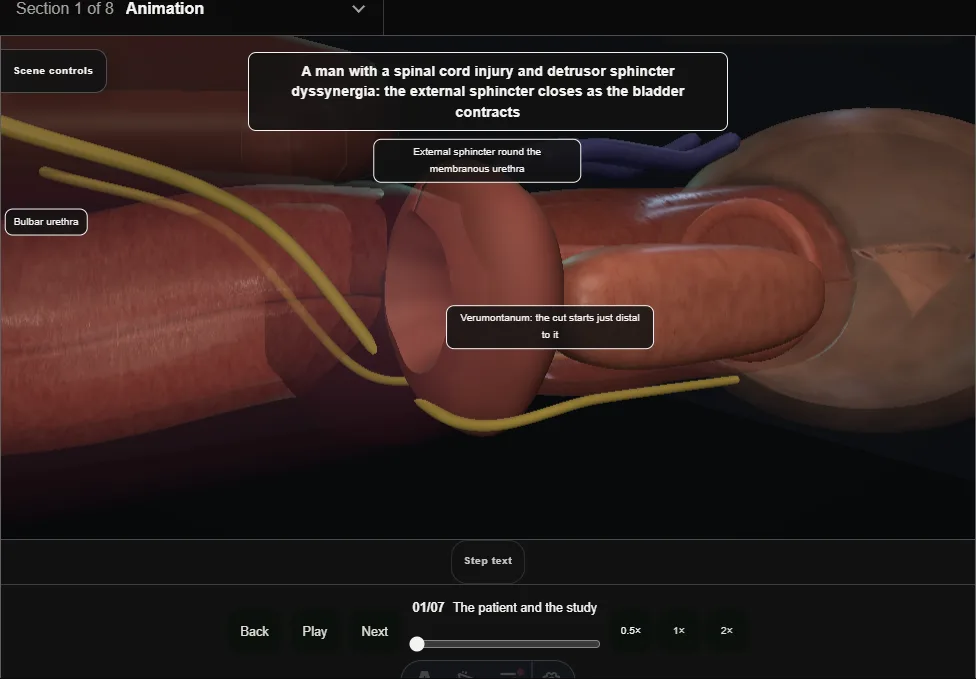
- A man with a spinal cord injury: the bladder contracts at high pressure and the external sphincter closes against it.
- He will use a sheath catheter; the outlet is to be opened for good.
- 02
The level of the obstruction
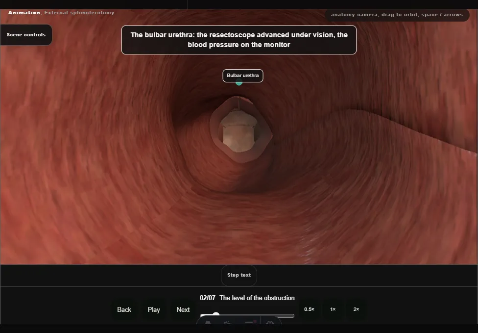
- The resectoscope in under vision with the blood pressure watched: the sphincter ring, the verumontanum on the floor, and the neck and bladder checked as the other levels of resistance.
- 03
The line at twelve o'clock
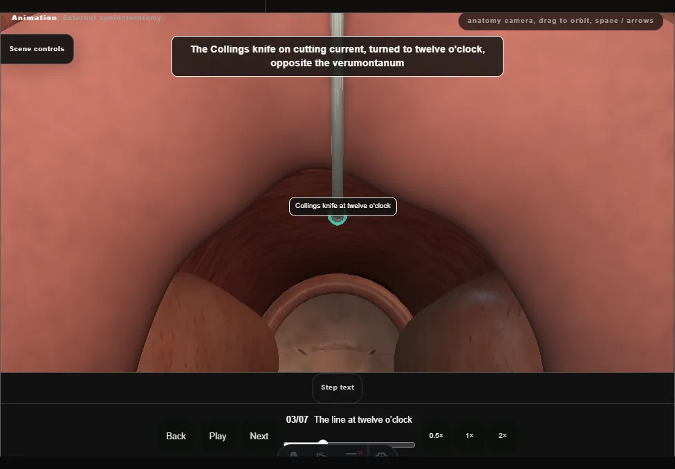
- The Collings knife turned to twelve o'clock: from just distal to the verumontanum, through the sphincter, to the bulbar urethra.
- 04
The incision
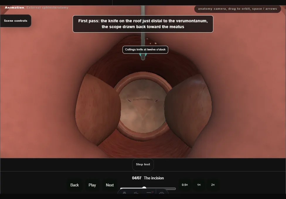
- The knife drawn from the verumontanum toward the meatus, through the ring and on into the bulbar urethra, then back to the start for a second pass on the same line.
- 05
Through the full thickness
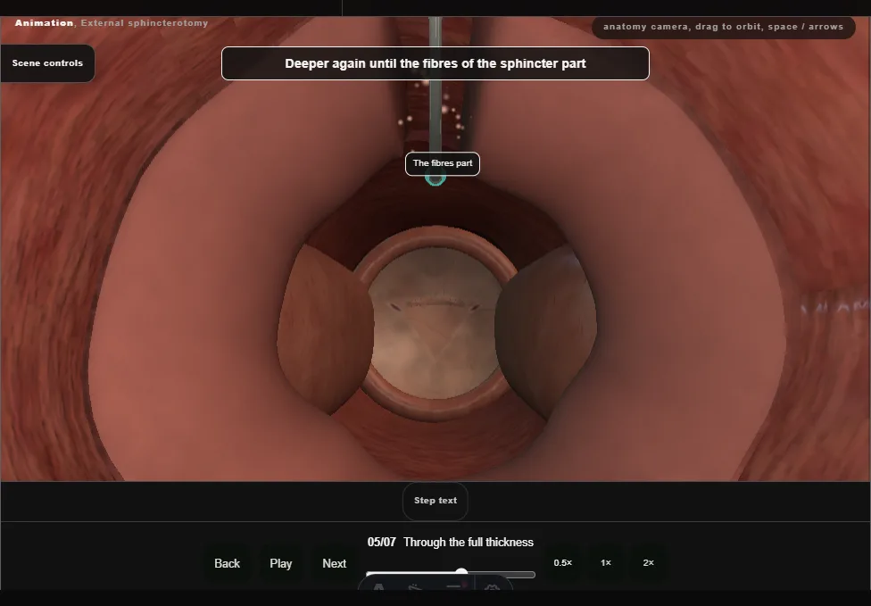
- Deeper until the fibres part and the ring opens, and no deeper: the venous plexus over the sphincter and the corpus spongiosum beyond it are not entered.
- 06
Haemostasis and drainage
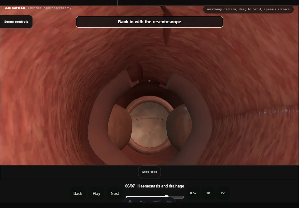
- Bleeding points touched with the button electrode; the verumontanum and the neck intact; with the scope out, the full bladder pressed gives a free stream.
- 07
Catheter, sheath and follow-up
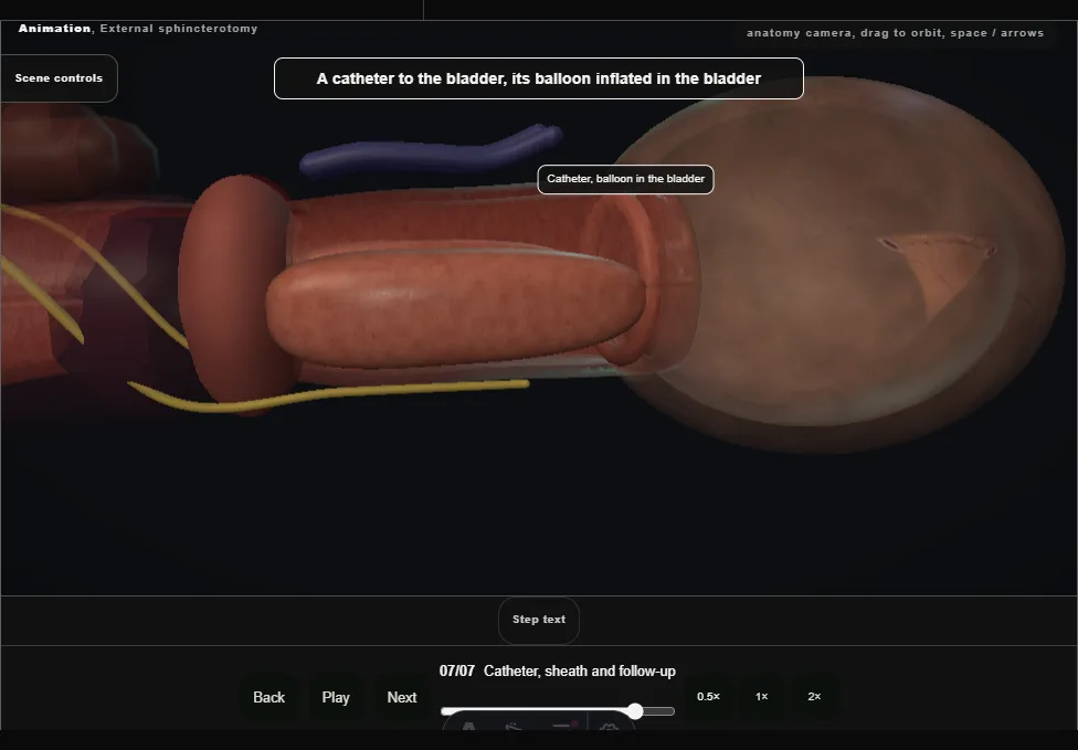
- A catheter for a day or two, then a sheath catheter to a leg bag.
- Follow-up is lifelong: video-urodynamics and the upper tracts on ultrasound.
Derived from endoscopy/SphincterotomyScene.tsx at build time: every step title and caption above is the authored
ProcedureDef text, reproduced verbatim, and this page shares its readable-points renderer with the animation
itself. 7 of 7 steps have a captured frame.
Text fingerprint a00608e4560c15a2. A caption edit without a re-capture fails the production
check, so the sheet and the animation cannot drift apart silently.