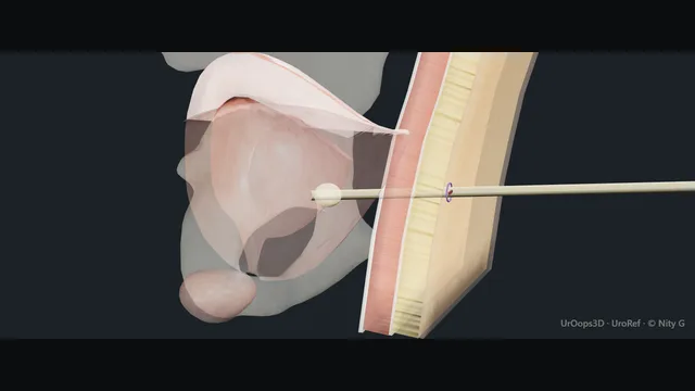
Wire-guided suprapubic catheter insertion using Mediplus S-Cath: bedside balloon test and full deflation, confirmed needle urine return, floppy-tip-first wire, introducer and retained outer sheath, then catheter urine return before balloon inflation.
The owner-specified sequence is shown with a sagittal teaching cutaway.
Cross-section of the lower abdomen along the midline track. Schematic, not to scale; relationships only.
A cross-section of the lower abdomen: the abdominal wall in front, the well-filled bladder dome tented up against it, bowel riding above the dome and the pubic symphysis below.
The track is a short line from skin two to three centimetres above the pubis into the dome; every complication in this scene lives in the gap that opens when the bladder is not full.
›Bladder dome - the target: well filled, it tents up to the abdominal wall and pushes bowel cranially.
›Bowel over the dome - the danger neighbour, interposed whenever the bladder is empty or adherent after previous surgery.
›Pubic symphysis - the landmark: entry two to three centimetres above it, in the midline.
›The track - a short straight line from skin to dome: midline, slight caudal aim, no blind depth.
Balloon integrity test + full deflationCheck the kit: Mediplus S-Cath, catheter size, intact sterile packaging, expiry and balloon volume printed on the valve. In the owner-specified technique shown here, test balloon integrity at the bedside, then deflate the balloon completely before insertion. This preparatory test is separate from inflation after intravesical catheter placement; it is recorded as the owner's technique, rather than a requirement stated in the manufacturer's insertion guide.
Confirm bladder fillingThe bladder is confirmed full before access: assess filling and the intended route with ultrasound. A filled dome approaches the abdominal wall and displaces bowel away from the route. Re-check filling and access rather than assuming a safe window; if a safe route cannot be demonstrated, stop and obtain senior help or use an appropriate alternative approach. where it bites: An unfilled bladder or an uncertain access window requires reassessment before puncture.
Landmark + ultrasoundMark the midline entry above the pubic symphysis. Ultrasound confirms the filled dome beneath the mark, assesses depth and checks that bowel is clear of the planned route. Prior surgery may alter these relationships; the scene is a teaching cutaway, not a patient-specific placement guide.
Local anaesthetic + needle urine returnInfiltrate local anaesthetic along the planned track and make the skin nick for the selected kit. Advance the access needle along the confirmed route while aspirating: urine in the syringe is the early access cue. Urine returns through the needle at this stage. Detach the syringe while holding the needle steady. Absent aspirate leaves position unconfirmed; stop and reassess rather than advancing a wider instrument. where it bites: No confirmed needle urine return: stop and reassess position.
Guidewire: floppy tip firstPass the guidewire through the needle, floppy tip first, to the first black mark in the Mediplus insertion guide. Hold the wire as the needle is withdrawn over the wire. The wire remains in place to preserve access; do not force it against resistance.
Introducer in; inner component outAdvance the sheath-dilator over the wire to the second black mark. The inner dilator and guidewire are removed together. What remains in place is the outer peel-away sheath, with urine at its outlet. A thumb over the sheath limits loss of bladder filling while the catheter is prepared.
Catheter, urine return, balloon, sheath removalPass the catheter through the sheath to the position described by the selected kit. Confirm urine draining through the catheter and intravesical position. The balloon is then inflated using the volume printed on its valve; the catheter size and balloon volume are not assumed here. Stop for uncertain position or resistance. Withdraw the outer sheath along the catheter until outside the abdomen, then its two halves are pulled apart and removed. where it bites: Confirm intravesical catheter position before balloon inflation.
Secure + aftercareSeat the balloon gently and secure the catheter without tension. An optional silk drain stitch is shown schematically, without prescribing an unconfirmed suture size or number of throws; fixation must not occlude the catheter. Confirm free drainage. Record the catheter and kit, balloon volume, insertion date and aftercare/change plan, and explain when to seek help for poor drainage or other problems.
The spatial relationships the animation is built to make automatic. Before you read each one, ask yourself: at this point in the operation, what is about to go wrong, and what is my first move? The save for each sits in the Complications tab.
RiskThe unfilled bladder - bowel sits between the wall and an empty bladder; filling is the safety of the whole procedure.
RiskBowel adherent to the dome - previous lower abdominal surgery: the blind trocar is exchanged for an open or image-guided technique.
RiskThe intramural balloon - inflate only inside the bladder; a balloon in the wall creates a false track and a leak.
Trigger each of these live in the Animation tab, then read the save: the first move, the escalation trigger, and the line that goes in the op note.
Bowel injury
the trocar passes through a bowel loop interposed between the wall and the bladder.
The save
Recognise it: faeculent or gaseous return, unexpected resistance, or later faecaluria and pneumaturia.first move
Do not remove the catheter blindly if transfixion is suspected: it may be tamponading the injury.
Resuscitate, start antibiotics, and image with a contrast CT to define the relationships of catheter, bladder and bowel.
Urgent senior urological and general surgical review: management depends on the bowel injury, imaging and clinical condition; do not assume that bladder-injury guidance applies to bowel injury.
Peritonism, intraperitoneal injury or deterioration → laparotomy or laparoscopy with repair and possible diversion.escalation trigger
Op note: technique, depth of insertion, aspirate findings, imaging, and the joint plan with general surgery.document
Catheter in the wall / extraperitoneal leak
the balloon inflates in the abdominal wall, or the track runs outside the bladder: a false track that leaks.
The save
Suspect it when urine does not return, drainage stays poor, or urine leaks around the catheter.first move
Never inflate a balloon against resistance: resistance requires stopping and reassessing position.
Image the track: a cystogram or CT catheter study shows the tip and the leak.
Deflate and re-site over a wire or by formal re-insertion with the bladder refilled; open technique if the track is lost.escalation trigger
Op note: balloon volume and position, imaging performed, re-siting technique and drainage confirmed.document
Haematuria / clot retention
tract bleeding or mucosal injury fills the catheter with clot and obstructs drainage.
The save
Irrigate the catheter manually to clear clot and restore drainage; most tract ooze settles.first move
Check the coagulation screen, anticoagulant use and haemoglobin; correct what is correctable.
Maintain irrigation until the return clears; exchange for a three-way catheter if continuous irrigation is needed.
Clot that cannot be cleared, or obstruction with a distending bladder → cystoscopy, evacuation and diathermy in theatre.escalation trigger
Op note: bleeding source, irrigation volumes, transfusion or correction given, plan for cystoscopic control.document
When the case turns: the problem, then the next move.
ActionNo urine on the test puncture - stop: re-image and refill the bladder; never advance the trocar hoping for urine.
ActionFlow stops after removal of the inner dilator - the tip may lie in the tract or against the wall: reposition under imaging before inflating anything.
ActionCatheter will not drain after placement - flush gently and check the balloon position on imaging; do not force irrigation against a blocked catheter.
ActionLeak around the catheter - check balloon position and volume; a false track needs imaging and re-siting, not a bigger catheter.
The examiner stems this scene prepares you for. Try to answer out loud first, then reveal, and learn the discriminator, the detail that turns a pass into a strong pass.
QWhen is suprapubic catheterisation contraindicated?reveal answer
The contraindications all follow from one spatial fact: bowel lies between the abdominal wall and an empty bladder.
An unfilled bladder, or one whose filling cannot be confirmed by palpation or ultrasound, is the primary contraindication.
Previous lower abdominal or pelvic surgery contraindicates the blind technique, because bowel may be adherent to the dome and to the wall; the same caution applies to a distended bowel or ileus.
Coagulopathy and anticoagulation that cannot be corrected are relative contraindications, as for any puncture.
A known bladder tumour is a specific caution, because track seeding is a described concern and an alternative route is preferred where one exists.
A small contracted bladder, an inability to lie flat and an uncooperative patient all make the procedure harder and riskier.
Where doubt exists, the answers are imaging guidance, an experienced operator or a different drainage strategy.
Strong pass
Say both
Framing every contraindication around the bowel
Wall and bladder relationship rather than reciting a list
Examiner note
Framing every contraindication around the bowel, wall and bladder relationship rather than reciting a list.
QLong-term drainage: suprapubic or urethral catheter?reveal answer
For long-term drainage the suprapubic catheter is usually the better tolerated option.
It bypasses the urethra, so it avoids urethral erosion, meatal stricture and the discomfort of a tube through a sensitive passage;
it is easier to change and to manage for patients and carers;
it drains dependently when the patient sits;
and it is concealed by clothing.
The urethral catheter remains first choice for short-term drainage, for accurate urine output monitoring in the acutely unwell, and when the bladder cannot be safely filled or the abdomen is inaccessible.
Both routes share the long-term burdens: bacteriuria with episodic symptomatic infection, encrustation and blockage, bladder spasms and leakage, and the need for a scheduled change regimen.
The decision is made with the patient: comfort, dignity, sexuality and the practicalities of managing the device weigh as much as the urology.
Strong pass
Say both
Naming urethral erosion and stricture as the specific long-term cost of the urethral route
Framing the choice as patient-centred
Examiner note
Naming urethral erosion and stricture as the specific long-term cost of the urethral route, and framing the choice as patient-centred.
QYou suspect the trocar has gone through bowel during insertion. What now?reveal answer
The first move is not to remove anything.
Suspicion is raised by faeculent or gaseous return through the trocar, by unexpected resistance and a wrong feel on insertion, and later by faecaluria, pneumaturia or peritonitis.
If transfixion of bowel by the catheter is suspected, the catheter stays: it may be tamponading the injury, and removing it blindly can open two holes and contaminate two cavities.
The patient is resuscitated, antibiotics are started, and a contrast CT defines the relationships of catheter, bladder and bowel.
General surgical review follows.
Management is individualised by the urological and general surgical teams after imaging and assessment; bowel injury must not be treated as a simple extraperitoneal bladder leak.
Intraperitoneal injury, peritonism or deterioration means laparotomy or laparoscopy with repair and possible diversion.
Strong pass
Say all three
Leaving a possibly transfixing catheter in place
Imaging before acting
Urgent joint assessment of the suspected bowel injury
Examiner note
Leaving a possibly transfixing catheter in place, imaging before acting, and urgent joint assessment of the suspected bowel injury.
QWhat are the late complications of a long-term suprapubic catheter?reveal answer
The late problems are infection, stones and mucosal change.
Catheter-associated bacteriuria is near universal, with episodic symptomatic infection that is treated when symptomatic rather than with routine antibiotics for cloudy urine.
Encrustation by biofilm and struvite leads to blockage and even to a retained, calcified catheter; a regular change schedule, good hydration and attention to residual volume prevent it.
Bladder stones form on persistent debris and foreign material.
Long-term catheterisation, particularly for years and in the neurogenic bladder, is associated with chronic inflammation and squamous metaplasia of the urothelium, with a recognised risk of squamous carcinoma, which is the rationale for annual review: symptom check, catheter history, examination and consideration of surveillance cystoscopy or imaging per local policy.
The patient should know the warning signs: a blocked catheter, new bleeding, pain or a palpable suprapubic mass.
Strong pass
Say both
Naming squamous change and the rationale for annual review
Rather than listing only blockage and infection
Examiner note
Naming squamous change and the rationale for annual review, rather than listing only blockage and infection.
Say it aloud, then reveal. Reading is not remembering.
01The relationshipSay the critical spatial relationship out loud, then reveal.reveal
Memory frame
A cross-section of the lower abdomen
Why?
A cross-section of the lower abdomen: the abdominal wall in front, the well-filled bladder dome tented up against it, bowel riding above the dome and the pubic symphysis below. The track is a short line from skin two to three centimetres above the pubis into the dome; every complication in this scene lives in the gap that opens when the bladder is not full.
02The first moveBowel injury. Your first move?reveal
First move
Recognise it: faeculent or gaseous return, unexpected resistance, or later faecaluria and pneumaturia.
03The escalation triggerBowel injury. What makes you call for help?reveal
Escalate when
Peritonism, intraperitoneal injury or deterioration → laparotomy or laparoscopy with repair and possible diversion.
04The discriminatorWhat turns a pass into a strong pass?reveal
Strong pass
Say both
Framing every contraindication around the bowel
Wall and bladder relationship rather than reciting a list
Examiner note
Framing every contraindication around the bowel, wall and bladder relationship rather than reciting a list.
05The pitfallWhere does this operation bite first?reveal
Pitfall
The unfilled bladder
Why?
bowel sits between the wall and an empty bladder; filling is the safety of the whole procedure.
Revision practice with self-marked answers, not an assessment of clinical competence. Kept in
this browser only; it does not sync between devices.