UrOops3D, danger-zone atlas

Schematic model on the radical pelvis atlas; right side drawn.

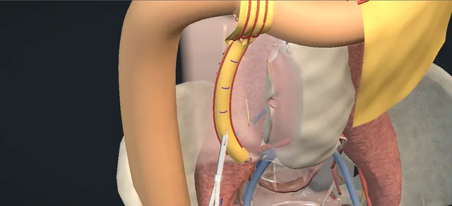
Operative animation, Ureterolysis with omental wrapping

Ureterolysis: the ureter freed from the plaque and wrapped in omentum

Indications

  • Ureteric obstruction from retroperitoneal fibrosis that medical treatment has not relieved.
  • A kidney that depends on a stent or a nephrostomy, or a stent that fails or is not tolerated.
  • Tissue is needed and the release is done at the same operation.

Alternatives

  • Medical treatment with drainage by stents or nephrostomies: the first line.
  • Laparoscopic or robot-assisted ureterolysis.
  • Intraperitoneal placement of the ureter without a wrap.
  • The ureter kept retroperitoneal but lateral, the peritoneum fixed to psoas medial to it.

Risks

  • Bleeding, including from the cava under the plaque.
  • Infection.
  • Ureteric injury, urine leak or a later stricture.
  • Ileus, bowel obstruction or injury to an abdominal organ.
  • Obstruction again; a stent or a further operation.

The operation for a ureter trapped in retroperitoneal fibrosis that drainage and medical treatment have not freed.

The ureter is found above the plaque, the plaque is biopsied, the ureter is released from above downwards with its adventitia on it, laid lateral in the peritoneal cavity and wrapped in a flap of omentum.

The lesson is what lies under the plaque, what keeps the ureter alive, and what keeps the plaque from taking it back.

Advanced tier FRCS retroperitoneal fibrosis, ureteric obstruction, reconstruction Interactive 3D

Operative visualisation

Schematic scene preview: Ureterolysis with omental wrapping
Preparing the 3D anatomy…

UrOops3D · UroRef · © Nity G

How to use this module. Play the nine steps, or use Back and Next. Watch the ureter run into the plaque, the trough open from above downwards, the ureter lift out, and the omentum come down on its arcade and close round it. The triggers show a torn cava, an injured ureter, a malignant biopsy and obstruction again.

Click me next Section 2 of 8 Anatomy The structures this operation works between, and the one it must not cross.

The structures in the scene

Ureterolysis with omental wrapping: how the structures relateThe right retroperitoneum from in front, colon reflected, kidney above and brim below. Right ureter, freed from the plaque: drawn into it; released from above down Plaque across the great vessels. Right ureter, freed from the plaque: cava deep and medial along the release Cava under the plaque (danger). Right ureter, freed from the plaque: kept on the ureter as it is freed Adventitia: the ureter’s blood supply (danger). Right ureter, freed from the plaque: left in front of it, off the plaque Posterior peritoneum, closed behind. Right ureter, freed from the plaque: wrapped round its whole circumference Omental flap as a loose sleeve. Right ureter, freed from the plaque: the sleeve lives on it; no twist Right gastroepiploic arcade (danger).Right ureter, freedfrom the plaquePlaque across thegreat vessels!Cava under theplaque!Adventitia: theureter’s bloodsupplyPosteriorperitoneum, closedbehindOmental flap as aloose sleeve!Right gastroepiploicarcadedrawn into it;released from abovedowncava deep andmedial along thereleasekept on the ureteras it is freedleft in front ofit, off the plaquewrapped round itswhole circumferencethe sleeve lives onit; no twistTargetDangerLandmarkContext
The right retroperitoneum from in front, colon reflected, kidney above and brim below. Schematic, not to scale; relationships only.
  • The plaque sits on the front of the aorta and the cava, runs down over the bifurcation on to the common iliac vessels, and spreads sideways over both ureters, which are often drawn toward the midline inside it.
  • Above the plaque each ureter is wide and lies in normal tissue, and that is where it is found.
  • The right ureter is released from above downwards with the cava, then the right common iliac vessels, beneath the plaque on its medial side.
  • It is then left lateral, in front of the closed peritoneum, inside a sleeve of omentum that hangs on the right gastroepiploic artery.
  • ›The plaque - white, hard and flat across the front of the aorta and the cava and down over the bifurcation, spreading sideways to take in both ureters; biopsied before anything is released.
  • ›Right ureter - wide above the plaque, narrow and pale inside it, often drawn toward the midline; freed from above downwards, then left lateral in the peritoneal cavity.
  • ›Ureteric adventitia - carries the ureter’s longitudinal blood supply; the plane of the release is between it and the scar.
  • ›Inferior vena cava and right common iliac vessels - under the plaque: the cava above the bifurcation and the common iliac vessels below it lie deep and medial to the right ureter along the whole release.
  • ›Aorta - encased with the cava; the left ureter lies beside it in the same plaque.
  • ›Gonadal vein - crosses in front of the ureter above the plaque and may be caught in the same scar; it is a small vein beside the field and is told apart from the cava, which is under the plaque.
  • ›White line of Toldt and the right colon - the line is opened and the colon reflected medially to reach the plaque; the colon is returned when the peritoneum is closed.
  • ›Posterior peritoneum - closed behind the freed ureter, which leaves the ureter in the peritoneal cavity and the plaque behind it.
  • ›Greater curvature and omentum - the omentum is freed off the curvature and divided at its middle into a right and a left flap.
  • ›Right gastroepiploic artery - runs along the greater curvature in the omentum; the only supply of the right flap.
  • ›Left ureter - the same disease and the same operation on the other side, on the left flap.
Click me next Section 3 of 8 Steps The operation in order, with the landmark that guards each move.

Operative steps

  1. Stents, then the plaque An adult with retroperitoneal fibrosis and both ureters obstructed. Cystoscopy first: a stent or an open-ended ureteric catheter is passed up each ureter to help find the ureters, and a urethral catheter is left. Midline transperitoneal laparotomy. The ascending and descending colon are mobilised along the white line of Toldt, a self-retaining retractor is set, the abdomen is examined for visible malignant disease and the bowel is packed away. A white, hard plaque lies across the cava and the aorta, and both ureters disappear into it.
    where it bites: the cava lies under the plaque on the right and the duodenum crosses the top of the field; both are identified before the dissection starts.
  2. Find the ureter above The dilated ureter is identified above the plaque. It is dissected carefully, without injury to its vascular adventitia, and encircled with a vessel loop. With the stent inside it as the guide, it is traced down to the dense scar.
    where it bites: the gonadal vein crosses in front of the ureter here, and the ureter’s blood supply runs in the adventitia that is being kept.
  3. Biopsy Before any release, a biopsy of the fibrotic tissue beside the ureter is taken and sent for histology. The operation goes on.
    where it bites: a deep bite over the cava or into the ureter makes the biopsy the first complication of the operation.
  4. Release, from above downwards The ureter is released from the encasing fibrosis by sharp and blunt dissection, from cranial to caudal, until it is entirely free. Haemostasis is secured with diathermy throughout the dissection.
    where it bites: the cava, and below the bifurcation the right common iliac vessels, lie deep and medial to the right ureter all the way down; the scissors stay on the ureter’s anterior surface, and a ureter stripped of its adventitia has lost its blood supply.
  5. A living ureter Once it is entirely free, the ureter should fill with urine and keep a pink colour along the whole freed length, which is the book’s test of patency and viability.
    where it bites: colour and filling are judged before the ureter is covered; afterwards it cannot be seen.
  6. Intraperitonealise The ureter is positioned laterally without tension and the posterior peritoneum is closed behind it with a running absorbable stitch, so that the ureter is left in the peritoneal cavity. Some surgeons stop here: intraperitoneal placement without a wrap is the operation Batra and colleagues report. The book’s own variation keeps the ureter retroperitoneal but lateral, with the overlying peritoneum secured to psoas medial to it.
    where it bites: the closure goes behind the ureter; a stitch round or through it, or a tight edge where the ureter passes through the peritoneum, obstructs it again.
  7. Raise the omental flap The omentum is mobilised off the stomach with the right and left gastroepiploic arteries preserved. It is then divided at about the middle of the greater curvature, with a sealing device or between clips, which gives two well-vascularised flaps, one for each ureter.
    where it bites: the gastroepiploic arcade is the flap’s only blood supply; it stays on the omentum, and the pedicle is laid without twist or pull.
  8. Wrap and drain The ureter is wrapped circumferentially in its flap and the flap is secured with several interrupted absorbable stitches. The abdomen is irrigated and checked for bleeding. A closed drain is placed in the retroperitoneum and brought out through a separate stab, and the vessels and the viscera are inspected before closure.
    where it bites: a tight sleeve or a twisted pedicle replaces one cause of obstruction with another.
  9. The other side, and after The left ureter is released, placed and wrapped in the same way at the same operation. In the book both ureters are released before either is placed and wrapped; the module follows the right side through to the end. Carletti and colleagues, operating robotically, staged the two sides. The wound is closed in layers. The operation treats the obstruction. The fibrosis is a medical disease, and its treatment and follow-up imaging continue.
    where it bites: the other kidney has the same disease; in bilateral disease a release on one side leaves it at risk.
Click me next Section 4 of 8 Danger zones Where this operation bites, and why it bites there.
Critical relationship

Danger zones

The spatial relationships the animation is built to make automatic. Before you read each one, ask yourself: at this point in the operation, what is about to go wrong, and what is my first move? The save for each sits in the Complications tab.

  • RiskThe cava and the common iliac vessels under the plaque - beside the right ureter for the whole length of the release, and deeper: the cava above the bifurcation, the right common iliac vessels below it. The scissors work on the ureter’s anterior surface and nowhere medial to it.
  • RiskThe ureter’s adventitia - stripped with the scar, the ureter loses its blood supply; a pale stretch that does not fill is the sign.
  • RiskA plaque that is not benign - the laparotomy looks for visible malignant disease and the biopsy is taken before the release; its result decides what follows.
  • RiskThe gastroepiploic pedicle - the flap lives on it; a twist, a pull or a clip across the arcade kills the flap.
  • RiskThe peritoneal edge and the sleeve - a stitch through the ureter, a tight gap in the peritoneum or a tight wrap obstructs the ureter that has just been freed.
  • RiskThe disease itself - the operation frees the ureter and does nothing to the fibrosis; medical treatment and imaging follow-up go on afterwards.
Practise it Take me into the scene Opens the animation at a danger moment and asks you to predict the mechanism and your first move before it reveals.
Click me next Section 5 of 8 Complications The save for each one: the first move, the escalation trigger, the op-note line.

Complications and immediate saves

Trigger each of these live in the Animation tab, then read the save: the first move, the escalation trigger, and the line that goes in the op note.

Caval injury

  • dark blood wells up in the trough from its medial side during the release: the cava, which runs under the plaque beside the right ureter, is torn.
The save
  1. Stop. Press a swab on the bleeding point and hold it; no blind clamp or stitch into the pool. first move
  2. Tell the anaesthetist, confirm blood is available, and get suction, light and a second pair of hands.
  3. Bleeding that pressure does not hold, or a tear that cannot be seen, is a call for the vascular surgeon now. escalation trigger
  4. Once it is controlled and exposed the tear is repaired under vision; the release goes on only if the plane is safe, and stopping with the kidneys drained is a reasonable outcome.
  5. Op note: the site of the tear, how it was controlled and repaired, the blood loss, and whether the release was completed. document

Ureteric injury

  • a hole in the ureteric wall with urine in the field, or a length left dusky because its adventitia came off with the plaque.
The save
  1. Stop the dissection at that point and define the injury: its level, its length, and whether the stent is seen through it. first move
  2. A clean hole is closed with fine interrupted absorbable stitches over the stent.
  3. A long devascularised or badly damaged length is a reconstruction problem, planned with a reconstructive colleague before more is cut. escalation trigger
  4. The repair is covered by the omental wrap and drained, and the stent stays.
  5. Op note: the level and length of the injury, the repair, the stent and the drain. document

Malignant biopsy

  • the histology of the wedge is malignancy.
  • The book sends the biopsy and carries on, so the result usually comes after the operation.
The save
  1. If the result is known in theatre, the release is not started and both kidneys stay drained by their stents. first move
  2. Enough tissue is taken for a full diagnosis, from a safe part of the mass.
  3. A result that comes after the operation goes to the cancer multidisciplinary meeting with the kidneys still drained. escalation trigger
  4. A nephrostomy replaces a stent that fails.
  5. Op note: the biopsy site, what was and was not done, and the drainage left. document

Obstruction again

  • months later the kidney dilates again or the stent cannot come out: the ureter is caught in fibrosis again, or it was narrowed or devascularised at the operation.
The save
  1. Drain the kidney: a stent, or a nephrostomy if a stent will not pass or does not drain. first move
  2. Image the ureter and the plaque, and check renal function and the inflammatory markers.
  3. An obstructed kidney that is infected, or a single kidney, is drained the same day. escalation trigger
  4. The medical treatment is taken back to the physician; a further operation is a decision for a specialist team.
  5. Record the side, the level, the drainage and the plan agreed with the medical team. document
Click me next Section 6 of 8 Troubleshooting When the case turns: the problem, then the next move.

Intra-operative troubleshooting

When the case turns: the problem, then the next move.

  • ActionThe ureter cannot be found in the scar - go back to where it is dilated above the plaque and follow the stent down by feel; the stent or ureteric catheter is placed for this.
  • ActionThe plaque will not come off the vessels safely - stop. Carletti and colleagues abandoned four of 21 planned robotic releases for dense fibrosis or vascular risk; a kidney left drained is better than a torn vessel.
  • ActionThe omentum will not reach, or has gone at earlier surgery - leave the ureter in the peritoneal cavity with the peritoneum closed behind it, or cover it with a local peritoneal flap; both are reported.
  • ActionThe freed ureter stays pale or does not fill - look for a kink or a tight peritoneal edge, then for a stripped or damaged length; a dead length is excised and reconstructed before anything is wrapped round it.
  • ActionThe second side - the book describes the operation as bilateral at one sitting; a planned second stage is the alternative Carletti and colleagues used.
  • ActionWhen the stents come out - the book does not say. Reported practice is a first attempt at four to six weeks, after a retrograde study shows the ureter open (Carletti and colleagues), or removal at four weeks (Simone and colleagues).
Click me next Section 7 of 8 FRCS viva Say it out loud, then mark yourself against the model answer.

FRCS viva questions

The examiner stems this scene prepares you for. Try to answer out loud first, then reveal, and learn the discriminator, the detail that turns a pass into a strong pass.

QA patient has bilateral hydronephrosis from retroperitoneal fibrosis and has been stented. When would you offer ureterolysis?reveal answer
  • This is a medical disease with a urological complication.
  • Fenaroli and colleagues’ review says treatment is mainly conservative, with glucocorticoids or other immunosuppressants plus drainage of the kidneys, and that surgical ureterolysis is left for refractory cases.
  • So I would offer the operation to the patient whose ureters stay obstructed despite that treatment.
  • In the Guy’s series of 50 open ureterolyses with omental wrap, the indications were dependence on a nephrostomy, stent failure with persistent hydronephrosis, severe stent symptoms and, in a few, patient choice.
  • O’Brien and Fernando argue from that series that ureterolysis should be considered in anyone who does not respond quickly to standard treatment.
  • I would also want the diagnosis secure: if the imaging is atypical I would want tissue, and an operation gives me that.
  • I would discuss the alternatives, which are continued stents or nephrostomies and a laparoscopic or robotic release in a centre that does them, and I would plan it with the physician who is treating the fibrosis.
Strong pass

Say both

  • Saying that first-line treatment is medical with drainage
  • Naming the surgical indications with the series they come from
Examiner note
Saying that first-line treatment is medical with drainage, and naming the surgical indications with the series they come from.
QTalk me through an open ureterolysis with omental wrapping.reveal answer
  • I start with a cystoscopy and place a stent in each ureter, because the stents help me find the ureters.
  • Through a midline laparotomy I mobilise the colon along the white line of Toldt, look for visible malignant disease and pack the bowel.
  • I find the ureter above the plaque where it is dilated, dissect it with its adventitia on it and pass a vessel loop round it, then follow it down to the scar.
  • I take a biopsy of the fibrotic tissue beside the ureter and send it.
  • I then release the ureter by sharp and blunt dissection from cranial to caudal, remembering that on the right the cava, and lower down the common iliac vessels, are under the plaque on its medial side.
  • When the ureter is free I check that it fills and stays pink.
  • I do the same on the other side.
  • I lay each ureter lateral without tension and close the posterior peritoneum behind it.
  • I mobilise the omentum off the stomach, keeping the gastroepiploic arteries on it, divide it at the middle of the greater curvature into two flaps, wrap each ureter circumferentially and secure each flap with interrupted absorbable stitches.
  • I leave a retroperitoneal drain and close.
Strong pass

Finding the ureter above the plaque, the biopsy before the release, the cranial to caudal direction, and the check of colour and filling before the ureter is covered.

QAfter the ureter is free, what stops the plaque taking it back, and what is the evidence?reveal answer
  • I move the ureter away from the plaque and put healthy tissue between them.
  • The open operation as Armenakas describes it places the ureter laterally in the peritoneal cavity, closes the peritoneum behind it and then wraps it in an omental flap;
  • its stated variation keeps the ureter retroperitoneal and lateral with the peritoneum secured to psoas.
  • All of these are reported.
  • O’Brien and Fernando wrapped in omentum and had 47 of 50 patients free of stents at twelve months.
  • Batra and colleagues intraperitonealised the ureter without a wrap in nine renal units and reported durable relief.
  • Carletti and colleagues used an omental wrap when the omentum reached without tension and a peritoneal flap when it did not.
  • These are small single-centre series and I know of no trial comparing the methods, so my choice follows what I have been trained to do and what the omentum allows on the day.
Strong pass

Knowing that wrap, intraperitoneal placement alone and lateral retroperitoneal placement are all reported, on small single-centre series and no comparative trial.

QDuring the release on the right there is sudden dark bleeding from the medial side. What do you do?reveal answer
  • I assume it is the cava, because it runs under the plaque just medial to the right ureter.
  • I stop dissecting, press a swab on the point and hold it, and I do not put a clamp or a stitch blindly into blood.
  • I tell the anaesthetist, make sure blood is available, and get better suction, light and an assistant.
  • If pressure holds it I take time to expose the tear properly and repair it under vision.
  • If it does not hold, or I cannot see the tear, I call the vascular surgeon at once and keep the pressure on until they arrive.
  • Afterwards I decide whether it is safe to go on.
  • I would be content to stop with the kidneys drained: in one robotic series about a fifth of planned releases were abandoned for dense fibrosis or vascular risk.
  • I record the site, the repair, the blood loss and how far the release got.
Strong pass

Pressure first and help early, no blind clamp, and the readiness to abandon the release with the kidney drained.

Click me next Section 8 of 8 Evidence The guidance each of these answers is built on.

Evidence and guidance

Operative Dictations in Urologic Surgery
Armenakas NA, Fracchia JA, Golan R. Ureterolysis (with omental flaps). Chapter 55. Wiley, 2019
The essential steps and their order: stents or ureteric catheters first, midline transperitoneal exposure with both colons mobilised, the dilated ureter found above and looped, the biopsy, release from cranial to caudal, the test of filling and colour, the other side, lateral placement with the peritoneum closed behind, omental flaps on the gastroepiploic arteries, the wrap and the drain. The variations (stents at the surgeon’s discretion; lateral retroperitoneal placement) and the complication list. Catheter sizes, suture gauges and the incision are kept off screen.
Open guideline
Current Rheumatology Reports
Fenaroli P, Maritati F, Vaglio A. Into clinical practice: diagnosis and therapy of retroperitoneal fibrosis. Curr Rheumatol Rep 2021;23:18. Abstract read 13 October 2026
A review. Imaging is the main diagnostic tool, with retroperitoneal biopsy reserved for atypical cases; treatment is mainly conservative, with medical therapies (glucocorticoids and immunosuppressants) plus urological interventions; surgical ureterolysis is left for refractory cases.
Open guideline
BJU International
O’Brien T, Fernando A. Contemporary role of ureterolysis in retroperitoneal fibrosis: treatment of last resort or first intent? An analysis of 50 cases. BJU Int 2017;120:556-561. Abstract read 13 October 2026
One centre, 50 patients, open ureterolysis with omental wrap, at least a year of follow-up. Indications: nephrostomy dependence, stent failure, severe stent symptoms, patient choice. 47 of 50 free of stents at twelve months; 11 of 50 had a further procedure, seven of them a nephrectomy; serious complications in 12 per cent. The authors’ own figures, from a specialist team, and their own argument that ureterolysis be considered in patients who do not respond quickly to standard treatment.
Open guideline
Urologia
Batra VS, Singh M, Kathuria S, et al. Minimally invasive ureterolysis and intraperitonealization of ureter for idiopathic retroperitoneal fibrosis; single center analysis of an erratic disease. Urologia 90(3):535-541, published online 2022. Abstract read 13 October 2026
One centre, retrospective: nine renal units in seven patients released laparoscopically or robotically and intraperitonealised without a wrap, median follow-up 60 months, symptoms resolved in all. The source for intraperitoneal placement alone.
Open guideline
BJUI Compass
Carletti F, Turcan A, Tamborino F, Morgantini LA, Salvodirocco L, Alkassis M, Dal Moro F, Crivellaro S. Single-port robot-assisted ureterolysis for retroperitoneal fibrosis: a less invasive path to functional recovery. BJUI Compass 2026;7(5):e70218. Full text read 13 October 2026
One surgeon, 16 patients and 21 renal units; bilateral disease staged. Four of 21 procedures abandoned for dense fibrosis or vascular risk. Omental wrap when the omentum reached without tension, a peritoneal flap otherwise. First stent removal attempted at four to six weeks after retrograde pyelography.
Open guideline
Urology
Simone G, Leonardo C, Papalia R, Guaglianone S, Gallucci M. Laparoscopic ureterolysis and omental wrapping. Urology 2008;72:853-858. Abstract read 13 October 2026
Six patients, laparoscopic release with an omental wrap; stents removed at four weeks. Quoted only for the timing of stent removal.
Open guideline

Curriculum. Supports learning relevant to the ISCP Urology Curriculum syllabus topic Ureteric reconstruction. Not a workplace-based assessment and not evidence of competence.

Two minutes

Close the lap: recall it before you leave.

Say it aloud, then reveal. Reading is not remembering.

  1. 01 The relationship Say the critical spatial relationship out loud, then reveal. reveal
    Memory frame

    The plaque sits on the front of the aorta and the cava, runs down over the bifurcation on to the common iliac vessels, and spreads sideways over both ureters, which are often drawn toward the midline inside it.

    Why?
    Above the plaque each ureter is wide and lies in normal tissue, and that is where it is found. The right ureter is released from above downwards with the cava, then the right common iliac vessels, beneath the plaque on its medial side. It is then left lateral, in front of the closed peritoneum, inside a sleeve of omentum that hangs on the right gastroepiploic artery.
  2. 02 The first move Caval injury. Your first move? reveal
    First move

    Stop.

    More detail
    Press a swab on the bleeding point and hold it; no blind clamp or stitch into the pool.
  3. 03 The escalation trigger Caval injury. What makes you call for help? reveal
    Escalate when

    Bleeding that pressure does not hold, or a tear that cannot be seen, is a call for the vascular surgeon now.

  4. 04 The discriminator What turns a pass into a strong pass? reveal
    Strong pass

    Say both

    • Saying that first-line treatment is medical with drainage
    • Naming the surgical indications with the series they come from
    Examiner note
    Saying that first-line treatment is medical with drainage, and naming the surgical indications with the series they come from.
  5. 05 The pitfall Where does this operation bite first? reveal
    Pitfall

    The cava and the common iliac vessels under the plaque

    Why?
    beside the right ureter for the whole length of the release, and deeper: the cava above the bifurcation, the right common iliac vessels below it. The scissors work on the ureter’s anterior surface and nowhere medial to it.

Revision practice with self-marked answers, not an assessment of clinical competence. Kept in this browser only; it does not sync between devices.