UrOops3D, danger-zone atlas

Schematic model.

Operative animation, Uterosacral vault suspension

Enterocele repair with uterosacral vault suspension

A transvaginal repair of vault prolapse with an enterocele. The prolapsed vault is brought out, the anterior wall is opened along the enterocele and the sac is freed to its neck and opened.

With the bowel held up on a pad, three sutures are passed through each uterosacral ligament, posteromedial to the ischial spine, and fixed to the pubocervical and rectovaginal fascia.

Tying them lifts the vault, and cystoscopy then shows efflux from both ureters and no suture in the bladder.

Advanced tier FRCS female urology, pelvic organ prolapse Interactive 3D

Operative visualisation

Schematic scene preview: Uterosacral vault suspension
Preparing the 3D anatomy…

UrOops3D · UroRef · © Nity G

How to use this module. Play the nine steps, or use Back and Next. Watch the vault come out, the sac open on the bowel, the bowel go up on its pad, the ureter shown lateral to each ligament and the vault rise as the sutures are tied. The triggers show a bowel injury, a kinked ureter and a suture in the bladder.

Click me next Section 2 of 8 Anatomy The structures this operation works between, and the one it must not cross.

The structures in the scene

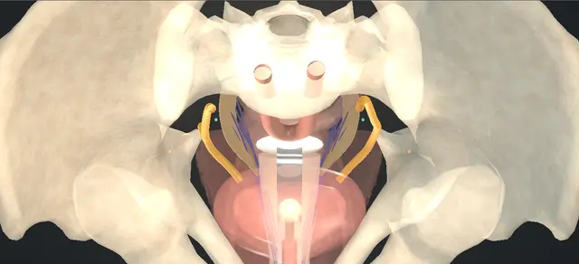
Uterosacral vault suspension: how the structures relateThe uterosacral ligament as the needle meets it, from the opened cul-de-sac. Uterosacral ligament: three sutures: felt posteromedial to it Ischial spine: the level of the lowest. Uterosacral ligament: three sutures: needle passed lateral to medial, away from it Ureter: lateral, closest near the vault (danger). Uterosacral ligament: three sutures: bites stay out of its wall Rectum: medial and behind (danger). Uterosacral ligament: three sutures: packed away before the first bite Small bowel: held up on a pad (danger). Uterosacral ligament: three sutures: one arm of each suture to each Pubocervical and rectovaginal fascia. Uterosacral ligament: three sutures: runs back to it from the side of the vault Sacrum: where the ligament ends.Uterosacral ligament:three suturesIschial spine: thelevel of the lowest!Ureter: lateral,closest near thevault!Rectum: medial andbehind!Small bowel: held upon a padPubocervical andrectovaginal fasciaSacrum: where theligament endsfelt posteromedialto itneedle passedlateral to medial,away from itbites stay out ofits wallpacked away beforethe first biteone arm of eachsuture to eachruns back to itfrom the side ofthe vaultTargetDangerLandmarkContext
The uterosacral ligament as the needle meets it, from the opened cul-de-sac. Schematic, not to scale; relationships only.
  • In lithotomy the vault hangs at the introitus with the pubis and urethra above it and the rectum below.
  • Inside, the uterosacral ligaments run back from the side of the vault to the sacrum, each medial and posterior to the ischial spine.
  • The ureter lies lateral to each ligament and closest to it near the vault, so the needle crosses the ligament from its lateral edge and stays at or above the spine.
  • ›Vaginal vault - prolapsed to the introitus at the start; brought out, opened and finally resuspended from both ligaments.
  • ›Enterocele sac - peritoneum herniating between the vagina and the rectum; isolated to its neck and opened at its far end.
  • ›Small bowel - lies in the sac; held up out of the pelvis on a moist pad before any suture is placed.
  • ›Pubocervical fascia - under the anterior wall; takes one arm of each suspension suture.
  • ›Rectovaginal fascia - under the posterior wall; takes the other arm.
  • ›Ischial spine - felt on each side; the landmark for the level of the lowest suture.
  • ›Uterosacral ligament - posteromedial to the spine, at four and eight o'clock; tightens with traction on the vault.
  • ›Ureter - lateral to the ligament on the side wall, closest to it near the vault; seen as efflux at the cystoscopy.
  • ›Bladder - kept empty through the dissection; inspected for a suture in its wall at the end.
  • ›Rectum - medial and posterior to the ligaments; kept out of every bite.
Click me next Section 3 of 8 Steps The operation in order, with the landmark that guards each move.

Operative steps

  1. The prolapse A woman with vault prolapse and an enterocele after hysterectomy, who has chosen a vaginal repair after counselling. Antibiotics, then dorsal lithotomy on padded stirrups. A urethral catheter drains the bladder and it is kept decompressed throughout. A weighted speculum is placed on the posterior wall and a self-retaining retractor is set. The anterior wall is infiltrated with local anaesthetic and adrenaline. With the bladder empty, the prolapsed vaginal wall is grasped between two tissue clamps and brought out through the introitus.
    where it bites: a full bladder lies against the anterior wall that is about to be opened.
  2. Open the wall A longitudinal midline incision is made in the anterior vaginal wall along the length of the enterocele. The vaginal wall is dissected off the underlying pubocervical fascia and off the enterocele sac, staying superficial.
    where it bites: a dissection that goes deep to the fascia reaches the bladder base; one that goes into the sac reaches bowel.
  3. Open the sac By sharp and blunt dissection the enterocele sac is isolated all round as far as its neck. It is opened at its far end and the peritoneal cavity is entered. Small bowel lies in the sac.
    where it bites: bowel may lie against the inside of the sac; it is opened under vision, away from any loop.
  4. Hold the bowel up A moist pad is placed through the opened sac and the abdominal contents are reduced ahead of it. A retractor is placed anteriorly over the pad to hold them out of the pelvis.
    where it bites: bowel left in the cul-de-sac lies where the ligament sutures will be placed.
  5. Find the ligaments The ischial spines are palpated on each side. With traction on the vault, each uterosacral ligament tightens and is identified posteromedial to the spine, at the four and eight o'clock positions on the pelvic side wall. The ureter runs on the pelvic side wall lateral to the ligament, about 2 cm from it at the level of the spine and closer still near the vault, where tension on the ligament pulls on it.
    where it bites: the ureter lies lateral to the ligament; a stitch placed too far lateral, or low near the vault, can take or kink it.
  6. Sutures through the ligaments Three double-armed interrupted sutures, of a slowly absorbable or a non-absorbable material, are passed from lateral to medial through each uterosacral ligament: the lowest at the level of the ischial spine and the others above it. Each is tagged and left untied.
    where it bites: each bite takes the ligament itself; a bite that wanders laterally risks the ureter and one that goes deep risks the rectum.
  7. Fix and tie One arm of each suture is passed through the pubocervical fascia and the other through the rectovaginal fascia at the apex. The pad and the retractor are removed and each suture is tied individually, which resuspends the vaginal apex. Care is taken to avoid injury to the bowel, the ureters and the bladder. The ends are cut.
    where it bites: a pad left behind, or a loop of bowel caught as the vault rises, is avoided by removing the pad before any suture is tied.
  8. Cystoscopy The urethral catheter is removed and cystourethroscopy is performed with 30-degree and 70-degree lenses. The urethra and bladder are intact, clear efflux is seen from both ureteric orifices and there is no suture in the bladder. The cystoscope is removed and the catheter replaced.
    where it bites: an orifice with no efflux after tying means a ureter caught or kinked until shown otherwise.
  9. Trim and close The edges of the vaginal epithelium are trimmed and the vaginal wall is closed in the midline with a running absorbable suture. The self-retaining retractor and the weighted speculum are removed and the catheter is connected to drainage.
    where it bites: trimming too much epithelium shortens and narrows the vagina.
Click me next Section 4 of 8 Danger zones Where this operation bites, and why it bites there.
Critical relationship

Danger zones

The spatial relationships the animation is built to make automatic. Before you read each one, ask yourself: at this point in the operation, what is about to go wrong, and what is my first move? The save for each sits in the Complications tab.

  • RiskUreter - lateral to the ligament and close to it; the usual reason for a dry orifice after tying.
  • RiskSmall bowel - in the sac when it is opened, and in the cul-de-sac until it is packed away.
  • RiskBladder - under the anterior dissection and near the anterior fascial arms; a suture through its wall is looked for at the end.
  • RiskRectum - medial to the ligaments and under the posterior fascia; kept out of every bite.
  • RiskLevel of the bites - at the spine and above it, in the ligament itself, not low near the vault where the ureter is closest.
Practise it Take me into the scene Opens the animation at a danger moment and asks you to predict the mechanism and your first move before it reveals.
Click me next Section 5 of 8 Complications The save for each one: the first move, the escalation trigger, the op-note line.

Complications and immediate saves

Trigger each of these live in the Animation tab, then read the save: the first move, the escalation trigger, and the line that goes in the op note.

Bowel injury

  • a loop of small bowel lying against the sac is cut as the sac is opened, or caught later by a suture.
  • The repair is not continued over an unrecognised hole.
The save
  1. Stop. Keep the injured loop in view and hold it out of the field. first move
  2. Inspect the whole loop and the neighbouring loops for a second injury.
  3. A serosal tear alone may be repaired directly; a full-thickness hole is not left to a vaginal closure.
  4. Any full-thickness injury, or any doubt, needs a general surgeon in theatre before the repair goes on. escalation trigger
  5. Op note: the loop, the site and size of the injury, how it was repaired and by whom, and the plan for observation. document

Ureteric kinking or obstruction

  • no efflux from one orifice at the cystoscopy after the sutures are tied: a suspension suture has taken or kinked the ureter, usually the most lateral, or the lowest, where the ureter is closest.
The save
  1. Remove the suspect suture on the dry side, the lowest and most lateral first, and look again for efflux. first move
  2. An intravenous dye before the second look makes the efflux easier to see.
  3. If efflux returns, the suture is placed again more medially and posteriorly in the ligament and the cystoscopy repeated.
  4. Still no efflux after removing the sutures on that side: retrograde study and a stent, and urological help in theatre. escalation trigger
  5. Op note: which suture was removed, efflux on both sides at the end, and any stent and its plan. document

Suture in the bladder

  • a loop of suture seen inside the bladder at the cystoscopy, from an anterior fascial arm or a bite that has passed through the bladder wall.
The save
  1. Identify which suture it is, then cut it and withdraw it. first move
  2. Place that arm again in the fascia, clear of the bladder, and tie it.
  3. Inspect the bladder again for the hole and for any other suture.
  4. A larger injury, or one near a ureteric orifice, needs a senior colleague before the operation goes on. escalation trigger
  5. Op note: the finding, what was done and how long the catheter stays. document
Click me next Section 6 of 8 Troubleshooting When the case turns: the problem, then the next move.

Intra-operative troubleshooting

When the case turns: the problem, then the next move.

  • ActionThe ligament cannot be felt - more traction on the vault with the clamps, toward the opposite side, tightens it. The spine is found first and the ligament sought posteromedial to it; a stitch is not placed on a guess.
  • ActionBowel keeps coming down past the pad - the pad is placed again higher with the patient tipped head down, and the anterior retractor seated over it again before any bite is taken.
  • ActionThe sac is hard to find in the dissection - it lies behind the apex, between vagina and rectum; a finger in the rectum shows where the rectal wall is, and the sac is the peritoneum above it.
  • ActionEfflux is hard to see from one orifice - indigo carmine or methylene blue given intravenously before the cystoscopy makes the efflux easier to see. A dry orifice is not left unexplained.
  • ActionThe ligaments are thin or will not hold - the described alternatives are suspension to the sacrospinous ligament or to the iliococcygeus fascia.
  • ActionThere is also stress incontinence or a rectocele - a mid-urethral sling or a rectocele repair may be performed at the same sitting.
Click me next Section 7 of 8 FRCS viva Say it out loud, then mark yourself against the model answer.

FRCS viva questions

The examiner stems this scene prepares you for. Try to answer out loud first, then reveal, and learn the discriminator, the detail that turns a pass into a strong pass.

QA woman has symptomatic vault prolapse years after a hysterectomy. What options would you discuss?reveal answer
  • I would go through both non-surgical and surgical options with her, using the NICE patient decision aid on surgery for vault prolapse, and I would be open about the possible complications and the lack of long-term evidence on how well the operations work.
  • For surgery, NICE recommends offering a choice of vaginal sacrospinous fixation with sutures or sacrocolpopexy with mesh, abdominal or laparoscopic;
  • if mesh were used, its details and outcomes must go into a national registry.
  • If she did not intend to have penetrative intercourse and her health made a longer operation risky, I would consider colpocleisis.
  • A uterosacral ligament suspension is another native-tissue vaginal operation, and I would tell her that it is not one of the two operations NICE lists for vault prolapse.
Strong pass

Say all three

  • Knowing that NICE offers sacrospinous fixation or sacrocolpopexy for vault prolapse
  • Considers colpocleisis for a frail woman
  • Does not list uterosacral suspension
Examiner note
Knowing that NICE offers sacrospinous fixation or sacrocolpopexy for vault prolapse, considers colpocleisis for a frail woman, and does not list uterosacral suspension.
QTalk me through a transvaginal enterocele repair with uterosacral ligament fixation.reveal answer
  • I drain the bladder with a catheter and keep it empty.
  • With a weighted speculum and a self-retaining retractor in place I infiltrate the anterior wall, grasp the prolapsed vault between two clamps and bring it out.
  • I open the anterior wall in the midline along the enterocele and dissect it off the pubocervical fascia and the sac, staying superficial.
  • I isolate the sac to its neck, open it at its far end and enter the peritoneum, then pack the bowel up with a moist pad and hold it with a retractor placed anteriorly.
  • I feel the ischial spines and find each uterosacral ligament posteromedial to the spine.
  • I pass three double-armed sutures through each ligament from lateral to medial, the lowest at the level of the spine, and take one arm of each through the pubocervical fascia and the other through the rectovaginal fascia.
  • I remove the pad and the retractor and tie each suture in turn, which lifts the vault.
  • I then do a cystoscopy for efflux on both sides and for a suture in the bladder, trim the epithelium and close the wall with a running absorbable suture.
Strong pass

Say all four

  • The order that keeps the bowel and the ureter safe: bowel packed before any bite
  • Bites lateral to medial at or above the spine
  • Pad out before tying
  • Cystoscopy after tying
Examiner note
The order that keeps the bowel and the ureter safe: bowel packed before any bite, bites lateral to medial at or above the spine, pad out before tying, and cystoscopy after tying.
QAfter tying the suspension sutures you see no efflux from the left ureteric orifice. What do you do?reveal answer
  • I would assume the left ureter has been caught or kinked by a suspension suture until I had shown otherwise, because that is the known hazard of the operation and the reason the cystoscopy comes after tying.
  • I would take down the left sutures, starting with the lowest and most lateral, and look again; an intravenous dye helps me see the efflux.
  • If efflux returned I would place that suture again further medially and posteriorly in the ligament, tie it and repeat the cystoscopy.
  • If there were still no efflux after removing the left sutures I would do a retrograde study and pass a stent, and I would ask for urological help in theatre.
  • I would record efflux on both sides before leaving theatre.
  • The anatomy explains it: in the cadaver dissections of Buller and colleagues the ureter lay 0.9 cm from the ligament at its cervical portion and 2.3 cm at its intermediate portion, over the ischial spine, and in the cadavers of Wieslander and colleagues the suspension sutures lay a mean of 14 mm from the ureter, with a range down to 0.
Strong pass

Removing the suspect suture first and looking again, and knowing why: the ureter is about 2 cm lateral to the ligament at the spine and closer near the vault.

QHow does uterosacral suspension compare with sacrospinous fixation?reveal answer
  • I would quote the OPTIMAL trial by Barber and colleagues, which randomised 374 women having vaginal surgery for apical prolapse and stress incontinence, all with a mid-urethral sling, at 9 centres in the United States, to uterosacral suspension or sacrospinous fixation.
  • At 2 years surgical success was 59.2 per cent after uterosacral suspension and 60.5 per cent after sacrospinous fixation, and serious adverse events were 16.5 per cent and 16.7 per cent; neither was significantly better.
  • I would add that these women had stress incontinence as well as prolapse, and that in the UK NICE lists sacrospinous fixation, not uterosacral suspension, as the native-tissue vaginal option for vault prolapse.
Strong pass

Say both

  • Quoting OPTIMAL accurately as no significant difference at 2 years in women who also had a sling
  • Setting it against the NICE choice for vault prolapse
Examiner note
Quoting OPTIMAL accurately as no significant difference at 2 years in women who also had a sling, and setting it against the NICE choice for vault prolapse.
QWhy is the bowel packed away before the ligament sutures, and why is the pad removed before they are tied?reveal answer
  • Once the sac is open, small bowel lies in the cul-de-sac exactly where I am passing a needle through the ligaments, so I hold it up on a moist pad with a retractor over it before the first bite.
  • I remove the pad and the retractor before I tie because tying lifts the vault up to the ligaments and closes the space the pad is lying in;
  • a pad left there would be retained, and a loop of bowel caught between the vault and the ligament would be at risk.
  • Bowel injury is on the list of complications of the operation, alongside ureteric and bladder injury.
Strong pass

Say both

  • Linking each move to the anatomy at that moment: bowel in the field while the needle passes
  • The space closing as the sutures are tied
Examiner note
Linking each move to the anatomy at that moment: bowel in the field while the needle passes, and the space closing as the sutures are tied.
Click me next Section 8 of 8 Evidence The guidance each of these answers is built on.

Evidence and guidance

Operative Dictations in Urologic Surgery
Armenakas NA, Fracchia JA, Golan R. Enterocele repair with uterosacral ligament fixation. Chapter 93. Wiley, 2019
Followed here: the catheter and the bladder kept decompressed, the weighted speculum and self-retaining retractor, the infiltration, the prolapsed wall grasped between two clamps and brought out, the midline incision along the enterocele, the wall dissected off the pubocervical fascia and the sac staying superficial, the sac isolated to its neck and opened distally, the moist pad and anterior retractor, the ischial spines palpated and the ligaments identified posteromedially at four and eight o'clock, three double-armed interrupted sutures passed lateral to medial through each ligament, one arm to the pubocervical and one to the rectovaginal fascia, the pad and retractor removed before each suture is tied, the cystoscopy (30-degree and 70-degree lenses, bladder and urethra intact, efflux from both sides, no intravesical suture), the trim and running closure, the variations (sacrospinous or iliococcygeus suspension, mesh augmentation, a concurrent sling or rectocele repair, an intravenous dye, a suprapubic catheter, a vaginal pack) and the complication list, from which the three error states are taken: bowel, ureteric and bladder injury. The chapter gives no management for its complications: the first, escalate and document steps and most troubleshooting entries are general surgical practice. It does not say whether the sac is closed or excised, nor where the ureter lies relative to the ligament; the ureter's relation is drawn from Buller et al. 2001 (lateral to the ligament, nearest at its cervical end). Its variation of a synthetic mesh to augment the repair is not drawn: this is a native-tissue repair. The scene is schematic, on the bones and bladder of the atlas, and no figure in it is a measurement.
Open guideline
NICE
Urinary incontinence and pelvic organ prolapse in women: management. NICE guideline NG123, recommendations 1.8.14 to 1.8.18, read 14 October 2026
Discuss non-surgical and surgical options for vault prolapse using a decision aid (1.8.14); discuss the possible complications and the lack of long-term evidence on effectiveness (1.8.15); offer a choice of vaginal sacrospinous fixation with sutures or sacrocolpopexy, abdominal or laparoscopic, with mesh (1.8.16); record mesh procedures in a national registry (1.8.17); consider colpocleisis for women who do not intend penetrative sex and whose physical condition increases operative risk (1.8.18). Uterosacral suspension does not appear in these recommendations.
Open guideline
JAMA
Barber MD, Brubaker L, Burgio KL, et al. Comparison of 2 transvaginal surgical approaches and perioperative behavioral therapy for apical vaginal prolapse: the OPTIMAL randomized trial. JAMA 2014;311(10):1023-34. doi 10.1001/jama.2014.1719
Abstract read via PubMed 14 October 2026. 374 women undergoing vaginal surgery, including a mid-urethral sling, for apical prolapse and stress incontinence at 9 US centres, randomised to sacrospinous fixation or uterosacral suspension. At 2 years: surgical success 59.2% after uterosacral suspension against 60.5% after sacrospinous fixation; serious adverse events 16.5% against 16.7%; neither significantly superior. The source of every figure in the viva.
Open guideline
Obstetrics and Gynecology
Buller JL, Thompson JR, Cundiff GW, et al. Uterosacral ligament: description of anatomic relationships to optimize surgical safety. Obstet Gynecol 2001;97(6):873-9. doi 10.1016/s0029-7844(01)01346-1
Abstract read via PubMed 14 October 2026. Fifteen female cadavers. Ureter to uterosacral ligament 0.9, 2.3 and 4.1 cm at the cervical, intermediate and sacral portions; the ischial spine lies beneath the intermediate portion; ligament tension is transmitted to the ureter, most near the cervix; the intermediate portion is suggested as the site for fixation. The source of the ureter's course in the scene and the distances in the viva.
Open guideline
American Journal of Obstetrics and Gynecology
Wieslander CK, Roshanravan SM, Wai CY, Schaffer JI, Corton MM. Uterosacral ligament suspension sutures: anatomic relationships in unembalmed female cadavers. Am J Obstet Gynecol 2007;197(6):672.e1-6. doi 10.1016/j.ajog.2007.08.065
Abstract read via PubMed 14 October 2026. Fifteen unembalmed cadavers: proximal suspension sutures a mean 14 mm from the ureter (range 0 to 33) and 10 mm from the rectal lumen; sutures can directly injure the ureters, rectum and side-wall neurovascular structures.
Open guideline

Curriculum. Supports learning relevant to female urology and pelvic organ prolapse in the ISCP Urology Curriculum. Not a workplace-based assessment and not evidence of competence.

Two minutes

Close the lap: recall it before you leave.

Say it aloud, then reveal. Reading is not remembering.

  1. 01 The relationship Say the critical spatial relationship out loud, then reveal. reveal
    Memory frame

    In lithotomy the vault hangs at the introitus with the pubis and urethra above it and the rectum below.

    Why?
    Inside, the uterosacral ligaments run back from the side of the vault to the sacrum, each medial and posterior to the ischial spine. The ureter lies lateral to each ligament and closest to it near the vault, so the needle crosses the ligament from its lateral edge and stays at or above the spine.
  2. 02 The first move Bowel injury. Your first move? reveal
    First move

    Stop.

    More detail
    Keep the injured loop in view and hold it out of the field.
  3. 03 The escalation trigger Bowel injury. What makes you call for help? reveal
    Escalate when

    Any full-thickness injury, or any doubt, needs a general surgeon in theatre before the repair goes on.

  4. 04 The discriminator What turns a pass into a strong pass? reveal
    Strong pass

    Say all three

    • Knowing that NICE offers sacrospinous fixation or sacrocolpopexy for vault prolapse
    • Considers colpocleisis for a frail woman
    • Does not list uterosacral suspension
    Examiner note
    Knowing that NICE offers sacrospinous fixation or sacrocolpopexy for vault prolapse, considers colpocleisis for a frail woman, and does not list uterosacral suspension.
  5. 05 The pitfall Where does this operation bite first? reveal
    Pitfall

    Ureter

    Why?
    lateral to the ligament and close to it; the usual reason for a dry orifice after tying.

Revision practice with self-marked answers, not an assessment of clinical competence. Kept in this browser only; it does not sync between devices.