UrOops3D, danger-zone atlas

Schematic model.

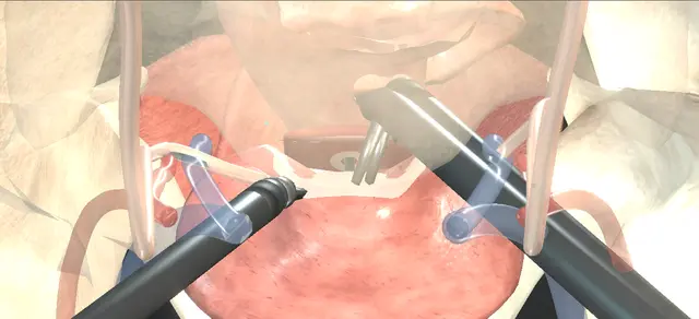
Operative animation, Robotic vesicovaginal fistula repair

Robotic vesicovaginal fistula repair: two lines, kept apart

Indications

  • Vesicovaginal fistula high at the vaginal vault after hysterectomy
  • A fistula that has persisted after an earlier vaginal repair

Alternatives

  • Transvaginal layered (flap-splitting) repair
  • Transvesical repair through a separate cystotomy
  • Open abdominal repair
  • Continuous catheter drainage for a small, recent fistula

Risks

  • Bleeding
  • Infection
  • Injury to bowel or another abdominal organ
  • Ureteric injury or obstruction
  • Ileus
  • A fistula that persists or recurs

Transperitoneal robotic repair of a fistula between the back wall of the bladder and the vaginal vault after hysterectomy, in a woman whose vaginal repair has failed. Cystoscopy marks both ureters and the tract with catheters before docking.

The bladder is separated from the vagina round the tract, the fistula is excised, the vagina is closed in one layer and the bladder in two, the closure is tested, and omentum is anchored between the two lines.

Advanced tier FRCS female urology, urinary fistula, robotic reconstruction Interactive 3D

Operative visualisation

Schematic scene preview: Robotic vesicovaginal fistula repair
Preparing the 3D anatomy…

UrOops3D · UroRef · © Nity G

How to use this module. Play the eight steps, or use Back and Next. Watch catheters pass up both ureters and through the tract, the bladder parted from the vagina, the fistula cut out, two lines closed across each other and omentum laid on the repair. The triggers show a caught orifice, a leak and a bowel injury.

Click me next Section 2 of 8 Anatomy The structures this operation works between, and the one it must not cross.

The structures in the scene

Robotic vesicovaginal fistula repair: how the structures relateThe fistula at the vault from the robot, and what lies round it. Fistula: bladder back wall to vaginal vault: opens on its back wall above the trigone Bladder: closed in two layers. Fistula: bladder back wall to vaginal vault: opens on the anterior wall of the vault Vagina: closed in one running layer. Fistula: bladder back wall to vaginal vault: lie close below it; bites stay clear Ureteric orifices, catheterised (danger). Fistula: bladder back wall to vaginal vault: pass beside the dissection plane Ureters, lateral to the plane (danger). Fistula: bladder back wall to vaginal vault: covers the repair, anchored to the vagina Omentum, laid between the two lines. Fistula: bladder back wall to vaginal vault: above and behind; first port and retraction Small bowel and rectum (danger).Fistula: bladder backwall to vaginal vaultBladder: closed intwo layersVagina: closed inone running layer!Ureteric orifices,catheterised!Ureters, lateral tothe planeOmentum, laidbetween the twolines!Small bowel andrectumopens on its backwall above thetrigoneopens on theanterior wall ofthe vaultlie close below it;bites stay clearpass beside thedissection planecovers the repair,anchored to thevaginaabove and behind;first port andretractionTargetDangerLandmarkContext
The fistula at the vault from the robot, and what lies round it. Schematic, not to scale; relationships only.
  • From the camera above the umbilicus, the drained bladder lies in front, the vaginal vault behind it and the rectum behind that.
  • The fistula runs from the back wall of the bladder, just above the trigone, to the anterior wall of the vault.
  • Each ureter passes down lateral to the plane between them to an orifice close below the fistula.
  • Once separated, the vaginal opening faces the console and the bladder opening faces away from it.
  • ›Fistula tract - joins the back wall of the bladder, above the trigone, to the vaginal vault; intubated at cystoscopy and excised.
  • ›Ureteric orifices - below the fistula on the trigone; catheterised before the repair so they are known throughout.
  • ›Ureters - run down lateral to the vesicovaginal plane to enter the bladder base; kept out of the dissection.
  • ›Vaginal vault - the closed top of the vagina after hysterectomy; lifted by a weighted retractor so its anterior wall can be found from above.
  • ›Bladder back wall - separated from the vagina round and beyond the tract; closed in two layers.
  • ›Peritoneum over the vesicovaginal space - the first incision of the console work.
  • ›Omentum - brought down on its blood supply and anchored between the two lines.
  • ›Rectum and small bowel - behind the vault and above the pelvis; bowel falls away with the head-down tilt.
Click me next Section 3 of 8 Steps The operation in order, with the landmark that guards each move.

Operative steps

  1. Cystoscopy and catheters Antibiotics, general anaesthesia, lithotomy on padded stirrups, arms tucked, steep head down. At cystoscopy both ureteric orifices are seen with clear efflux. A guidewire, then an open-ended catheter, is passed up each ureter to the renal pelvis under fluoroscopy. The fistula is intubated with a fine catheter brought out through the vagina. A urethral catheter drains the bladder and the ureteric catheters are secured to it.
    where it bites: the orifices lie below and close to a vault fistula; the catheters mark them for the rest of the operation.
  2. Ports and docking Pneumoperitoneum, then the camera port above the umbilicus and a look round the abdomen for injury, adhesions and landmarks. Under vision, a robotic port at each rectus margin a little below the camera, a third robotic port medial to one anterior superior iliac spine, an assistant port medial to the other and a second assistant port in an upper quadrant. The robot is docked.
    where it bites: the first port goes in blind or nearly so; bowel held by adhesions under the umbilicus is where it is injured.
  3. Vesicovaginal plane The peritoneum over the vesicovaginal space is incised sharply. A weighted retractor in the vagina shows the anterior vaginal wall. The bladder is dissected off the vagina toward the fistula until the catheter is seen crossing the tract, then all round the tract until it is separate from the vagina, and on beyond it so both organs close without tension.
    where it bites: each ureter runs lateral to this plane toward its orifice; the dissection stays in the midline between bladder and vagina.
  4. Excise the fistula The tract catheter is withdrawn. The fistula is widely excised to healthy margins of vagina and of bladder, and the tissue is sent for histology. Two separate openings are left: one in the anterior vaginal wall, one in the back wall of the bladder.
    where it bites: healthy margins, but the bladder opening is close to the orifices; the catheters show how much room there is.
  5. Close the vagina The vaginal opening is closed in a single running layer of absorbable suture, side to side.
    where it bites: the vaginal line is placed so it will not lie on the bladder line.
  6. Close the bladder The bladder opening is closed in two layers of absorbable suture, the first up and down, at an angle to the vaginal line, and the second over it. The ureteric catheters mark each orifice while the bites are placed.
    where it bites: a bite carried too far toward the trigone catches an orifice; the catheter on that side is the check.
  7. Leak test With the urethral catheter in the bladder, the bladder is filled with saline to confirm a watertight closure. The field is inspected for bleeding and injury, and again with the insufflation pressure reduced.
    where it bites: a leak found now is closed now, before the flap hides the line.
  8. Interpose and finish Omentum is mobilised, brought down into the pelvis and laid between the bladder and vaginal suture lines, and anchored to the vagina over the repair. A drain is placed in the pelvis through a port site. The robot is undocked, and the ports and the vaginal retractor come out under vision. The urethral and ureteric catheters stay.
    where it bites: the flap is there to keep the two lines apart; it must reach without tension.
Click me next Section 4 of 8 Danger zones Where this operation bites, and why it bites there.
Critical relationship

Danger zones

The spatial relationships the animation is built to make automatic. Before you read each one, ask yourself: at this point in the operation, what is about to go wrong, and what is my first move? The save for each sits in the Complications tab.

  • RiskBowel at entry - adhesions after hysterectomy can hold small bowel under the umbilicus in the line of the first port.
  • RiskUreters beside the plane - lateral to the vesicovaginal dissection on each side; catheterised before docking.
  • RiskOrifices near the bladder line - the lower bites of the bladder closure lie close to them.
  • RiskLines on top of each other - vagina side to side, bladder up and down, with omentum between.
  • RiskAn untested closure - the saline fill is done before the flap covers the bladder line.
Practise it Take me into the scene Opens the animation at a danger moment and asks you to predict the mechanism and your first move before it reveals.
Click me next Section 5 of 8 Complications The save for each one: the first move, the escalation trigger, the op-note line.

Complications and immediate saves

Trigger each of these live in the Animation tab, then read the save: the first move, the escalation trigger, and the line that goes in the op note.

Ureteric orifice caught in a stitch

  • a bite of the bladder closure carried on to an orifice; ureteric injury or obstruction is a listed complication of this operation.
The save
  1. Stop, and check the ureteric catheter on that side: if it no longer slides, the stitch has caught the orifice. first move
  2. Take the stitch out and place it again clear of the orifice, with the catheter in view.
  3. If the ureter still cannot be shown to be free, call senior help; reimplantation may be needed. escalation trigger
  4. Leave the ureteric catheter in on that side and image the upper tract before it is removed.
  5. Op note: where the orifices lay, what was caught, what was done and the plan for the catheters. document

Leak at the test

  • saline escapes from the bladder line at the fill; a persistent or recurrent fistula is a listed complication, and the test is there to find a leak in theatre.
The save
  1. Find the point of leak with the bladder full; do not empty it until the point is seen. first move
  2. Reinforce the line at that point and fill again until the closure holds.
  3. Tissue that will not hold a stitch, or a leak that recurs, needs senior help before the flap is laid. escalation trigger
  4. Keep the bladder drained continuously afterwards until healing is confirmed.
  5. Op note: each fill, where it leaked, what was done and the final result. document

Bowel injury at entry

  • a loop of small bowel held under the umbilicus by adhesions is caught by the first port; intra-abdominal organ injury is a listed complication.
The save
  1. Stop. Leave the port where it is and inspect the whole loop under vision. first move
  2. Judge the depth: a serosal tear or a full-thickness hole, and whether there is soiling.
  3. A full-thickness injury needs a general surgical colleague in theatre for the repair. escalation trigger
  4. Decide with that colleague whether the fistula repair goes ahead in a soiled field.
  5. Op note: the site, the depth, the repair, who did it and the plan; the patient is told afterwards. document
Click me next Section 6 of 8 Troubleshooting When the case turns: the problem, then the next move.

Intra-operative troubleshooting

When the case turns: the problem, then the next move.

  • ActionThe plane between bladder and vagina cannot be found - the weighted retractor in the vagina pushes the vault up so its anterior wall can be seen and felt from above; the tract catheter shows where the fistula is.
  • ActionThe relation of the fistula to the orifices is unclear from outside - a separate cystotomy, the transvesical variation the chapter names, shows the tract and the orifices from inside the bladder.
  • ActionBowel keeps falling into the pelvis - more head-down tilt; an extraperitoneal approach is the chapter's variation that avoids the intraperitoneal contents, at the cost of working space and with port placement critical.
  • ActionNo omentum is available - the chapter allows a peritoneal flap, mesenteric fat or pericolic fat as the interposed tissue.
  • ActionDrainage after the operation is a worry - a suprapubic catheter can be placed as well as the urethral catheter; the chapter names it to improve drainage and to help the later voiding trial.
  • ActionIs a routine stent needed in every pelvic operation to prevent this? - no. The European guideline advises against routine prophylactic ureteric stents in routine gynaecological surgery; in this repair the catheters are placed because the fistula lies near the orifices.
Click me next Section 7 of 8 FRCS viva Say it out loud, then mark yourself against the model answer.

FRCS viva questions

The examiner stems this scene prepares you for. Try to answer out loud first, then reveal, and learn the discriminator, the detail that turns a pass into a strong pass.

QA woman has continuous leakage after a hysterectomy and her vaginal repair has failed. How do you plan her repair?reveal answer
  • I would confirm the diagnosis and map the fistula first: examination, cystoscopy with a coloured fill, and cross-sectional imaging with a late excretory phase to exclude a ureteric fistula, which the European guideline recommends when the diagnosis is difficult.
  • I would time the repair to when inflammation and infection have settled, keep her bladder drained and look after her skin meanwhile.
  • For a fistula that has persisted after a failed repair, the guideline recommends an abdominal repair, open, laparoscopic or robotic, according to the skills available;
  • that recommendation is weak and rests on series, not trials.
  • For a high fistula fixed at the vault I would offer a robotic transperitoneal repair with omental interposition.
  • I would warn her of bleeding, infection, injury to bowel or another organ, ureteric injury, ileus and persistence of the fistula.
Strong pass

Say both

  • Excluding a ureteric fistula before the repair
  • Knowing that the abdominal route for a persistent fistula is a weak recommendation
Examiner note
Excluding a ureteric fistula before the repair, and knowing that the abdominal route for a persistent fistula is a weak recommendation.
QTalk me through a robotic repair.reveal answer
  • Lithotomy and steep head down.
  • I start with cystoscopy, see both orifices and pass an open-ended catheter over a wire up each ureter.
  • I intubate the fistula with a fine catheter and bring it out through the vagina, then leave a urethral catheter.
  • I place the camera port above the umbilicus, inspect the abdomen, watch each of the other ports in and dock.
  • I incise the peritoneum over the vesicovaginal space, with a weighted retractor in the vagina, and separate the bladder from the vagina until I see the catheter crossing the tract, then all round it and beyond.
  • I excise the fistula to healthy margins and send it for histology.
  • I close the vagina in one running layer and the bladder in two, at an angle to it, test the bladder with saline, anchor omentum to the vagina between the lines and leave a drain.
Strong pass

Say all four

  • The order the operative text gives: catheters before docking
  • The tract found by its catheter
  • The vagina in one layer and the bladder in two
  • The test before the flap
Examiner note
The order the operative text gives: catheters before docking, the tract found by its catheter, the vagina in one layer and the bladder in two, the test before the flap.
QWhat makes this repair hold?reveal answer
  • I would name four things.
  • Separation well beyond the fistula, so that each organ closes without tension.
  • Excision back to healthy tissue, though the European guideline notes a single randomised trial in which trimming the fistula edge made no difference to success, so I would not take more than I need near the orifices.
  • Two closures that do not lie on one another, with tissue between them; the guideline says there is no high-level evidence that a flap improves outcome, and it recommends interposition for radiation fistulae.
  • And drainage: it recommends continuous bladder drainage until healing is confirmed, about ten to fourteen days for a simple post-surgical fistula on expert opinion.
  • A systematic review of omentum in robotic reconstruction by Anderson and colleagues, built on small series, found recurrence rates of 0 to 6.7 per cent.
Strong pass

Say both

  • Separating what the guideline supports from what is practice: drainage until healing is recommended
  • The flap is unproven but standard here
Examiner note
Separating what the guideline supports from what is practice: drainage until healing is recommended, the flap is unproven but standard here.
QAt the end of the bladder closure the left ureteric catheter will not move. What do you do?reveal answer
  • I would assume a stitch has caught the orifice until shown otherwise.
  • I take out the last bites near it, the lower bites of the second layer first, and try the catheter again.
  • If it slides, I place those bites again clear of the orifice with the catheter in view.
  • If it still will not move, I would not close over it and hope; I call senior help, and if the orifice cannot be freed the ureter may need reimplanting.
  • I leave that catheter in and image the upper tract before it comes out, and I record exactly what was found and done.
Strong pass

Say both

  • Using the catheter as the test during the closure
  • Rather than finding the obstruction afterwards
Examiner note
Using the catheter as the test during the closure, rather than finding the obstruction afterwards.
Click me next Section 8 of 8 Evidence The guidance each of these answers is built on.

Evidence and guidance

Operative Dictations in Urologic Surgery
Armenakas NA, Fracchia JA, Golan R. Robotic-assisted laparoscopic vesicovaginal fistula repair. Chapter 104. Wiley, 2019
The six essential steps and their order, the template dictation followed step by step (cystoscopy with bilateral open-ended ureteric catheters and the fistula intubated out through the vagina, the port positions, the peritoneal incision with a weighted vaginal retractor, wide excision to healthy margins with histology, the vagina in one running layer and the bladder in two, the saline test, interposition anchored to the vagina, the drain), the variations and the complication list. The chapter does not give the orientation of the two lines; drawing them at an angle is general practice. The first, escalate and document steps are general surgical practice. The scene is schematic.
Open guideline
EAU Guidelines
Non-neurogenic Female LUTS, 2026: Disease management, section 4.8 Urinary fistula, read 14 October 2026
Abdominal repair for high fistulae fixed at the vault; laparoscopic and robotic repair reported in small series; one randomised trial with no difference from trimming the fistula edge; no high-level evidence that interposition improves outcome; continuous drainage until healing is confirmed, about 10 to 14 days for simple fistulae (weak, expert opinion); abdominal repair for persistent fistulae (weak); interposition for radiation fistulae (weak); no routine prophylactic ureteric stents in routine gynaecological surgery (strong); imaging when the diagnosis is difficult or a ureteric fistula is suspected (weak).
Open guideline

World Journal of Urology
Anderson C et al. Use of omentum during robotic-assisted reconstructive urological surgery: a systematic review of the current literature. World J Urol 2024;42:620
Abstract read 14 October 2026. Thirteen retrospective studies and case series, seven of them robotic vesicovaginal fistula repair; recurrence rates 0% to 6.7% across the procedures reviewed; the authors note the evidence rests on small series.
Open guideline

Curriculum. Supports learning relevant to the ISCP Urology Curriculum syllabus topic Female urology. Not a workplace-based assessment and not evidence of competence.

Two minutes

Close the lap: recall it before you leave.

Say it aloud, then reveal. Reading is not remembering.

  1. 01 The relationship Say the critical spatial relationship out loud, then reveal. reveal
    Memory frame

    From the camera above the umbilicus, the drained bladder lies in front, the vaginal vault behind it and the rectum behind that.

    Why?
    The fistula runs from the back wall of the bladder, just above the trigone, to the anterior wall of the vault. Each ureter passes down lateral to the plane between them to an orifice close below the fistula. Once separated, the vaginal opening faces the console and the bladder opening faces away from it.
  2. 02 The first move Ureteric orifice caught in a stitch. Your first move? reveal
    First move

    Stop, and check the ureteric catheter on that side: if it no longer slides, the stitch has caught the orifice.

  3. 03 The escalation trigger Ureteric orifice caught in a stitch. What makes you call for help? reveal
    Escalate when

    If the ureter still cannot be shown to be free, call senior help; reimplantation may be needed.

  4. 04 The discriminator What turns a pass into a strong pass? reveal
    Strong pass

    Say both

    • Excluding a ureteric fistula before the repair
    • Knowing that the abdominal route for a persistent fistula is a weak recommendation
    Examiner note
    Excluding a ureteric fistula before the repair, and knowing that the abdominal route for a persistent fistula is a weak recommendation.
  5. 05 The pitfall Where does this operation bite first? reveal
    Pitfall

    Bowel at entry

    Why?
    adhesions after hysterectomy can hold small bowel under the umbilicus in the line of the first port.

Revision practice with self-marked answers, not an assessment of clinical competence. Kept in this browser only; it does not sync between devices.