UrOops3D, danger-zone atlas

Schematic model.

Operative animation, Augmented anastomotic urethroplasty

Augmented anastomotic urethroplasty: ventral anastomosis, dorsal oral graft

A transecting repair for a bulbar stricture that cannot be excised and joined end to end without tension.

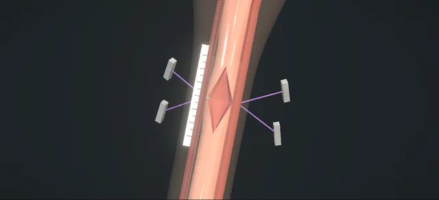
The scar is excised, both ends are spatulated dorsally, the ventral walls are joined in two layers and the dorsal gap is filled with an oral mucosal graft quilted to the corpora.

The lesson is which wall is joined, which is augmented, how far the urethra may be mobilised and what each suture layer takes.

Advanced tier FRCS reconstruction, urethral stricture, andrology Interactive 3D

Operative visualisation

Schematic scene preview: Augmented anastomotic urethroplasty
Preparing the 3D anatomy…

UrOops3D · UroRef · © Nity G

How to use this module. Play through the nine steps, or use Back and Next. Watch the scar leave the field, both ends opened along their dorsal walls, the ventral edges meet with a diamond open behind them, and the graft quilted to the corpora. The triggers show a narrowed repair, an over-long dissection and the mouth.

Click me next Section 2 of 8 Anatomy The structures this operation works between, and the one it must not cross.

The structures in the scene

Augmented anastomotic urethroplasty: how the structures relateThe perineum from the foot of the table, down onto the bulbar urethra. Bulbar urethra at the stricture: lies under it; the tunica beneath is thin Bulbospongiosus, divided in the midline. Bulbar urethra at the stricture: lies on them; freed off their tunica Corpora cavernosa: the bed for the graft. Bulbar urethra at the stricture: released distally no further than this Suspensory ligament: the distal limit (danger). Bulbar urethra at the stricture: kept when the bulb is freed behind Bulbar arteries, into the bulb from behind (danger). Bulbar urethra at the stricture: the wall toward the surgeon; edge to edge Ventral wall: joined in two layers. Bulbar urethra at the stricture: the wall against the corpora; a diamond gap Dorsal wall: opened, filled by the graft.Bulbar urethra at thestrictureBulbospongiosus,divided in themidlineCorpora cavernosa:the bed for thegraft!Suspensory ligament:the distal limit!Bulbar arteries,into the bulb frombehindVentral wall: joinedin two layersDorsal wall: opened,filled by the graftlies under it; thetunica beneath isthinlies on them; freedoff their tunicareleased distallyno further thanthiskept when the bulbis freed behindthe wall toward thesurgeon; edge toedgethe wall againstthe corpora; adiamond gapTargetDangerLandmarkContext
The perineum from the foot of the table, down onto the bulbar urethra. Schematic, not to scale; relationships only.
  • From the foot of the table in lithotomy, the bulbar urethra runs in the midline of the perineum under bulbospongiosus, the bulb toward the anus and the penile urethra toward the scrotum.
  • The corpora cavernosa lie dorsal to it, deep to the surgeon; their tunica is the surface the urethra is freed from and the graft is quilted to.
  • The bulb is held posteriorly by its midline attachments, with a bulbar artery entering on each side.
  • After the excision the two ends are opened along their dorsal walls and drawn together.
  • Ventrally, toward the surgeon, edge meets edge.
  • Dorsally, against the corpora, a diamond is left for the graft.
  • ›Bulbospongiosus - divided in the midline and dissected off the bulb; closed with a running suture at the end.
  • ›Corpus spongiosum and its tunica albuginea - thin over the bulb; the outer layer of the ventral anastomosis is placed in it.
  • ›The stricture - the scarred segment; transected and excised whole.
  • ›Corpora cavernosa - dorsal to the urethra; the urethra is freed from their tunica and the graft is quilted onto them.
  • ›Suspensory ligament of the penis - the distal limit of the dorsal dissection.
  • ›Posterior midline attachments of the bulb - divided to free the proximal urethra.
  • ›Bulbar arteries - enter the bulb from behind; preserved during the proximal dissection.
  • ›Urethral mucosa - the inner layer of the ventral anastomosis takes it alone.
  • ›Buccal mucosa and the parotid duct - the graft comes from the inner cheek; the duct opens at the level of the upper second molar.
Click me next Section 3 of 8 Steps The operation in order, with the landmark that guards each move.

Operative steps

  1. Perineal exposure Intravenous antibiotics, general anaesthesia, then high dorsal lithotomy in well-padded stirrups, with the radiographs in the room. Under loupe magnification a vertical midline perineal incision runs from the base of the scrotum to just above the anus. Colles fascia and the subcutaneous tissue are opened with diathermy to expose bulbospongiosus. The muscle is divided sharply in the midline and dissected off the corpus spongiosum. A self-retaining retractor is set.
    where it bites: the tunica albuginea of the corpus spongiosum is thin over the bulb and is perforated by careless dissection of the muscle.
  2. The stricture A catheter is advanced from the urethral meatus until it stops. That is the distal end of the stricture and the level at which the urethra will be dissected. The site and length of the scar are known from the imaging. Where there is doubt that the stricture suits this repair, the urethra is opened vertically and the length of the stricture assessed before the urethra is cut across.
    where it bites: transection commits the operation; the length is settled before it, on the imaging or through a vertical urethrotomy.
  3. Transect and excise The urethra is dissected circumferentially just distal to the stricture and freed dorsally from the corporal bodies. It is cut across sharply at the level of the stricture and the whole fibrotic scar is excised. Stay sutures are placed at three and nine o'clock on both healthy ends for traction. Bougies à boule calibrate each end progressively.
    where it bites: the excision is complete only when both ends are healthy urethra that accepts the bougie.
  4. Inspect and mobilise Cystourethroscopy through the proximal end confirms that there is no proximal urethral or bladder pathology. With sharp and blunt dissection the distal bulbar urethra is released from the tunica of the corpora cavernosa as far as the suspensory ligament of the penis. Proximally the bulb is freed from its posterior midline attachments. The bulbar arteries are preserved and haemostasis is secured with diathermy.
    where it bites: in a sexually active man, dissection distal to the suspensory ligament may shorten the penis and may cause chordee.
  5. Spatulate and measure With the mobilisation complete, the distal and proximal ends are spatulated dorsally. The ends are brought together ventrally and the length of the dorsal defect that remains is measured. The graft is harvested next and tailored to that measurement.
    where it bites: tension on the ventral join: the ends must lie together ventrally before the defect is measured.
  6. Oral graft harvest The mouth is opened with an oral retractor, the tongue is held aside and stay sutures are placed at the commissure. The graft is marked on the inner cheek and the parotid duct is identified at the level of the upper second molar, away from the harvest. The marked mucosa is hydrodissected with lidocaine and adrenaline, incised with a No. 15 blade and dissected off buccinator with fine scissors. Haemostasis is secured with diathermy and the site is packed with soaked gauze, to heal by secondary intention. The graft is defatted and kept in saline.
    where it bites: the parotid duct opens beside the harvest; it is identified before the mucosa is incised and seen intact before the retractor comes out.
  7. Graft on the corpora The tailored graft is placed dorsally and spread-fixed onto the corporal bodies with several quilting sutures. The proximal and distal apical edges of the graft are then sutured to the corresponding spatulated dorsal wall of the corpus spongiosum with interrupted full-thickness sutures.
    where it bites: a free graft takes only where it is held flat and still against its bed.
  8. Ventral anastomosis Ventrally the urethral ends are anastomosed in two layers. The inner layer takes the urethral mucosa only; the outer layer takes the tunica albuginea of the corpus spongiosum. The lateral edges of the corpus spongiosum are then secured to the lateral edges of the graft, incorporating the underlying tunica of the corpora, with a running suture on each side. Before the repair is completed a urethral catheter is passed easily into the bladder. A haemostatic sealant may be added to reinforce the suture lines.
    where it bites: the spongiosal blood supply: the inner layer takes mucosa only and the outer layer the tunica.
  9. Layered closure With a tension-free anastomosis and a dry field, the retractor is removed and the wound irrigated with saline. Bulbospongiosus is closed with a running suture. Colles fascia and the subcutaneous tissue are closed separately with running sutures, and the skin with interrupted sutures. A dressing, gauze swabs and a supporter limit swelling. The patient is returned to the supine position and the catheter is connected to a drainage bag and taped to the abdominal wall.
    where it bites: dead space in the perineal wound: Colles fascia and the subcutaneous tissue are closed as separate layers to obliterate it.
Click me next Section 4 of 8 Danger zones Where this operation bites, and why it bites there.
Critical relationship

Danger zones

The spatial relationships the animation is built to make automatic. Before you read each one, ask yourself: at this point in the operation, what is about to go wrong, and what is my first move? The save for each sits in the Complications tab.

  • RiskThe thin tunica over the bulb - perforated while the muscle is dissected off the corpus spongiosum.
  • RiskThe suspensory ligament - the distal limit of mobilisation; dissection beyond it may shorten the penis and may cause chordee.
  • RiskThe bulbar arteries - at the proximal dissection, where the bulb is freed from its posterior attachments.
  • RiskThe spongy tissue at the ventral join - the inner layer takes mucosa and the outer layer tunica; the spongiosal blood supply lies between them.
  • RiskThe parotid duct - at the level of the upper second molar, beside the harvest.
  • RiskThe lithotomy position - neuropraxia after high lithotomy; the stirrups are well padded.
Practise it Take me into the scene Opens the animation at a danger moment and asks you to predict the mechanism and your first move before it reveals.
Click me next Section 5 of 8 Complications The save for each one: the first move, the escalation trigger, the op-note line.

Complications and immediate saves

Trigger each of these live in the Animation tab, then read the save: the first move, the escalation trigger, and the line that goes in the op note.

Recurrent stricture

  • the lumen narrows again at the anastomosis or along the graft, and the stream slows.
The save
  1. Confirm it objectively: a flow rate, then a urethrogram or cystoscopy to show where the narrowing is and how long. first move
  2. Separate a short ring at the anastomosis from a narrowing along the graft.
  3. A short, veil-like recurrence may be treated endoscopically.
  4. A long recurrent stricture is referred for redo urethroplasty with an oral mucosal graft, which the European guideline recommends for redo surgery. escalation trigger
  5. Record the original technique, where the recurrence lies and the flow rate before and after each intervention. document

Penile shortening and chordee

  • the penis is shorter or bends on erection after transection and a mobilisation carried distal to the suspensory ligament.
The save
  1. Prevent it at the mobilisation: in a sexually active man the dissection stops at the suspensory ligament. first move
  2. Consent covers shortening and chordee, reduced penile sensitivity and erectile or ejaculatory change.
  3. At follow-up, ask about length, curvature on erection and erectile function.
  4. Curvature that prevents intercourse, or erectile dysfunction that persists, is referred to andrology. escalation trigger
  5. Record the distal extent of the dissection in the operation note, and what the man reports at each review. document

Oral harvest-site complications

  • bleeding, injury to the parotid duct, oral pain and numbness, tightness of the mouth, infection or a cheek granuloma after the harvest.
The save
  1. Prevent duct injury: identify the papilla at the upper second molar before the mucosa is incised, and inspect it again before the retractor is removed. first move
  2. Bleeding from the donor bed is controlled with diathermy and a pack soaked in lidocaine and adrenaline before the patient leaves theatre.
  3. Pain, numbness and tightness are explained before surgery. Whether the donor site is closed is left to the surgeon: the guideline finds no clear difference.
  4. Suspected injury to the parotid duct is referred to the maxillofacial team. escalation trigger
  5. Record the side, the size of the graft, where the duct lay relative to the harvest and the state of the donor bed at the end. document
Click me next Section 6 of 8 Troubleshooting When the case turns: the problem, then the next move.

Intra-operative troubleshooting

When the case turns: the problem, then the next move.

  • ActionIt is not clear on the table that the stricture suits this repair - open the urethra vertically and assess the length of the stricture before transecting it.
  • ActionThe stricture is in the mid or distal bulbar urethra - retract bulbospongiosus inferiorly instead of dividing it.
  • ActionThe stricture is longer and involves the most proximal bulbar urethra - the same repair may be used for longer strictures of the most proximal bulbar segment; the graft is tailored to the longer defect.
  • ActionThe ventral edges will not lie together - complete the mobilisation within its limits: distally to the suspensory ligament, proximally off the posterior midline attachments. The anastomosis is completed only if it is free of tension.
  • ActionThe plan is a ventral graft - the mirror operation: spatulate ventrally, join the dorsal walls with full-thickness interrupted sutures, suture the graft into the ventral diamond and close the corpus spongiosum over it with superficial sutures.
  • ActionOozing from the suture lines at the end - a haemostatic sealant may be used with the anastomotic sutures to reinforce the repair.
Click me next Section 7 of 8 FRCS viva Say it out loud, then mark yourself against the model answer.

FRCS viva questions

The examiner stems this scene prepares you for. Try to answer out loud first, then reveal, and learn the discriminator, the detail that turns a pass into a strong pass.

QWhat is an augmented anastomotic urethroplasty, and where does it sit among the bulbar repairs?reveal answer
  • It is a transecting bulbar urethroplasty in which the scar is excised, one wall of the urethra is joined end to end and the opposite wall is augmented with a graft.
  • I would describe the form the operative text dictates: both ends spatulated dorsally, a ventral anastomosis in two layers and an oral mucosal graft quilted to the corpora in the dorsal diamond.
  • The same text gives the mirror operation with a ventral graft, and lists the repair for bulbar strictures of 3 cm or less.
  • Guralnick and Webster used it for strictures too long for excision and primary anastomosis.
  • In their 29 men the mean stricture length on urethrography was 1.5 cm, and 27 were stricture-free at a mean of 28 months.
  • The European guideline says the technique has been challenged.
  • In a single-centre retrospective series of 507 dorsal graft urethroplasties for long bulbar strictures, Redmond and colleagues found it independently associated with recurrence, with a hazard ratio of 4.8 against a non-transecting dorsal onlay.
  • The guideline recommends free graft urethroplasty for bulbar strictures not amenable to excision and anastomosis.
  • It adds a weak recommendation for an augmented non-transecting repair where there is a short, nearly obliterative segment.
  • So I would transect only when a segment has to be excised.
Strong pass

Say both

  • Naming which wall is joined and which is grafted
  • Setting the operation against the guideline's preference for a repair that does not transect
Examiner note
Naming which wall is joined and which is grafted, and setting the operation against the guideline's preference for a repair that does not transect.
QTalk me through the operation.reveal answer
  • High lithotomy in padded stirrups, antibiotics given, the imaging in the room.
  • I make a vertical midline perineal incision from the base of the scrotum to just above the anus and open Colles fascia with diathermy.
  • I divide bulbospongiosus in the midline, dissect it off the spongiosum without breaching its tunica and set a self-retaining retractor.
  • I pass a catheter from the meatus to the stricture, dissect the urethra circumferentially just distal to it and free it from the corpora.
  • I transect at the stricture and excise the whole scar.
  • I place stays at three and nine o'clock on both ends, calibrate with bougies à boule and pass a cystoscope proximally.
  • I mobilise distally off the corpora no further than the suspensory ligament, and proximally off the posterior midline attachments, preserving the bulbar arteries.
  • I spatulate both ends dorsally, bring them together ventrally and measure the dorsal defect.
  • The oral graft is harvested, defatted and tailored.
  • I spread it on the corpora with quilting sutures and suture its apices to the spatulated dorsal wall of each end.
  • I join the ends ventrally in two layers, mucosa and then the tunica of the spongiosum, and run each lateral spongiosal edge to the graft edge with the corporal tunica beneath.
  • I pass a catheter into the bladder before completing the repair, then close bulbospongiosus, Colles fascia, the subcutaneous tissue and the skin in layers.
Strong pass

Say all three

  • The order: excise and calibrate before mobilising
  • Spatulate and measure before tailoring the graft
  • The catheter passed before the repair is completed
Examiner note
The order: excise and calibrate before mobilising, spatulate and measure before tailoring the graft, and the catheter passed before the repair is completed.
QWhy two layers ventrally, and does it matter where the graft goes?reveal answer
  • The ventral join has to be watertight and still leave the spongiosum perfused.
  • The inner layer takes the urethral mucosa alone and the outer layer the tunica albuginea of the corpus spongiosum.
  • The reason given is that this limits injury to the spongiosal blood supply.
  • The graft is a free graft, so I would fix it to the corporal bodies with quilting sutures and run the spongiosal edges to its margins with a bite of corporal tunica, so that it lies flat on its bed.
  • On position, the European guideline's summary is that the location of the graft has no impact on patency.
  • It recommends a dorsal, dorsolateral or ventral approach according to surgical practice, expertise and the findings at operation.
  • The guideline cites a randomised trial of 80 men comparing ventral with dorsal onlay, and non-randomised series.
  • I would therefore describe the dorsal position as the one I am trained in and make no claim that it is better.
Strong pass

Say both

  • Giving the stated reason for the outer layer taking tunica only
  • Making no claim of a patency advantage for a dorsal graft
Examiner note
Giving the stated reason for the outer layer taking tunica only, and making no claim of a patency advantage for a dorsal graft.
QWhere is the distal limit of your mobilisation, and why?reveal answer
  • The suspensory ligament of the penis.
  • To bring the ventral edges together I need length, and I get it by releasing the distal bulbar urethra from the corpora and the bulb from its posterior midline attachments.
  • The chapter sets the suspensory ligament as the distal limit in a sexually active man and names what lies beyond it: penile shortening and chordee.
  • Proximally I keep the bulbar arteries.
  • The European guideline notes that complete transection of the corpus spongiosum disrupts its antegrade blood flow, which is my reason for protecting what enters the bulb.
  • If the edges do not meet within those limits I would not force them: the anastomosis has to be free of tension.
Strong pass

Say both

  • Naming the suspensory ligament as the limit
  • Linking it to shortening and chordee
Examiner note
Naming the suspensory ligament as the limit and linking it to shortening and chordee.
QWhat do you tell the patient about complications, and how do you follow him up?reveal answer
  • I would go through the chapter's list.
  • It names bleeding, infection, urine leak, post-void dribbling, graft sacculation, penile shortening and chordee, reduced penile sensitivity, erectile or ejaculatory dysfunction, neuropraxia from the position, urethrocutaneous fistula, problems at the mouth and recurrent stricture.
  • I would put numbers only on what has been measured.
  • In Guralnick and Webster's 29 men, 13 reported post-void dribbling, 5 subjective shortening, 2 a pseudodiverticulum and 1 new erectile dysfunction.
  • The Scandinavian randomised trial compared transecting anastomotic repair with buccal grafting for strictures of 2 cm or less.
  • That is a different operation which shares the transection: more men reported a shortened penis and reduced glans filling after transection, with no difference in erectile function scores.
  • Redmond's series found no difference in new erectile dysfunction between augmented anastomotic and dorsal onlay repairs.
  • The operative text gives no catheter duration.
  • I would leave the catheter according to my unit's protocol and image the repair before it comes out, as the European guideline recommends.
  • Guralnick and Webster performed urethrography at three weeks and three months.
  • After that, follow-up is symptoms and a flow rate, with a urethrogram or cystoscopy if either changes.
Strong pass

Say both

  • Quoting each figure with the series it came from
  • Saying that the transection data come from a different operation
Examiner note
Quoting each figure with the series it came from, and saying that the transection data come from a different operation.
Click me next Section 8 of 8 Evidence The guidance each of these answers is built on.

Evidence and guidance

Operative Dictations in Urologic Surgery
Armenakas NA, Fracchia JA, Golan R. Augmented anastomotic urethroplasty. Chapter 58. Wiley, 2019
The source of the sequence drawn and of every step, danger and troubleshooting line: the ten essential steps, the dorsal onlay dictation, the four variations and the complication list. Book figures kept off screen: the indication (bulbar strictures of 3 cm or less), a 20 Fr catheter to find the stricture, calibration to 30 Fr and 1 cm of spatulation. Also off screen: 4-0 chromic stays and quilting sutures, 5-0 polydioxanone for the repair and an 18 or 20 Fr catheter. The chapter gives no catheter duration and describes no drain; neither is stated or drawn. It is silent on what to do when the edges will not lie together. The module follows the dictation's order, with the harvest after the defect is measured, not the order of the essential-steps list.
Open guideline
Operative Dictations in Urologic Surgery
Armenakas NA, Fracchia JA, Golan R. Buccal mucosal graft harvest. Chapter 60. Wiley, 2019
The harvest shown in step 6 and the oral complication list. Its sequence: retractor and commissure stays, the parotid duct found, hydrodissection with lidocaine and adrenaline, a No. 15 blade, fine scissors off buccinator, diathermy and a soaked pack, secondary intention, defatting and saline. Primary closure is its named variation. The 2 cm graft width is kept off screen. The suture size of the commissure stays and the concentration of the local anaesthetic do appear on screen, on the instrument tags of the harvest shared with the bulbar urethroplasty module.
Open guideline
EAU Guidelines
Urethral Strictures: Disease Management in Males, sections 6.2.1, 6.3.2.b.1.b, 6.3.2.e.2, 6.3.2.e.3 and 6.3.2.h, read 13 October 2026
Augmented anastomotic repair is described for strictures just too long for tension-free excision and anastomosis, and for longer strictures with a short nearly obliterative segment. Its use has been challenged by Redmond. Recommendations: free graft urethroplasty for strictures not amenable to excision and anastomosis (strong); augmented non-transecting repair in short, nearly obliterative segments (weak); a dorsal, dorsolateral or ventral graft by practice, expertise and findings (strong), graft location having no impact on patency; oral graft urethroplasty for a long redo stricture (strong); internal urethrotomy or dilatation for a short, veil-like recurrent stricture after bulbar urethroplasty (weak). Complete transection is said to disrupt antegrade spongiosal blood flow. The guideline also quotes a patency range for the transecting repair. Its numbered reference for that range could not be matched to a named series, so the range is not used here.
Open guideline
EAU Guidelines
Urethral Strictures: Perioperative Care of Urethral Surgery, section 10.3 Catheter management, read 13 October 2026
Recommends a form of validated urethrography after urethroplasty to assess for urinary extravasation before the catheter is removed (strong). It gives no single catheter duration: a catheter is commonly left for two to three weeks.
Open guideline
EAU Guidelines
Urethral Strictures: Tissue Transfer, sections 9.3 and 9.5, read 13 October 2026
Buccal mucosa is the most commonly used graft; the randomised trials of closing against not closing the donor site allow no clear recommendation, and the decision is left to the treating surgeon.
Open guideline
The Journal of Urology
Guralnick ML, Webster GD. The augmented anastomotic urethroplasty: indications and outcome in 29 patients. J Urol 2001;165:1496-501
Abstract read through PubMed, 13 October 2026. A retrospective series from one centre, 1990 to 1999: 9 roof-strip repairs with a ventral onlay and 20 floor-strip repairs with a dorsal onlay. The onlays were pedicled skin flaps in 7 and grafts in 22. Mean stricture length was 1.5 cm on urethrography, and 27 of 29 men were stricture-free at a mean of 28 months. Complications: post-void dribbling in 13, subjective shortening in 5, pseudodiverticulum in 2, new erectile dysfunction in 1. Urethrography was performed at three weeks and three months.
Open guideline
The Journal of Urology
Redmond EJ, Hoare DT, Rourke KF. Augmented anastomotic urethroplasty is independently associated with failure after reconstruction for long bulbar urethral strictures. J Urol 2020;204:989-95
Abstract read through PubMed, 13 October 2026. A retrospective review at one institution of 507 bulbar urethroplasties with a dorsal buccal graft: 221 transecting augmented anastomotic and 286 non-transecting dorsal onlay, mean follow-up 78.9 months. On multivariate analysis the augmented anastomotic repair was associated with recurrence (hazard ratio 4.8); new erectile dysfunction did not differ between the groups.
Open guideline
European Urology
Nilsen OJ et al. To transect or not transect: results from the Scandinavian Urethroplasty Study. Eur Urol 2022;81:375-82
Abstract read through PubMed, 13 October 2026. A multicentre randomised trial of 151 men with bulbar strictures of 2 cm or less: transecting excision and primary anastomosis against buccal mucosal grafting. More penile complications after transection, in particular a shortened penis and reduced glans filling; no difference in erectile function scores. It did not study the augmented anastomotic repair; it is cited for the effect of transection.
Open guideline

Curriculum. Supports learning relevant to the ISCP Urology Curriculum syllabus topic Urethral stricture disease. Not a workplace-based assessment and not evidence of competence.

Two minutes

Close the lap: recall it before you leave.

Say it aloud, then reveal. Reading is not remembering.

  1. 01 The relationship Say the critical spatial relationship out loud, then reveal. reveal
    Memory frame

    From the foot of the table in lithotomy, the bulbar urethra runs in the midline of the perineum under bulbospongiosus, the bulb toward the anus and the penile urethra toward the scrotum.

    Why?
    The corpora cavernosa lie dorsal to it, deep to the surgeon; their tunica is the surface the urethra is freed from and the graft is quilted to. The bulb is held posteriorly by its midline attachments, with a bulbar artery entering on each side. After the excision the two ends are opened along their dorsal walls and drawn together. Ventrally, toward the surgeon, edge meets edge. Dorsally, against the corpora, a diamond is left for the graft.
  2. 02 The first move Recurrent stricture. Your first move? reveal
    First move

    Confirm it objectively: a flow rate, then a urethrogram or cystoscopy to show where the narrowing is and how long.

  3. 03 The escalation trigger Recurrent stricture. What makes you call for help? reveal
    Escalate when

    A long recurrent stricture is referred for redo urethroplasty with an oral mucosal graft, which the European guideline recommends for redo surgery.

  4. 04 The discriminator What turns a pass into a strong pass? reveal
    Strong pass

    Say both

    • Naming which wall is joined and which is grafted
    • Setting the operation against the guideline's preference for a repair that does not transect
    Examiner note
    Naming which wall is joined and which is grafted, and setting the operation against the guideline's preference for a repair that does not transect.
  5. 05 The pitfall Where does this operation bite first? reveal
    Pitfall

    The thin tunica over the bulb

    Why?
    perforated while the muscle is dissected off the corpus spongiosum.

Revision practice with self-marked answers, not an assessment of clinical competence. Kept in this browser only; it does not sync between devices.