
UrOops3D, danger-zone atlas

Schematic child bladder: every dimension is an authoring estimate.

Operative animation, Extravesical ureteric reimplantation

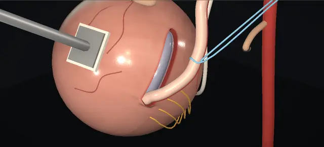
Extravesical ureteric reimplantation: the Lich-Gregoir detrusor trough, left side

Indications

  • Persistent high-grade vesicoureteric reflux.
  • Recurrent upper tract infection despite antibiotic prophylaxis, or where prophylaxis is not taken reliably.
  • The family’s preference for definitive correction after counselling.

Alternatives

  • Continued antibiotic prophylaxis and surveillance.
  • Endoscopic subureteric injection, offered for the lower grades of reflux.
  • An intravesical repair (Cohen or Politano-Leadbetter), which may be considered when both sides reflux.
  • Laparoscopic or robot-assisted extravesical repair in centres with established experience.

Risks

  • Bleeding and infection.
  • Urinary retention after the catheter is removed.
  • Ureteric obstruction from a kink or a tight tunnel.
  • Persistent or recurrent reflux, and further upper tract infection.
  • Injury to the vas in a boy or to the round ligament in a girl.

Open antireflux surgery done wholly from outside the bladder, shown here on the left in a boy with persistent high-grade reflux.

The obliterated umbilical artery is tied and divided, the bladder is rolled away from that side, and the ureter is followed down to its junction with the bladder, the dissection kept on the ureteric wall.

The detrusor is then incised along the ureter’s course down to the mucosa, which bulges into the gap and stays whole. The muscle edges are undermined, the ureter is laid in the trough, the bladder is drained and the muscle is closed loosely over the ureter.

The bladder is never opened and the orifice is never moved. The lesson is where the nerve branches lie at the junction, what must stay intact in the floor of the trough, and the last look with the bladder filled again.

Advanced tier FRCS paediatric urology, vesicoureteric reflux, reconstruction Interactive 3D

Operative visualisation

Schematic scene preview: Extravesical ureteric reimplantation
Preparing the 3D anatomy…

UrOops3D · UroRef · © Nity G

How to use this module. Play the eight steps, or use Back and Next. Watch the artery tied and divided, the bladder rolled away, the detrusor part down to a bulging mucosa, and the muscle closed over the ureter. Two triggers show a tight tunnel and scissors among the nerve branches.

Click me next Section 2 of 8 Anatomy The structures this operation works between, and the one it must not cross.

The structures in the scene

Extravesical ureteric reimplantation: how the structures relateOutside the bladder on the left: the distal ureter and what lies round it. Distal ureter, down to its junction: lies medial to the origin of Obliterated umbilical artery. Distal ureter, down to its junction: run beside its junction; stay on its wall Nerve branches entering the bladder (danger). Distal ureter, down to its junction: is laid under; closed loosely over it Detrusor: two leaves over the ureter. Distal ureter, down to its junction: rests on; never breached Mucosa: the unbroken floor (danger). Distal ureter, down to its junction: still ends at; never moved Ureteric orifice, inside, untouched. Distal ureter, down to its junction: is crossed by, close to the bladder Vas, or round ligament in a girl (danger).Distal ureter, down toits junctionObliteratedumbilical artery!Nerve branchesentering the bladderDetrusor: two leavesover the ureter!Mucosa: the unbrokenfloorUreteric orifice,inside, untouched!Vas, or roundligament in a girllies medial to theorigin ofrun beside itsjunction; stay onits wallis laid under;closed loosely overitrests on; neverbreachedstill ends at;never movedis crossed by,close to thebladderTargetDangerLandmarkContext
Outside the bladder on the left: the distal ureter and what lies round it. Schematic, not to scale; relationships only.
  • Three views are used.
  • Through the wound the head is at the top and the patient’s right is on the surgeon’s left.
  • For the work beside the bladder the abdominal wall is left out and the field is seen from the patient’s left.
  • The bladder has been rolled away, so its posterolateral wall faces the viewer, with the ureter coming down from the pelvic brim at the top to the junction at the bottom.
  • The trough runs up the bladder wall from the junction.
  • Once the retraction is released the repair faces the back of the pelvis, so the final inspection is shown from behind.
  • ›Obliterated umbilical artery - a fibrous cord in the space lateral to the bladder, running forward from the side wall. The ureter lies medial to its origin, so the cord is the landmark that leads to the ureter.
  • ›Ureter and its vessels - freed along its lateral side; the vessels that reach it from the medial side stay with it.
  • ›Ureterovesical junction and intramural ureter - where the ureter passes through the detrusor. The intramural ureter is separated from the muscle all round before the trough is made.
  • ›Terminal nerve branches - enter the bladder at the level of the junction; the dissection keeps to the ureter to spare them.
  • ›Detrusor and mucosa - two layers. The incision goes through serosa and muscle and stops at the mucosa, which is left whole as the floor of the trough.
  • ›Ureteric orifice - inside the bladder and never seen during the operation; it is left exactly where it was.
  • ›Vas, or round ligament - in a boy the vas, and in a girl the round ligament, lies beside the distal ureter and is at risk there.
  • ›Peritoneum - swept cephalad off the bladder and left closed.
  • ›Abdominal wall - skin, Scarpa’s fascia, the rectus sheath opened in an arc clear of the inguinal canals, then the recti parted in the midline with the inferior epigastric vessels beneath them.
Click me next Section 3 of 8 Steps The operation in order, with the landmark that guards each move.

Operative steps

  1. Fill the bladder and expose it The child lies supine with the hips abducted. A urethral catheter is passed, the bladder is partly filled with saline and the catheter is clamped. A small curved transverse incision is made along the skin lines a fingerbreadth above the pubic symphysis and carried down to the rectus sheath, which is opened in the same arc, clear of the inguinal canals. The recti are separated in the midline and retracted laterally. The anterior bladder wall is exposed and the peritoneum is swept cephalad. A self-retaining ring retractor is set within the true pelvis with a moist swab under each blade.
    where it bites: The inferior epigastric vessels lie beneath the recti as they are retracted.
  2. The obliterated artery, then the ureter The obliterated umbilical artery is identified between the bladder and the pelvic side wall on the side of the reflux. It is dissected free, tied twice and divided between the ties. The bladder is then retracted gently downward and toward the midline, and the ureter is identified medial to the origin of the obliterated artery.
    where it bites: The ureter is close by, on the medial side of this artery’s origin.
  3. Mobilise the ureter to the junction The ureter is dissected down to the ureterovesical junction along its lateral side, so that the blood supply reaching it from the medial side is kept. Close to the junction the scissors stay on the ureter itself. The ureter is encircled with a sling and gently lifted. By blunt and sharp dissection round its whole circumference the intramural ureter is then separated from the detrusor.
    where it bites: The terminal nerve branches enter the bladder at this level, and a dissection that strays from the ureter divides them. In a boy the vas crosses the ureter close by.
  4. Incise the detrusor A full-thickness incision is made in the detrusor from the junction cephalad, following the ureter, through serosa and muscle. The mucosa is left intact and bulges into the gap. The ureteric orifice is not disturbed. The tunnel this makes should be five times as long as the ureter is wide.
    where it bites: A breach of the mucosa or of the orifice.
  5. Undermine the muscle edges The incised detrusor is undermined off the mucosa on both sides. This raises two leaves of muscle and makes a trough wide enough to accommodate the ureter.
    where it bites: The mucosa must stay whole while the muscle is lifted off it.
  6. Lay the ureter in and drain the bladder The distal ureter is positioned in the trough on the mucosa and the sling is taken off. The bladder is then drained through the catheter.
  7. Close the detrusor over the ureter The two leaves of detrusor are approximated over the ureter with interrupted absorbable stitches, from the proximal end to the distal. The closure is loose, and the opening at its proximal end is left wide enough for the ureter to enter freely.
    where it bites: A closure that grips the ureter obstructs it.
  8. Release, refill, inspect, close Retraction on the bladder is released and the bladder is again partly distended with saline. The ureter is inspected for a straight course, without kinking or compression. Haemostasis is secured with diathermy. The ring retractor is removed and the wound is closed in layers: the recti, the rectus sheath, Scarpa’s fascia and the skin. The urethral catheter is connected to a drainage bag.
    where it bites: A kink or compression that shows only once the bladder is filled.
Click me next Section 4 of 8 Danger zones Where this operation bites, and why it bites there.
Critical relationship

Danger zones

The spatial relationships the animation is built to make automatic. Before you read each one, ask yourself: at this point in the operation, what is about to go wrong, and what is my first move? The save for each sits in the Complications tab.

  • RiskBeside the junction - the terminal nerve branches enter the bladder here; scissors that leave the ureter divide them.
  • RiskThe floor of the trough - mucosa only. It must stay whole, and so must the orifice under its lower end.
  • RiskThe entry to the tunnel - where the ureter passes under the most proximal stitch; a tight closure or a sharp bend here obstructs it.
  • RiskThe medial side of the ureter - its blood supply; the dissection is on the lateral side.
  • RiskThe vas - in a boy it crosses the ureter close to the bladder.
  • RiskUnder the recti - the inferior epigastric vessels.
Practise it Take me into the scene Opens the animation at a danger moment and asks you to predict the mechanism and your first move before it reveals.
Click me next Section 5 of 8 Complications The save for each one: the first move, the escalation trigger, the op-note line.

Complications and immediate saves

Trigger each of these live in the Animation tab, then read the save: the first move, the escalation trigger, and the line that goes in the op note.

Ureteric obstruction

  • the ureter angled or squeezed where it enters or lies in the tunnel; the reason it is inspected with the bladder filled again.
The save
  1. On the table: if the ureter is kinked or compressed on refilling, take down the stitch responsible and close again loosely. first move
  2. Refill and inspect once more before the wound is closed.
  3. After surgery, loin pain, fever, vomiting or a falling urine output prompts an ultrasound for new or worsening hydronephrosis.
  4. Worsening dilatation with pain, infection or a rising creatinine needs drainage and senior paediatric urology review. escalation trigger
  5. Op note: the refill inspection and what was seen. document

Urinary retention

  • the child cannot void once the catheter is out.
  • The dissection at the junction lies beside the nerve branches entering the bladder, and damage to them is the suspected cause.
  • The EAU guideline reports a higher risk of temporary retention when both sides are repaired this way at one sitting.
The save
  1. In theatre: at the junction keep the dissection on the ureter. first move
  2. If the child cannot void after the catheter is removed, drain the bladder again by catheter.
  3. Retention that persists after a further trial without the catheter goes to senior paediatric urology review. escalation trigger
  4. Op note: one side or both, and how the junction was dissected. document

Persistent or recurrent reflux

  • reflux continues or returns on the operated side, with or without further upper tract infection.
  • The tunnel is the antireflux mechanism, so its length and its intact mucosal floor matter.
The save
  1. In theatre: make the trough long enough for the tunnel the ureter needs, and keep the mucosa whole beneath it. first move
  2. Follow the child up clinically and with ultrasound.
  3. Recurrent febrile infection after the repair goes back to the paediatric urology team for a planned decision. escalation trigger
  4. Op note: the length of the trough against the width of the ureter, and that the mucosa stayed intact. document

Injury to the vas or the round ligament

  • either structure can be divided or caught in the dissection beside the distal ureter.
The save
  1. Identify the vas before any cord beside the distal ureter is tied or divided. first move
  2. A divided vas is declared at once to the senior surgeon and is not closed over. escalation trigger
  3. Op note: the vas seen and preserved, or the injury and what was done about it. document
Click me next Section 6 of 8 Troubleshooting When the case turns: the problem, then the next move.

Intra-operative troubleshooting

When the case turns: the problem, then the next move.

  • ActionThe ureter cannot be found - find the obliterated artery first, lateral to the bladder; the ureter lies medial to its origin.
  • ActionThe mucosa is breached while the detrusor is divided or undermined - a small hole is closed with a fine absorbable stitch before the ureter is laid in.
  • ActionThe trough will not take the ureter - undermine the muscle edges further on both sides until the trough accommodates it.
  • ActionThe ureter kinks when the bladder is filled again - take down the stitch at that point and close again loosely; then refill and look once more.
  • ActionBoth sides reflux - the same operation can be done on the other side at the same sitting. The European guideline notes a higher risk of temporary retention after simultaneous bilateral extravesical repair and says an intravesical repair may be considered.
  • ActionFine structures are hard to see in a small pelvis - optical magnification with surgical loupes.
  • ActionHow much of the intramural ureter to free - its mobilisation can be limited, as in the original Gregoir description, which simplifies the operation.
  • ActionThe patient is a girl - the round ligament takes the place of the vas as the structure at risk beside the distal ureter.
Click me next Section 7 of 8 FRCS viva Say it out loud, then mark yourself against the model answer.

FRCS viva questions

The examiner stems this scene prepares you for. Try to answer out loud first, then reveal, and learn the discriminator, the detail that turns a pass into a strong pass.

QDescribe an extravesical (Lich-Gregoir) ureteric reimplantation.reveal answer
  • It is an open antireflux operation done from outside the bladder, which I do not open.
  • With the child supine I partly fill the bladder through a catheter and clamp it.
  • Through a small transverse suprapubic incision I open the rectus sheath, part the recti, sweep the peritoneum up and place a ring retractor.
  • On the side of the reflux I find the obliterated umbilical artery between the bladder and the side wall, tie and divide it, retract the bladder gently down and medially, and find the ureter medial to the origin of that artery.
  • I free the ureter to the junction on its lateral side, keeping to its wall near the bladder to spare the nerve branches entering there, pass a sling round it and separate the intramural ureter from the detrusor.
  • I then incise the detrusor along the ureter’s course down to the mucosa, leaving the mucosa and the orifice intact, and undermine the muscle on both sides to make a trough wide enough for the ureter.
  • I lay the ureter in, drain the bladder, and close the muscle loosely over it with interrupted absorbable stitches from the proximal end to the distal.
  • Finally I release the retraction, fill the bladder part-way again and check that the ureter runs straight without a kink or compression before I close.
Strong pass

Say all three

  • Saying that the bladder is not opened and the orifice is not moved
  • Naming the obliterated artery as the guide to the ureter
  • Describing the inspection after refilling
Examiner note
Saying that the bladder is not opened and the orifice is not moved, naming the obliterated artery as the guide to the ureter, and describing the inspection after refilling.
QWhy does this operation stop reflux, and how long should the tunnel be?reveal answer
  • The ureter normally runs obliquely through the bladder wall and then under the mucosa with muscle behind it, so a rise in bladder pressure presses that segment flat against its backing.
  • In reflux that segment is too short.
  • The European guideline states the principle of every open repair as lengthening the intramural ureter by embedding it under the mucosa.
  • Here I do that from outside: the ureter lies on intact mucosa in a trough of detrusor and the muscle is closed over it, so the tunnel is as long as the closed trough.
  • The operative text I follow asks for a ratio of tunnel length to ureteric width of five to one.
  • A 2024 review of megaureter surgery attributes that ratio to Paquin and records the view that the full ratio may not be necessary in megaureters, so I quote it as the traditional rule.
Strong pass

Giving the mechanism as a length of ureter between mucosa and muscle, quoting the ratio and saying whose rule it is.

QWhy is bilateral extravesical reimplantation approached with caution?reveal answer
  • Because of urinary retention.
  • The nerve branches to the bladder enter it close to the ureterovesical junction, which is exactly where I dissect, and the operative text tells me to stay close to the ureter at that level to avoid them.
  • A dissection study in adult cadavers by Leissner and colleagues found the main part of the pelvic plexus dorsal and medial to the junction, concluded that dissection close to the ureter avoids it, and gave nerve damage as the suspected cause of retention after bilateral extravesical surgery.
  • My reasoning is that damage on one side leaves the other side working, whereas an operation on both sides at one sitting puts both at risk.
  • The European guideline says that simultaneous bilateral extravesical repair carries an increased risk of temporary postoperative retention and that an intravesical repair may be considered for bilateral reflux.
  • It repeats the caution for bilateral robot-assisted extravesical repair.
  • So for one side I am comfortable with this operation, and for both I would discuss an intravesical repair with the family.
  • If a child does go into retention I drain the bladder by catheter.
Strong pass

Say both

  • Linking retention to the nerve branches at the junction
  • Quoting the guideline’s position on simultaneous bilateral repair
Examiner note
Linking retention to the nerve branches at the junction, and quoting the guideline’s position on simultaneous bilateral repair.
QYou refill the bladder at the end and the ureter kinks where it enters the tunnel. What do you do?reveal answer
  • I do not close the wound.
  • A kink or compression at the tunnel will obstruct the kidney, and the reason I refill the bladder is to find it now.
  • I take down the stitch at that point, lay the ureter straight again and close the muscle more loosely, leaving room round the ureter where it enters.
  • Then I fill the bladder again and look once more: I want a straight course into the tunnel with no kink and no compression.
  • I record the inspection in the operation note.
  • After surgery, pain, fever or a falling urine output on that side would make me ask for an ultrasound, and worsening hydronephrosis would need drainage.
Strong pass

Say both

  • Treating the inspection after refilling as the test of the repair
  • Revising the closure on the table
Examiner note
Treating the inspection after refilling as the test of the repair, and revising the closure on the table.
QWhat do you check before choosing an extravesical repair, and what are the alternatives?reveal answer
  • The European guideline says that when an extravesical repair is planned, cystoscopy should be done beforehand to assess the bladder mucosa and the position and configuration of the ureteric orifices.
  • That matters to me because I will not see the inside of the bladder during the operation.
  • The alternatives are continued prophylaxis and surveillance;
  • endoscopic injection, which the guideline offers for the lower grades of reflux;
  • and the intravesical repairs, of which it calls the cross-trigonal operation the most popular and reliable, with later endoscopic access to the ureter as its main concern.
  • Laparoscopic and robot-assisted extravesical repair are offered in experienced centres, and the guideline does not recommend the laparoscopic approach as routine.
  • It describes the success of the surgical procedures as very high and similar.
Strong pass

Say both

  • Quoting cystoscopy before an extravesical repair
  • Explaining why it matters when the orifice is not seen at operation
Examiner note
Quoting cystoscopy before an extravesical repair, and explaining why it matters when the orifice is not seen at operation.
Click me next Section 8 of 8 Evidence The guidance each of these answers is built on.

Evidence and guidance

Operative Dictations in Urologic Surgery
Armenakas NA, Fracchia JA, Golan R. Chapter 123, “Ureteral Reimplantation: Extravesical Technique”. Wiley, 2019
Read 13 October 2026. Indications; the six essential steps (fill the bladder; expose and ligate the obliterated artery; mobilise the ureter to the junction close to its wall; a trough through serosa and muscle sparing mucosa and orifice; loose detrusor closure with a tunnel length to ureteric width of five to one; check for obstruction or kinking); the variations (loupes; limited mobilisation of the intramural ureter); the complication list; and the order of the dictation, which this page follows. The chapter ligates the obliterated artery with ties and does not state that it is divided; its incision length, suture sizes and bladder capacity formula are not reproduced here. The family’s preference after counselling, among the indications, is written from general practice.
Open guideline
EAU Guidelines
Paediatric Urology, chapter 17 Vesicoureteric reflux: 17.3.2.b open surgical techniques, 17.3.2.c laparoscopy and robot-assisted, 17.4 recommendations (web edition, read 13 October 2026)
All open techniques lengthen the intramural ureter by submucosal embedding. Cystoscopy before a planned extravesical (Lich-Gregoir) repair, to assess the mucosa and the orifices. In bilateral reflux an intravesical repair may be considered, because simultaneous bilateral extravesical repair carries an increased risk of temporary postoperative retention. The laparoscopic approach is not recommended as routine; caution with bilateral robot-assisted repair. Reimplantation is offered for persistent high-grade reflux.
Open guideline
Current Urology Reports
Boswell TC. Advancements in surgical management of megaureters. Curr Urol Rep 2024;25(9):215-223
A review of megaureter surgery. It attributes the tunnel length to ureteric diameter ratio of five to one to Paquin and records the view, citing Kalayeh et al. 2021, that the full ratio may not be necessary in megaureters. Cited only for the standing of that ratio; it is not a study of this operation.
Open guideline
The Journal of Urology
Leissner J, Allhoff EP, Wolff W, Feja C, Höckel M, Black P, Hohenfellner R. The pelvic plexus and antireflux surgery: topographical findings and clinical consequences. J Urol 2001;165(5):1652-1655
Abstract read on PubMed, 13 October 2026; the full paper was not opened. A dissection study in adult cadavers. The main part of the pelvic plexus lay dorsal and medial to the ureterovesical junction, with bundles ending at the distal ureter, trigone and rectum. The authors conclude that careful dissection close to the ureter avoids injury to the plexus, and they suggest that bilateral antireflux surgery be staged unless the nerves can be preserved. Neural damage is given as the suspected cause of retention after bilateral extravesical surgery. Its measurement is in adults and is not reproduced here.
Open guideline

Curriculum. Supports learning relevant to the ISCP Urology Curriculum syllabus topic Paediatric urology: vesicoureteric reflux. Not a workplace-based assessment and not evidence of competence.

Two minutes

Close the lap: recall it before you leave.

Say it aloud, then reveal. Reading is not remembering.

  1. 01 The relationship Say the critical spatial relationship out loud, then reveal. reveal
    Memory frame

    Three views are used.

    Why?
    Through the wound the head is at the top and the patient’s right is on the surgeon’s left. For the work beside the bladder the abdominal wall is left out and the field is seen from the patient’s left. The bladder has been rolled away, so its posterolateral wall faces the viewer, with the ureter coming down from the pelvic brim at the top to the junction at the bottom. The trough runs up the bladder wall from the junction. Once the retraction is released the repair faces the back of the pelvis, so the final inspection is shown from behind.
  2. 02 The first move Ureteric obstruction. Your first move? reveal
    First move

    On the table: if the ureter is kinked or compressed on refilling, take down the stitch responsible and close again loosely.

  3. 03 The escalation trigger Ureteric obstruction. What makes you call for help? reveal
    Escalate when

    Worsening dilatation with pain, infection or a rising creatinine needs drainage and senior paediatric urology review.

  4. 04 The discriminator What turns a pass into a strong pass? reveal
    Strong pass

    Say all three

    • Saying that the bladder is not opened and the orifice is not moved
    • Naming the obliterated artery as the guide to the ureter
    • Describing the inspection after refilling
    Examiner note
    Saying that the bladder is not opened and the orifice is not moved, naming the obliterated artery as the guide to the ureter, and describing the inspection after refilling.
  5. 05 The pitfall Where does this operation bite first? reveal
    Pitfall

    Beside the junction

    Why?
    the terminal nerve branches enter the bladder here; scissors that leave the ureter divide them.

Revision practice with self-marked answers, not an assessment of clinical competence. Kept in this browser only; it does not sync between devices.