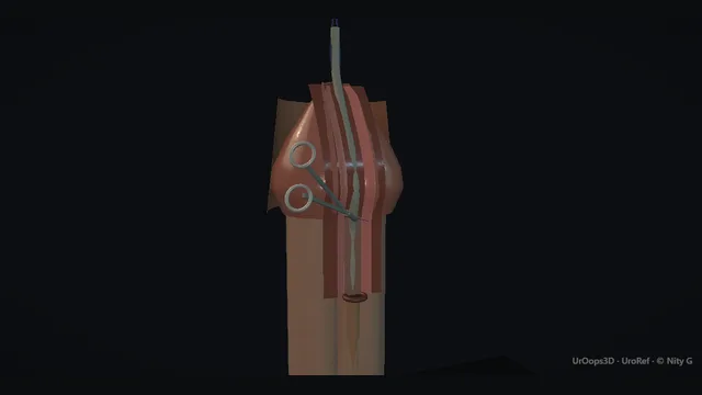
A tubularised incised plate repair taught from the ventral surface: the ectopic meatus marks where the urethral plate begins, the plate runs as a bare midline strip from there onto the glans, and the whole neourethra is rolled from that strip over a stent.
Every common complication, fistula, stenosis and breakdown, happens on the lines this repair is sewn along.
Core tierFRCS paediatric urology, genital surgeryInteractive 3D
The ventral midline strip of the penis, meatus to glans tip. Schematic, not to scale; relationships only.
Everything sits on the ventral surface in one midline strip.
The ectopic meatus lies on the ventral shaft; from it the urethral plate runs forward onto the undersurface of the glans between the two glans wings, and the dorsal hood overhangs above because ventral skin never formed.
Chordee bows the distal shaft ventrally towards that same strip, and the dorsal 12 o'clock line, where residual bend is plicated, is the opposite point of the same axis.
The repair tubularises the ventral strip and wraps the dorsal hood around to replace the missing ventral skin, so the operation relocates dorsal skin to a ventral defect along that single midline plane.
›Urethral plate - the bare midline strip from ectopic meatus onto the glans; the entire neourethra is tubularised from it.
›Glans wings - the two halves of the glans lateral to the plate; elevated off it, then folded back over the finished tube.
›Ectopic meatus - the ventral opening proximal to the glans tip; its position sets distal versus proximal disease and the length of the repair.
›Hooded prepuce - dorsally redundant, ventrally deficient foreskin; the skin bank that is rotated ventrally for cover and the reason circumcision before repair is forbidden.
›Dorsal 12 o'clock line - the midline plication site for residual chordee, with the dorsal neurovascular bundle immediately either side.
AssessmentThree findings define the operation and each is recorded before incision: the meatal position on the ventral shaft, the degree of chordee demonstrated on artificial erection, and the quality of the hooded prepuce. The meatus marks where the urethral plate begins and sets distal versus proximal disease, which sets single-stage repair against staged reconstruction. The hooded prepuce is examined as the skin bank it is, deficient ventrally and redundant dorsally, and its adequacy for ventral cover is judged on the table. No circumcision and no discard of the hood happens before repair, because skin removed at the start is cover missing at the end. where it bites: never circumcise or discard the hood before repair: without it there is no ventral skin cover.
Chordee correctionThe penis is degloved to the root through a circumferential subcoronal incision, releasing the tethering skin and fascial layers, and the artificial erection test is repeated before anything further is decided. Skin release alone straightens a proportion of penises, and the re-test is what identifies them. Where bend persists, the dorsal tunica is plicated at 12 o'clock with small bites strictly in the midline, shortening the longer side to match the ventral. The erection test is repeated until the penis is straight, and the midline discipline is absolute because the dorsal neurovascular bundle runs immediately either side of that line. where it bites: plication is strictly midline at 12: the dorsal neurovascular bundle lies just either side of that line.
Glans wing elevationThe glans wings are dissected off the lateral edges of the urethral plate with fine scissors, in the plane immediately lateral to the strip, until they will mobilise later without tension. The plate itself is left as a bare midline strip running from the ectopic meatus onto the glans tip, untouched by the dissection. The depth of the wing dissection decides whether the wings will meet over the neourethra: wings left under-elevated will not meet, and wings closed under tension strangle the meatus. The unforgivable error is cutting into the strip itself, because the plate is the repair's only building material and a devascularised strip breaks down. where it bites: the dissection plane is lateral to the plate: cutting into the strip devascularises the repair's only building material.
Midline plate incisionA single incision is made along the midline of the plate from meatus to glans tip, deep through the full thickness of the strip, and the strip is seen to relax and widen as the incision opens. The widened edges become mobile enough to roll into a tube without being stretched across the gap. The cut is decisive and strictly midline: a hesitant, shallow or wandering incision neither relaxes the plate properly nor preserves its blood supply, and a lateral cut devascularises the strip entirely. Bleeding from the incised edges is controlled with fine precision, because over-cautery here is a burn delivered to the future neourethra. where it bites: strictly midline and decisively deep: hesitant lateral cuts both narrow and devascularise the plate.
Tubularisation over a stentA silastic stent is laid along the strip and the lateral plate edges are brought together over it with a fine running absorbable suture, starting distally where the meatus will be and working proximally. The suture line is checked for watertightness before moving on, because a leak found now is a stitch and a leak found later is a fistula. The tube must sit tension-free around the stent: closed tight over it, the tube strangles its own blood supply and parts. The stent stays to drain the neourethra while it heals, secured so that neither dressing nor movement can pull on it. where it bites: tension is the enemy: a tube closed tight over the stent strangles its own blood supply and parts.
Glansplasty + skin coverThe elevated glans wings are folded back over the neourethra and closed in front of it with interrupted sutures, coned to a meatus at the tip rather than cinched tight over it. The hooded prepuce is then rotated ventrally, often as two flaps, so the dorsal skin the child was born with becomes the ventral cover the repair needs. The design principle is layering: a second well-vascularised layer over the neourethral suture line is the barrier that stands between urine and a fistula. Skin closed under tension is not cover but a tourniquet, and the flaps are re-planned rather than pulled. where it bites: a second well-vascularised layer over the neourethral line is the barrier to fistula.
Dressing + stent careA firm dressing supports the repair without constricting it, and the stent is checked to drain freely before the child leaves theatre, with the dressing secured around it so nothing can pull on the tube. The ward instructions are specific: the stent stays patent and draining, the dressing stays dry, and the team is called if either fails. The stent is removed on the planned day, and the first void afterwards is watched for split stream or spraying before the family goes home. A blocked stent or a traction injury in the first days is how a well-sewn repair fails, which is why the plan for both is written before theatre ends. where it bites: a blocked stent or a traction injury in the first days is how a good repair fails.
The spatial relationships the animation is built to make automatic. Before you read each one, ask yourself: at this point in the operation, what is about to go wrong, and what is my first move? The save for each sits in the Complications tab.
RiskThe plate strip itself - the sole building material of the neourethra; lateral incisions or over-dissection devascularise it and the repair breaks down.
RiskThe dorsal 12 o'clock line - midline plication is safe precisely because the bundle lies either side of it; off-midline bites catch it.
RiskThe new meatus - the narrowest point of the reconstruction; tight glans closure or over-cautery here is the setup for meatal stenosis.
When the case turns: the problem, then the next move.
ActionPlate looks too narrow to tubularise - deepen the midline incision rather than cut laterally; the incised plate widens and softens, it does not need to be wider at the start.
ActionGlans wings will not meet over the neourethra - go back and elevate them further off the plate laterally; closing them under tension strangles the meatus.
ActionVentral skin short after cover - re-plan the flaps rather than pull them tight: a dorsal relaxation incision or Byars-type flap cover beats a closed-tight ventral line.
ActionStream sprays at the first void after stent removal - reassure and observe in the first instance while the meatus matures, but examine the meatus and review if it persists or narrows.
The examiner stems this scene prepares you for. Try to answer out loud first, then reveal, and learn the discriminator, the detail that turns a pass into a strong pass.
QHow do you classify hypospadias and why does the classification matter?reveal answer
Hypospadias is classified by the position of the meatus after chordee correction, not before, because the apparent meatal position migrates once the penis is straightened.
Distal hypospadias, with the meatus on the glans, coronal or subcoronal, accounts for the great majority and is amenable to a single-stage tubularised incised plate repair.
Proximal hypospadias, with the meatus on the penile shaft, at the penoscrotal junction or perineal, is a different proposition: chordee is more severe, the plate may be genuinely unhealthy, and ventral skin is materially short.
Proximal disease is where staged repair is considered, first straightening the penis and grafting the plate if it is poor, then tubularising at a second stage.
The classification matters because it predicts the difficulty of the repair, the realistic complication burden, and who should be doing it: distal disease sits within general paediatric urological practice, while proximal disease is best concentrated in experienced hands.
It also drives counselling: the family of a child with distal disease can be told the outlook is good with a single operation in most cases, whereas proximal disease carries a real possibility of staged surgery and revision.
Strong pass
Say both
Classifying by meatal position after straightening
Naming proximal disease as the indication for staged repair and subspecialist concentration
Examiner note
Classifying by meatal position after straightening, and naming proximal disease as the indication for staged repair and subspecialist concentration.
QDiscuss the principles of the tubularised incised plate repair.reveal answer
The tubularised incised plate repair rests on one observation: the urethral plate, even in proximal disease, is usually well vascularised and worth keeping.
The operation therefore preserves the plate rather than discarding it.
First, chordee is corrected, by degloving and, where needed, dorsal midline plication, because straightening changes the effective meatal position.
The glans wings are then elevated lateral to the plate.
The key step is the midline incision along the plate, deep through its full thickness: this relaxes the strip, widens it and makes the edges mobile, so that what looked like a flat gutter can be rolled into a tube.
Tubularisation follows over a stent, bringing the lateral plate edges together with a running absorbable suture, watertight and without tension.
The glans wings are folded back over the neourethra, and ventral cover is completed from the hooded prepuce, giving a second vascularised layer over the suture line.
The stent drains the repair while it heals and is removed on a planned day.
The design explains its complications as well: everything depends on the plate's blood supply, so lateral cuts, tension and over-cautery at the meatus translate directly into fistula, stenosis and breakdown.
Strong pass
Say both
Explaining that the midline incision both widens and relaxes the plate
That a second vascularised cover layer is deliberately interposed over the neourethral line
Examiner note
Explaining that the midline incision both widens and relaxes the plate, and that a second vascularised cover layer is deliberately interposed over the neourethral line.
QHow do you manage chordee during hypospadias repair?reveal answer
Chordee is assessed by artificial erection at the start of the operation and re-tested after each corrective step, because the correction is judged on the table or not at all.
Degloving the penis to its root releases tethering skin and the fascial layers beneath, and this alone corrects a proportion of bends.
After degloving, erection is repeated.
If bend persists, the next consideration is whether the urethral plate itself is short and tethered: in distal disease it usually is not, and residual bend is corrected dorsally by plication of the tunica at 12 o'clock, taking small bites strictly in the midline, clear of the neurovascular bundle that runs either side.
Plication shortens the dorsal side to match the ventral side.
In severe proximal disease, the plate may be the tether: the plate is then divided or excised, chordee corrected, and the urethra reconstructed in stages with graft.
The decision framework is therefore sequential: skin release first, dorsal plication for residual simple bend, plate division and staged reconstruction only when the plate itself is the problem.
Every decision is confirmed by repeating the erection test until the penis is straight, because residual chordee is the complication the patient notices most.
Strong pass
Say both
Repeating the artificial erection test after each manoeuvre
Naming plate division for a tethering plate as the point where single-stage repair converts to staged reconstruction
Examiner note
Repeating the artificial erection test after each manoeuvre, and naming plate division for a tethering plate as the point where single-stage repair converts to staged reconstruction.
QWhat do you tell the parents before a hypospadias repair?reveal answer
The conversation covers what will be done, what the repair can achieve, and what can go wrong.
The repair aims to bring the meatus to the glans tip, give a straight penis, and reconstruct a normal-looking shaft with a circumferential appearance after the hood is used for cover.
Timing is in infancy, when the penis is large enough to operate on and healing is favourable, and after the first months of life have passed.
I name the specific risks.
Fistula is the classic complication, a second urinary opening needing a further small procedure later.
Meatal stenosis presents as a narrowing of the new opening with a fine stream.
Breakdown of the repair can occur, particularly with tension or infection.
Residual or recurrent chordee may need further surgery.
I am explicit that revision surgery is a real possibility rather than a rare event, most often for fistula or meatal problems, and that distal repairs do better than proximal ones.
Practically, the family should expect a stent and dressing for a period after surgery, and they should understand the follow-up plan through childhood, because the result is judged at puberty, not at discharge.
No circumcision happens before repair, since the hood is the skin bank.
Strong pass
Say both
Framing revision as a realistic possibility rather than a footnote
Explaining that the hooded prepuce is deliberately preserved as the skin bank
Examiner note
Framing revision as a realistic possibility rather than a footnote, and explaining that the hooded prepuce is deliberately preserved as the skin bank.
Say it aloud, then reveal. Reading is not remembering.
01The relationshipSay the critical spatial relationship out loud, then reveal.reveal
Memory frame
Everything sits on the ventral surface in one midline strip.
Why?
The ectopic meatus lies on the ventral shaft; from it the urethral plate runs forward onto the undersurface of the glans between the two glans wings, and the dorsal hood overhangs above because ventral skin never formed. Chordee bows the distal shaft ventrally towards that same strip, and the dorsal 12 o'clock line, where residual bend is plicated, is the opposite point of the same axis. The repair tubularises the ventral strip and wraps the dorsal hood around to replace the missing ventral skin, so the operation relocates dorsal skin to a ventral defect along that single midline plane.
02The first moveUrethrocutaneous fistula. Your first move?reveal
First move
Recognise the split stream or leak from a separate punctum once the stent is out; do not instrument the child immediately.
03The escalation triggerUrethrocutaneous fistula. What makes you call for help?reveal
Escalate when
Recurrent fistula, breakdown or an associated meatal problem → specialist revision surgery with flap or graft cover.
04The discriminatorWhat turns a pass into a strong pass?reveal
Strong pass
Say both
Classifying by meatal position after straightening
Naming proximal disease as the indication for staged repair and subspecialist concentration
Examiner note
Classifying by meatal position after straightening, and naming proximal disease as the indication for staged repair and subspecialist concentration.
05The pitfallWhere does this operation bite first?reveal
Pitfall
The plate strip itself
Why?
the sole building material of the neourethra; lateral incisions or over-dissection devascularise it and the repair breaks down.
Revision practice with self-marked answers, not an assessment of clinical competence. Kept in
this browser only; it does not sync between devices.