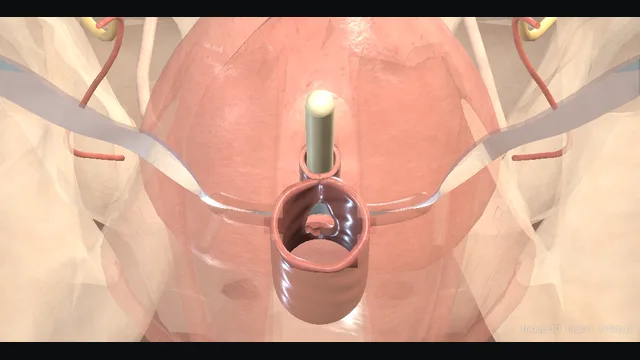
A tension-free tape passed from the inner thigh through the obturator foramen to lie flat under the mid-urethra. The case is won in the planes: para-urethral tunnels that stay lateral, a trocar that stays outside the bladder, and a tape that hugs rather than compresses.
Core tierFRCS continence, female pelvic surgeryInteractive 3D
Sagittal, the outside-in transobturator route to the mid-urethra. Schematic, not to scale; relationships only.
The sagittal map of the outside-in transobturator route: a short vertical urethra from bladder to perineum, the tape flat under its midpoint, and a curved corridor from each inner thigh through the obturator foramen to the para-urethral tunnel.
The bladder sits medial to that corridor and the obturator canal at the anterosuperior edge of the foramen.
The retropubic route is a separate corridor behind the pubis; a lateral deviation from it can reach the external iliac vessels, shown only in the complication view.
›Mid-urethra - the functional point of the tape: compression under stress happens here, which is why position, not tension, does the work.
›Obturator foramen and membrane - the corridor the trocar crosses from the inner thigh; the canal at its superomedial edge carries the vessels and nerve.
›Obturator internus and levator - the lateral wall of the passage; the trocar crosses the membrane and stays outside the levator.
›Bladder - medial to the trocar path; cystoscopy exists because the perforated bladder is silent.
›External iliac vessels - lateral to the expected retropubic corridor and threatened by lateral trocar deviation; they are not on the intended transobturator path.
Landmarks: the mid-urethraThe tape supports the mid-urethra, not the bladder neck, and the whole operation is laid out on the skin before prepping. The mid-urethral point is marked over the urethra at the level of the clitoral hood, the short vaginal incision line is drawn just beneath it, and the obturator skin entries are marked in the groin creases against the adductor longus tendons. Marking everything before prep means the landmarks survive tissue distension and the shift of the drapes, and the case proceeds by plan rather than by search. A tape placed at the bladder neck obstructs rather than supports, which is why the first minutes of this operation are spent drawing, not cutting. where it bites: a tape placed at the bladder neck obstructs rather than supports.
Vaginal incisionThe bladder is emptied with a catheter, which stays in until the cystoscopy. A short incision is made in the anterior vaginal wall at the marked mid-urethral level, and sharp dissection opens the plane between vaginal epithelium and urethra on either side. Scissors are held close to the vaginal flap and spread rather than cut, until the tissue accepts the spread without resistance and the glistening layer over the periurethral fascia is reached. Depth is judged by how the tissue behaves: a plane that bleeds or shows muscle fibre is too deep and threatens the urethra, while one that never separates is too shallow. Infiltration beneath the epithelium before the incision lifts this plane and reduces the bleeding that would otherwise obscure it. where it bites: cutting too deep breaches the urethra; too shallow never finds the plane.
Para-urethral tunnelsScissors are inserted lateral to the urethra on each side and spread through the perineal membrane toward the ipsilateral obturator foramen, creating the tunnel the trocar will follow. The spread is directed toward the foramen and away from the urethra, stopping against the resistance of the membrane with the tips felt against the expected line of the foramen. A well-made tunnel is blind but directional: it fixes the angle of the trocar pass before the trocar is anywhere near the field. Tunnels that drift medially or superiorly point the trocar at the bladder, which is why this small step decides where the big one lands. where it bites: tunnels that drift medially or superiorly point the trocar at the bladder.
Trocar passage (out-in)The curved trocar enters through the marked groin point and is guided by a finger in the vaginal incision, piercing the obturator membrane with a controlled give and hugging the bone as it turns medially toward the para-urethral tunnel. The tip is received in the tunnel rather than hunted for, and the pass stays outside the levator throughout so the pelvis itself is never entered. The trocar emerges through the tunnel carrying one end of the tape, and the mirror pass is repeated identically on the other side before anything is tensioned. Cystoscopy follows the completed bilateral outside-in passage and precedes final adjustment, trimming and closure. where it bites: medial deviation of the trocar tip is the mechanism of bladder perforation.
Tape under the mid-urethraThe tape is drawn through both tunnels until its midpoint sits flat under the mid-urethra, with its sleeves keeping it flat and the ends free in the groins. It rests loosely while cystoscopy checks the bladder and urethra; final adjustment and sleeve removal follow that integrity check. Tension is set by feel rather than force: an instrument placed between tape and urethra should pass freely, and the tape should rest without dimpling or bowstringing the urethra. A tape that compresses the urethra obstructs voiding without adding continence, which defeats the design of the whole operation. where it bites: a tape that compresses the urethra obstructs; tension-free is the whole design.
Cystoscopy and adjustmentAfter both outside-in passes, cystoscopy with a filled bladder inspects the dome, trigone and both lateral walls for perforation or intraluminal mesh and confirms both ureteric orifices are clear. The urethra is also inspected before the tape is finally adjusted, the sheaths removed and the ends cut below skin. The vaginal incision is closed with absorbable suture, and a pack or catheter is left only if the dissection or a recognised injury demanded it. Skipping the cystoscopy converts a small, manageable intra-operative finding into an unrecognised perforation or intravesical tape that presents later as stones and infection. Before discharge the patient voids and the residual is checked; retention is a catheter and a review, not a wait. where it bites: skipping cystoscopy can leave a perforation or intravesical tape unrecognised.
The spatial relationships the animation is built to make automatic. Before you read each one, ask yourself: at this point in the operation, what is about to go wrong, and what is my first move? The save for each sits in the Complications tab.
RiskMid-urethra, not the bladder neck - the tape position that restores the stress mechanism without obstructing voiding.
RiskObturator canal - vessels and nerve at the anterosuperior foramen; the trocar crosses the membrane below and lateral to it.
RiskMedial deviation into the bladder - the trocar aims at the para-urethral tunnel on its own side, never across the midline; cystoscopy follows completed sling passage before final adjustment.
RiskThe retropubic space (route contrast) - the intended TVT corridor stays behind the pubis beside the bladder; lateral deviation can reach the external iliac vessels.
Trigger each of these live in the Animation tab, then read the save: the first move, the escalation trigger, and the line that goes in the op note.
Bladder perforation
the trocar tip drifts medially and enters the bladder; the injury is silent unless looked for.
The save
Perform cystoscopy after sling passage and before final adjustment: the perforation you look for is the one you can manage.first move
Withdraw the trocar and re-pass it in the correct plane, lateral to the urethra and hugging the obturator membrane.
Drain the recognised breach with a catheter according to the injury and local protocol.
Tape seen through the mucosa → reposition or remove at the same anaesthetic; tape left in the bladder stones and infects.escalation trigger
Op note: side and site of perforation, trocar re-passed, catheter plan and follow-up.document
Retropubic vascular injury (TVT contrast)
on the retropubic route, lateral trocar deviation can leave the intended corridor behind the pubis and reach the external iliac vessels; this is shown only as route contrast.
The save
Recognise the setup: retropubic route, lateral deviation, sudden pressure drop or expanding suprapubic mass.first move
Pressure where possible, and help early: open preparation with vascular input, never a blind clamp in the retropubic space.
Resuscitate in parallel: cross-match, bloods, anaesthetic senior present.
Haemodynamic instability or an expanding retropubic haematoma → laparotomy with vascular surgical support.escalation trigger
Op note: route used, the event, volumes and products given, and the definitive management plan.document
Mesh erosion / extrusion
a late complication in which tissue covering the tape breaks down, exposing mesh into the urethral or bladder lumen or through the vaginal wall.
The save
Suspect it with new voiding symptoms, de novo leakage, recurrent infection, discharge or dyspareunia after tape surgery.first move
Examine the vaginal wall with a speculum and perform cystoscopy for urethral and bladder erosion.
Small vaginal extrusion may be managed conservatively or with local closure; intraluminal tape may not.
Tape inside the urinary tract or symptomatic extrusion → excision with urethrolysis in a specialist centre, not office trimming.escalation trigger
Notes: tape type and route, symptoms, examination and cystoscopy findings, referral made.document
When the case turns: the problem, then the next move.
ActionTape looks tight at adjustment - loosen before closing: an instrument should pass freely between tape and urethra, and a cough test with a filled bladder where used should leak, not dribble.
ActionTrocar will not find the tunnel - guide the tip with a finger in the vaginal incision; withdraw and re-aim rather than force a wrong plane.
ActionScarred planes after previous pelvic surgery - abandon the tunnel and reconsider the route or refer; scarred planes move the danger rather than remove it.
ActionPerforation found on cystoscopy - withdraw, re-pass correctly, leave a catheter, and document; tape through the mucosa needs repositioning at the same sitting.
The examiner stems this scene prepares you for. Try to answer out loud first, then reveal, and learn the discriminator, the detail that turns a pass into a strong pass.
QRetropubic or transobturator tape: how do the routes differ in risk?reveal answer
Both routes place a tape under the mid-urethra but use different trocar corridors.
The retropubic route passes from the mid-urethra through the retropubic space behind the pubis to suprapubic skin: the intended corridor lies beside the bladder and bowel, and a lateral deviation can reach major pelvic vessels, including the external iliac vessels.
Its specific risks therefore include bladder perforation and rare but potentially catastrophic vascular or bowel injury.
The transobturator route passes laterally through the obturator foramen and stays outside the pelvic cavity: it has a lower bladder-perforation risk than the retropubic route but introduces route-specific groin pain and potential injury near the obturator canal.
Current EAU guidance reports better long-term patient-reported outcomes for the retropubic route, so route choice requires an explicit discussion of effectiveness, complications and alternatives rather than a claim that one route is universally safer.
Strong pass
Say all three
Naming the specific structure each route endangers: bladder
Bowel and iliacs behind the pubis
Obturator canal and groin pain through the foramen
Examiner note
Naming the specific structure each route endangers: bladder, bowel and iliacs behind the pubis; obturator canal and groin pain through the foramen.
QYou perforate the bladder with the trocar during a tape. What do you do?reveal answer
Perforation is why cystoscopy follows sling passage before final adjustment, because the injured bladder may otherwise be silent.
On cystoscopy the trocar or tape may be seen tenting or entering the mucosa.
The trocar comes out and is re-passed in the correct plane, lateral to the urethra and hugging the obturator membrane rather than drifting medially.
A small clean perforation is drained according to the recognised injury and local protocol.
Mesh that has passed through the bladder mucosa or wall cannot be left in that position: it is repositioned or removed at the same anaesthetic.
Failure to recognise the injury is the real danger, which is why a patient with unexpected pain, haematuria or a poor result after tape surgery is investigated rather than reassured.
Strong pass
Say both
Cystoscopy framed as mandatory rather than discretionary
Tape through mucosa as an indication for immediate repositioning
Examiner note
Cystoscopy framed as mandatory rather than discretionary, and tape through mucosa as an indication for immediate repositioning.
QWhy does the tape sit at the mid-urethra and not the bladder neck?reveal answer
The integral theory reframed stress incontinence as failure of mid-urethral support rather than bladder neck descent.
In health the pubo-urethral ligaments and the vaginal hammock let the mid-urethra kink against a rise in abdominal pressure; when that support is lost, pressure transmits equally to bladder and urethra and urine leaks.
A tape at the mid-urethra restores that kinking point: it works by position, dynamically and only under stress.
Supporting the bladder neck instead creates a static obstruction that impedes voiding without restoring the stress mechanism, which is why older bladder neck elevation procedures failed on both counts.
The practical consequence in theatre is that the tape is set loose, tension-free, with an instrument passing freely between tape and urethra: stress leakage stops because the tape compresses under a rise in pressure, not because it was tied tight.
Tape placed too high or tied too tight gives the worst of both worlds: obstruction without continence.
Strong pass
Say both
Explaining the dynamic kinking mechanism
Naming tension as the enemy of the design
Examiner note
Explaining the dynamic kinking mechanism and naming tension as the enemy of the design.
QHow does mesh erosion after a sling present?reveal answer
Erosion is a late diagnosis with two faces.
Vaginal extrusion presents with persistent discharge, spotting, dyspareunia or a partner reporting discomfort, and examination shows or feels an exposed tape edge through the vaginal epithelium;
it is uncommon and usually relates to healing or infection in the early months.
Urethral or bladder erosion presents differently: new voiding symptoms, obstructed flow or retention from a tape tightened by scarring, recurrent urinary infection, haematuria, or de novo leakage as the tape cuts into the wall, and cystoscopy shows tape inside the lumen.
Management is specialist: a small vaginal extrusion may be managed conservatively or with local closure, but intraluminal tape needs excision with urethrolysis, and reconstruction is best referred to a centre that does this work regularly.
Any woman with new voiding or pain symptoms after tape surgery deserves examination and cystoscopy rather than reassurance, because late diagnoses are made by looking, not by waiting.
Strong pass
Separating extrusion, an examination finding, from erosion, a cystoscopic finding, and referring rather than trimming in the office.
Say it aloud, then reveal. Reading is not remembering.
01The relationshipSay the critical spatial relationship out loud, then reveal.reveal
Memory frame
The sagittal map of the outside-in transobturator route
Why?
The sagittal map of the outside-in transobturator route: a short vertical urethra from bladder to perineum, the tape flat under its midpoint, and a curved corridor from each inner thigh through the obturator foramen to the para-urethral tunnel. The bladder sits medial to that corridor and the obturator canal at the anterosuperior edge of the foramen. The retropubic route is a separate corridor behind the pubis; a lateral deviation from it can reach the external iliac vessels, shown only in the complication view.
02The first moveBladder perforation. Your first move?reveal
First move
Perform cystoscopy after sling passage and before final adjustment: the perforation you look for is the one you can manage.
03The escalation triggerBladder perforation. What makes you call for help?reveal
Escalate when
Tape seen through the mucosa → reposition or remove at the same anaesthetic; tape left in the bladder stones and infects.
04The discriminatorWhat turns a pass into a strong pass?reveal
Strong pass
Say all three
Naming the specific structure each route endangers: bladder
Bowel and iliacs behind the pubis
Obturator canal and groin pain through the foramen
Examiner note
Naming the specific structure each route endangers: bladder, bowel and iliacs behind the pubis; obturator canal and groin pain through the foramen.
05The pitfallWhere does this operation bite first?reveal
Pitfall
Mid-urethra, not the bladder neck
Why?
the tape position that restores the stress mechanism without obstructing voiding.
Revision practice with self-marked answers, not an assessment of clinical competence. Kept in
this browser only; it does not sync between devices.