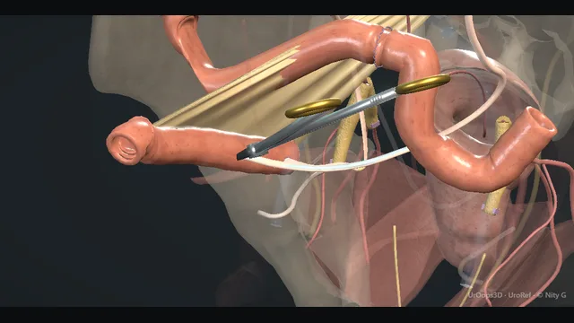
The pelvis is cleared as one specimen, bladder to prostate, with both ureters divided at the pelvic brim where they cross the iliac vessels, the posterior plane developed directly onto the rectum, and the dorsal venous plexus the last and bloodiest attachment at the apex.
The conduit is a short ileal segment on the right: its afferent limb receives both ureters over stents and its distal end is matured through a stoma site marked before the first incision.
Advanced tierFRCS oncology, pelvic surgeryInteractive 3D
Open pelvis as a funnel, the brim above and the apex below. Schematic, not to scale; relationships only.
The operation is a funnel.
Superiorly both ureters cross the iliac vessels at the pelvic brim and are divided there.
The dissection then narrows: posteriorly the plane on Denonvilliers' fascia drops behind the vesicles and prostate onto the rectal wall, laterally the pedicles run from the internal iliac branches to the bladder base in the same gutter as the obturator nerve, and inferiorly everything converges on the dorsal venous plexus at the apex.
The reconstruction sits outside that funnel: an ileal segment on the right, ureters swinging to its afferent limb, and its efferent end rising through the abdominal wall to the stoma site marked on day zero.
›Ureters at the pelvic brim - cross the iliac bifurcation directly over the vessels; divided here to give both length for the afferent limb and distance from the tumour.
›Posterior plane (Denonvilliers' fascia) - the avascular plane behind vesicles and prostate that drops to the apex; the rectum is its posterior wall.
›Lateral pedicles - the bladder's blood supply from the internal iliac branches; controlled along a line from the divided ureter to the apex.
›Dorsal venous plexus - the venous mesh at the apex, the bloodiest attachment of the specimen; sutured, not diathermied, when it tears.
›Ileal segment + stoma site - a short segment on a mobile mesentery, afferent limb receiving both ureters, efferent end spouted at the pre-marked site.
Access + stoma siteThe stoma site is marked with the patient standing, over the rectus muscle and clear of skin folds, scars and the belt line, so the appliance sits where the patient can see and manage it. The abdomen is then opened through a midline incision or by the robotic approach, and the bladder is assessed for resectability before the dissection commits. Marking before induction means the mark reflects the standing anatomy the appliance must live with, not the anatomy of a supine, anaesthetised abdomen. where it bites: a stoma sited on the ward with the patient supine is the complication that outlives the operation: parastomal hernia, leakage and appliance failure all follow a bad site.
Ureteric divisionBoth ureters are identified where they cross the common iliac vessels at the pelvic brim and freed with their strip of adventitia, divided between clips well clear of the bladder, and the distal ends sent for frozen section where the indication demands it. The crossing is dissected with the iliac vessels in view and clips applied only under direct vision, because the ureter lies directly on the iliac vein at this point. Division at the brim, rather than low over the bladder, gives clean margins and enough length for the ureters to reach the afferent limb without tension later. where it bites: the ureter crosses the iliac bifurcation directly over the vessels; traction that avulses the ureter or a clip applied blind bites the iliac vein.
Posterior planeThe peritoneum is incised behind the vasa and seminal vesicles and the plane is developed directly onto Denonvilliers' fascia, then swept down to the apex with the rectum in view throughout. The plane is avascular and drops almost without bleeding when the layer is right, and fat or bleeding means the plane has been lost, so the dissection returns to a clean area and re-enters under direct vision. In the female the same plane runs behind the uterus and vagina, with the anterior vaginal wall taken en bloc where the disease demands and the vault repaired as a planned part of the case. where it bites: the plane is avascular until a wrong-layer entry puts the instrument on the rectal wall; previous radiotherapy or a posterior tumour thins the margin to nothing.
Lateral pediclesThe lateral bladder pedicles are divided along a line from each divided ureter down toward the apex, stepping sequentially through the bladder blood supply from its internal iliac branches, with an energy device where the approach allows and clips beside structures that must not be exposed to heat. Each step is taken with the obturator nerve in mind as it runs in the same lateral gutter, so bites are shallow and applied only to tissue that has been skeletonised. Working from the pelvic brim downward in a fixed order keeps the field organised and prevents a pedicle stump being missed until it bleeds. where it bites: the obturator nerve shares the lateral gutter with the pedicle; the clip that is too deep takes the nerve with the pedicle.
Apex + dorsal venous plexusThe posterior attachments are divided and the urethra is freed at the apex, with the dorsal venous plexus ligated and controlled before the urethra is cut, so the last attachment of the specimen comes away without a venous tear. The catheter is withdrawn before the cut, and the specimen side of the urethra is clipped or tied as it is divided so nothing spills from the bladder into the pelvis. The urethral margin is sent where a neobladder is contemplated, because a positive margin changes the reconstruction. If the plexus tears it retracts into the pelvic floor, and the response is pressure, exposure and then a haemostatic suture, never blind diathermy into a pool. where it bites: the plexus retracts into the pelvic floor when torn; the save is a haemostatic suture, never blind diathermy into a pool.
LymphadenectomyThe node template is cleared to match the disease and the patient, running from the aortic bifurcation or the common iliacs down to the inguinal ligament and including the obturator and presacral packets as indicated. Each packet is swept off the vessels as a complete block with clips on lymphatic channels, and the obturator nerve is kept in view along the floor of the gutter. Presacral veins and obturator vessels bleed in awkward corners, so the template is cleared with the same discipline and exposure as the cystectomy itself, never as a rushed afterthought. where it bites: presacral veins and the obturator vessels bleed in awkward corners; the template is cleared with the same discipline as the cystectomy.
Ileal conduit + stomaA segment of ileum is selected on a healthy mesentery with a good arc of mobility and enough reach to bring the conduit to the marked stoma site without tension, then bowel continuity is restored and the mesenteric window is closed. Both ureters are joined to the afferent limb over stents, and the distal end of the conduit is brought out through the rectus at the pre-marked site and matured as a spouted bud. The bud is checked for a healthy pink colour before the drapes come down, because a dusky bud is revised now, in theatre, while salvage is easy. where it bites: a segment chosen under tension or on a marginal mesentery is the setup for ischaemia, retraction and stenosis of the whole conduit.
The spatial relationships the animation is built to make automatic. Before you read each one, ask yourself: at this point in the operation, what is about to go wrong, and what is my first move? The save for each sits in the Complications tab.
RiskRectum behind the posterior plane - one wrong layer deep from Denonvilliers' fascia; the injury that turns a cystectomy into a colorectal reconstruction.
RiskIliac vessels at the ureteric crossing - the ureter lies directly on them at the brim; traction and blind clips are the mechanism of an iliac vein injury.
RiskDorsal venous plexus at the apex - a venous mesh under the pubis that retracts when torn; pressure, exposure, then a haemostatic suture.
Trigger each of these live in the Animation tab, then read the save: the first move, the escalation trigger, and the line that goes in the op note.
Rectal injury
the posterior plane entered on the wrong layer, or a posterior tumour or previous radiotherapy leaving no plane at all; recognised by direct vision or by the appearance of gas or stool.
The save
Recognise it openly the moment the plane is lost: call it, improve the light and exposure, and control contamination.first move
Assess the injury: site, size, and whether the bowel was prepared, irradiated or devascularised.
Small clean injuries are repaired immediately in two layers with an omental or tissue interposition between the repair and the pelvis.
Larger, irradiated, contaminated or fragile-bowel injuries are repaired or resected and defunctioned with a loop stoma; a low injury near the anorectal junction gets colorectal input early.
Copious irrigation, pelvic drain, and antibiotics; watch post-operatively for pelvic sepsis and drain any collection radiologically.escalation trigger
Op note: site and size of injury, layers of repair, interposition used, diversion or not, and the plan for review.document
Uretero-ileal leak / stricture
the anastomosis fails early as a urine leak with high, creatinine-rich drain output, or late as a stenosis presenting with hydronephrosis and declining renal function.
The save
Send the drain fluid for creatinine: a level at serum-urine gradient confirms urine; a persistently high-output drain in the first days is the leak.first move
Keep the drain in place and imaging the kidney: controlled fistulae close on drainage alone in most cases.
Maintain conduit drainage and treat or prevent sepsis; image with contrast studies or cross-sectional imaging to map the leak.
Nephrostomy and antegrade stenting for a leak that will not close or an obstructed system.
Generalised peritonitis, worsening sepsis or a completely disrupted anastomosis → return to theatre for revision.escalation trigger
Op note and discharge summary: side, technique, stent plan, and the imaging surveillance for the late stricture.document
Apical bleeding: dorsal venous plexus
the plexus tears at the apex, retracts into the pelvic floor and bleeds briskly into the deepest corner of the operation exactly when the specimen is nearly free.
The save
Direct pressure with a pack or swab and do not keep lifting it to look; tell the anaesthetist and call for help early.first move
Improve the exposure before the second look: light, retraction, suction, and a clear field beat speed.
Control with a haemostatic suture taken deep to the retracted plexus; adjuncts and haemostatic agents supplement a stitch, they do not replace it.
If the field is unreachable, pack the pelvis, resuscitate, and plan a staged return once physiology allows.
Ongoing blood loss, coagulopathy or haemodynamic instability → definitive control in theatre with senior help, not repeated attempts.escalation trigger
Op note: vessel or plexus, method of control, estimated loss, blood products given.document
When the case turns: the problem, then the next move.
ActionThe ureter will not reach the afferent limb without tension - mobilise the ureter proximally keeping its strip of adventitia, and re-site the afferent limb; a tensioned anastomosis is a planned stricture.
ActionDusky stoma at the end of the operation - re-inspect before leaving theatre: a dark bud needs the segment re-mobilised or revised now, when salvage is cheap.
ActionPersistent high drain output after surgery - send drain fluid for creatinine and image the upper tracts; urine settles the question that output volume alone cannot.
ActionIleus beyond the expected course - exclude mechanical obstruction and collection with imaging, minimise opioids, and treat the cause rather than starving the patient longer.
The examiner stems this scene prepares you for. Try to answer out loud first, then reveal, and learn the discriminator, the detail that turns a pass into a strong pass.
QHow do you decide between an ileal conduit and a neobladder?reveal answer
Cystectomy first, reconstruction second: no reservoir rescues an incomplete cancer operation, so disease factors, patient factors and bowel factors all outrank preference.
I assess fitness, renal and liver function, hand function, dexterity, social support and the patient's own motivation.
A neobladder demands a patient who can void on a schedule, wake to empty, accept that retention may require self-catheterisation for life, and attend long-term follow-up;
a conduit demands none of those and is the default for the older or comorbid patient and for impaired renal function, because a long ileal reservoir absorbs ammonium and chloride and acidifies.
Tumour factors also decide: carcinoma in situ at the bladder neck or a positive urethral margin makes a neobladder oncologically unsound, as the urethra becomes its outlet.
Previous radiotherapy, inflammatory bowel disease and dense adhesions push toward a conduit.
I counsel both honestly: a conduit means an appliance, with parastomal hernia, stenosis and skin problems;
a neobladder means learned continence, nocturnal emptying, mucus, infection and retention in a meaningful minority.
The strongest, best-supported candidates may choose a neobladder; the conduit remains the safest reconstruction for most, and saying so plainly is part of the consent.
Strong pass
Say all three
Framing it as cancer clearance first
Physiology: renal function and urethral margin as hard limits
Motivation and support as the swing factors
Examiner note
Framing it as cancer clearance first, then physiology: renal function and urethral margin as hard limits, motivation and support as the swing factors.
QTake me through your enhanced recovery pathway for cystectomy.reveal answer
The pathway starts before admission: counselling with the stoma nurse, the stoma sited with the patient standing, prehabilitation with smoking cessation and exercise, carbohydrate loading, and avoidance of prolonged mechanical bowel preparation.
Thromboprophylaxis begins before induction.
Intra-operatively the aims are normothermia, goal-directed fluid, meticulous haemostasis and an analgesia strategy, regional or local, that minimises systemic opioids;
nasogastric tubes are omitted or removed at the earliest safe moment.
After surgery the discipline is early feeding on the day, early and repeated mobilisation, chewing gum, laxatives, drains out as soon as safe, and daily criteria for progression rather than tradition.
Stents and catheters are removed on a protocolised schedule, with imaging before and after as the unit standard.
The morbidity that enhanced recovery most influences is ileus, alongside pulmonary complications; the markers of a working pathway are shorter time to diet and to walking without a rise in readmission.
Audit belongs inside the pathway: length of stay, ileus, readmission and unplanned intervention are what I would measure and review at the morbidity meeting.
The principle to state is that this is a bundle: the elements support one another, and quietly dropping one degrades the rest.
Strong pass
Say both
Naming ileus as the target morbidity and audit as part of the pathway
Rather than reciting a checklist of individual elements
Examiner note
Naming ileus as the target morbidity and audit as part of the pathway, rather than reciting a checklist of individual elements.
QRank the bowel complications you worry about after cystectomy.reveal answer
Early, the hierarchy runs ileus, then leak at the bowel anastomosis, then conduit ischaemia, then early obstruction.
Ileus is the commonest significant morbidity: it is managed by patience, early enteral feeding, minimising opioids and, when it persists beyond the expected course, imaging to exclude mechanical obstruction and intra-abdominal collection.
A leak at the ileal-ileal anastomosis declares itself with pain, fever, rising inflammatory markers and change in drain output;
a contained leak drains, treats with antibiotics and nutrition and observes, while free leak with generalised peritonitis returns to theatre for washout, repair or diversion.
Conduit ischaemia is the feared early problem: a dusky stoma on the first day is reviewed at the bedside repeatedly and any frankly necrotic segment returns to theatre before it perforates, with revision of the conduit or formal diversion if it cannot be salvaged.
Obstruction, internal hernia and volvulus around the mesenteric window present later, which is why the window is closed.
I also keep the urinary complications in a separate lane: a urine leak at the uretero-ileal anastomosis gives high, creatinine-rich output and is managed by drainage and stenting, with the late stricture dealt with endoscopically or surgically.
Late, the problems are parastomal hernia, stenosis and metabolic acidosis.
Strong pass
Say both
Separating the urinary from the faecal lanes
Treating conduit ischaemia as a next-day theatre problem rather than a wound review
Examiner note
Separating the urinary from the faecal lanes, and treating conduit ischaemia as a next-day theatre problem rather than a wound review.
QYou enter the posterior plane and the rectum is injured. What happens next?reveal answer
I suspect it the moment the plane stops being avascular, or gas and stool appear, and I confirm by direct vision; the unrecognised injury is what becomes the fistula.
Once recognised, the whole team should know: anaesthetist informed, antibiotics considered, and the field prepared for repair.
I assess the extent and the context: site, size, whether the bowel was prepared, whether the pelvis was irradiated, and how contaminated the field is.
A small, clean injury on prepared bowel in a fit patient is repaired at once in two layers with an omental or tissue interposition between the repair and the pelvis, and the operation continues.
A larger defect, a devascularised segment, heavy contamination, previous radiotherapy or a fragile patient changes the plan: repair or resection as required plus a defunctioning loop stoma, and colorectal input early for low injuries near the anorectal junction, where fistula risk is highest.
I irrigate copiously, drain the pelvis and watch for pelvic sepsis, draining collections radiologically.
The chronic rectourethral or perineal fistula is the late legacy of a missed or broken-down repair and is a specialist reconstruction.
The op note records site, size, layers, interposition and diversion.
Strong pass
Say both
Making the decision turn on context: clean and prepared versus irradiated and contaminated
Naming interposition and diversion as the two manoeuvres that protect the repair
Examiner note
Making the decision turn on context: clean and prepared versus irradiated and contaminated, and naming interposition and diversion as the two manoeuvres that protect the repair.
Say it aloud, then reveal. Reading is not remembering.
01The relationshipSay the critical spatial relationship out loud, then reveal.reveal
Memory frame
The operation is a funnel.
Why?
Superiorly both ureters cross the iliac vessels at the pelvic brim and are divided there. The dissection then narrows: posteriorly the plane on Denonvilliers' fascia drops behind the vesicles and prostate onto the rectal wall, laterally the pedicles run from the internal iliac branches to the bladder base in the same gutter as the obturator nerve, and inferiorly everything converges on the dorsal venous plexus at the apex. The reconstruction sits outside that funnel: an ileal segment on the right, ureters swinging to its afferent limb, and its efferent end rising through the abdominal wall to the stoma site marked on day zero.
02The first moveRectal injury. Your first move?reveal
First move
Recognise it openly the moment the plane is lost: call it, improve the light and exposure, and control contamination.
03The escalation triggerRectal injury. What makes you call for help?reveal
Escalate when
Copious irrigation, pelvic drain, and antibiotics; watch post-operatively for pelvic sepsis and drain any collection radiologically.
04The discriminatorWhat turns a pass into a strong pass?reveal
Strong pass
Say all three
Framing it as cancer clearance first
Physiology: renal function and urethral margin as hard limits
Motivation and support as the swing factors
Examiner note
Framing it as cancer clearance first, then physiology: renal function and urethral margin as hard limits, motivation and support as the swing factors.
05The pitfallWhere does this operation bite first?reveal
Pitfall
Rectum behind the posterior plane
Why?
one wrong layer deep from Denonvilliers' fascia; the injury that turns a cystectomy into a colorectal reconstruction.
Revision practice with self-marked answers, not an assessment of clinical competence. Kept in
this browser only; it does not sync between devices.