UrOops3D, danger-zone atlas

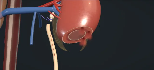
Schematic model on the kidney atlas, drawn from the front as a cutaway.

Operative animation, Ureterocalicostomy

Ureterocalicostomy: the ureter to the lower calyx

Indications

  • A failed pyeloplasty or endopyelotomy with no usable pelvis left.
  • Pelviureteric obstruction with a small intrarenal pelvis, a dilated lower calyx and thin parenchyma over it.
  • Traumatic avulsion of the pelviureteric junction.

Alternatives

  • Redo pyeloplasty, when there is enough pelvis to rebuild.
  • Endopyelotomy for a short, selected failure.
  • A laparoscopic or robot-assisted ureterocalicostomy in centres that offer it.
  • Nephrectomy for a poorly functioning kidney when the other kidney is normal.

Risks

  • Bleeding from the cut surface of the kidney.
  • Urine leak and a urinoma.
  • Narrowing at the join, with obstruction returning.
  • Infection; ileus or bowel obstruction.

The operation for the obstructed kidney that has no pelvis worth rebuilding: after a failed pyeloplasty the pelvis is small, scarred and inside the kidney, while the lower calyx is dilated under thin parenchyma.

The lower pole is amputated to lay that calyx open, and the healthy ureter is spatulated and sewn to it, mucosa to mucosa, over a stent. The lesson is why parenchyma is taken back from the calyx, and what keeps the join wide.

Advanced tier FRCS reconstruction, PUJ obstruction, ureter Interactive 3D

Operative visualisation

Schematic scene preview: Ureterocalicostomy
Preparing the 3D anatomy…

UrOops3D · UroRef · © Nity G

How to use this module. Play the eight steps, or use Back and Next. Watch the pole come off along the scored line, the calyx open on the cut face, the ureter spatulated and laid over the mouth, and the two rows either side of the stent. The triggers show bleeding, a narrowed join and a leak.

Click me next Section 2 of 8 Anatomy The structures this operation works between, and the one it must not cross.

The structures in the scene

Ureterocalicostomy: how the structures relateThe lower pole from the front, after the amputation. Lower calyx mouth on the cut face: sewn mucosa to mucosa, wider bites on the calyx Healthy ureter, spatulated on the kidney side. Lower calyx mouth on the cut face: laid open by taking the whole pole away Lower pole parenchyma, amputated. Lower calyx mouth on the cut face: the cut that opens it stays below them Lower segmental vessels above the line (danger). Lower calyx mouth on the cut face: the ureter reaches it alive only with this kept Periureteric adventitia and its vessels (danger). Lower calyx mouth on the cut face: lies well below it, the lowest point of the kidney Renal hilum, in front of the scarred pelvis (danger). Lower calyx mouth on the cut face: replaces it as the outlet of the kidney Scarred junction, tied and left.Lower calyx mouth onthe cut faceHealthy ureter,spatulated on thekidney sideLower poleparenchyma,amputated!Lower segmentalvessels above theline!Periuretericadventitia and itsvessels!Renal hilum, infront of the scarredpelvisScarred junction,tied and leftsewn mucosa tomucosa, wider biteson the calyxlaid open by takingthe whole pole awaythe cut that opensit stays below themthe ureter reachesit alive only withthis keptlies well below it,the lowest point ofthe kidneyreplaces it as theoutlet of thekidneyTargetDangerLandmarkContext
The lower pole from the front, after the amputation. Schematic, not to scale; relationships only.
  • Seen from the front, the scarred junction sits at the hilum with nothing usable above it, and the healthy ureter runs down medial to the lower pole.
  • The amputation plane crosses the lower pole below the hilum and opens the dilated calyx in the middle of the cut face.
  • The ureter is carried up and laterally to that mouth, so the new outlet of the kidney is its lowest point.
  • ›Lower pole parenchyma - thin over the dilated calyx; amputated below the hilum so that the calyx lies open on a flat cut face.
  • ›Dilated lower calyx - the new outlet of the kidney: dependent, wide, and lined by urothelium to sew to.
  • ›Small intrarenal pelvis and scarred junction - what makes a redo pyeloplasty impossible; left in place, its stump tied.
  • ›Healthy proximal ureter - divided at its highest viable point, carried up on its adventitia, spatulated on the side that faces the kidney.
  • ›Periureteric adventitia - carries the ureter’s blood supply; it stays on the ureter for the whole mobilised length.
  • ›Lower segmental vessels - enter the parenchyma in front of the hilum above the line of the cut; the line stays below them.
  • ›Renal hilum - in front of and medial to the scarred pelvis; the amputation is planned to stay well clear of it.
  • ›Renal capsule - scored first, then drawn loosely over a bolster at the end.
  • ›Gerota’s fascia and perirenal fat - opened posteriorly for the exposure; fat is laid over the repair and the fascia closed loosely.
  • ›Pleura and the eleventh intercostal bundle - the dangers of the flank incision, on the way in and at closure.
Click me next Section 3 of 8 Steps The operation in order, with the landmark that guards each move.

Operative steps

  1. Expose the lower pole An adult with recurrent obstruction after a pyeloplasty: the pelvis is small, intrarenal and scarred, and the lower calyx is dilated under thin parenchyma. The patient lies on the right side with the left flank up, and the incision runs in the eleventh interspace from the mid-axillary line toward the lateral border of rectus. The peritoneum is swept medially, the colon reflected off Gerota’s fascia, and the fascia opened posteriorly. The lower pole, the renal pelvis and the proximal ureter are dissected and mobilised until the site of obstruction and the healthy ureter below it are both in view.
    where it bites: the pleura and the neurovascular bundle under the eleventh rib on the way in; at the kidney, the hilar vessels lie in front of and medial to the scarred pelvis.
  2. Divide the ureter The ureter is traced down from the scar to healthy ureter and divided at its most superior viable point. Its adventitia, and the blood supply that runs in it, is kept on the ureter for the whole length that is mobilised. A stay stitch tags the healthy end so that the ureter is never held by forceps again. The junction stump is closed with an absorbable tie.
    where it bites: stripping the adventitia to gain length devascularises the end that is about to be sewn.
  3. Amputate the lower pole The lower pole parenchyma is excised to expose the lower pole calyx: a lower pole heminephrectomy, not a nephrotomy into the calyx. The line is scored on the capsule with diathermy and the parenchyma divided along it, well below the hilum, until the dilated calyx lies open on the cut face. The amputation is generous.
    where it bites: the lower segmental vessels: a line carried too high takes the blood supply of parenchyma that is being kept.
  4. Haemostasis The vessels on the cut face are controlled one at a time with figure-of-eight absorbable stitches until the parenchyma is dry. The dictation uses no clamp on the renal artery. Each stitch is placed in parenchyma, clear of the mouth of the calyx, so that the opening the ureter will be sewn to is left exactly as wide as the amputation made it.
    where it bites: a haemostatic bite that crosses the mouth of the calyx narrows the join before it is made.
  5. Spatulate the ureter The healthy ureter is brought to the cut face on its stay stitch. It must lie there without tension: if it does not, more ureter is mobilised with its adventitia, or the kidney is brought down. The end is spatulated on its lateral aspect, the side that faces the kidney, for a generous length.
    where it bites: a short spatulation or a ureter under tension invites a stricture.
  6. Posterior row The join is made mucosa to mucosa with interrupted fine absorbable stitches: calyceal wall, then ureter. The stitches are placed further apart on the calyx than on the ureter, which spreads the spatulated ureter round the larger opening and leaves the repair widely patent. The posterior row is placed first, while the front of the join is open and every bite can be seen from inside.
    where it bites: a bite that takes parenchyma and misses the calyceal mucosa leaves raw tissue in the suture line.
  7. Stent and anterior row With the posterior row tied, a JJ stent is passed through the open join: the upper coil into the calyx, the lower end down the ureter to the bladder. The anterior row is then completed over the stent. Kochakarn and colleagues added a nephrostomy in every patient; the dictation relies on the stent and the drain.
    where it bites: the stent goes across while the front is open and both ends can be seen into place.
  8. Bolster, capsule and drain An absorbable gelatin sponge bolster is laid in the parenchymal defect, round the join, and the renal capsule is approximated loosely over it with interrupted stitches, its two edges meeting at a seam. Available perirenal fat is secured over the repair and Gerota’s fascia closed loosely. A drain is placed in the infrarenal space, away from the join, and brought out through a separate stab. The flank is closed in layers without catching the intercostal bundle. A nephropexy, fixing the capsule caudally to the retroperitoneal muscles, is the dictation’s variation for a repair that needs steadying.
    where it bites: the capsule is drawn loosely: a tight closure over the join compresses it, and a drain lying on the suture line holds it open.
Click me next Section 4 of 8 Danger zones Where this operation bites, and why it bites there.
Critical relationship

Danger zones

The spatial relationships the animation is built to make automatic. Before you read each one, ask yourself: at this point in the operation, what is about to go wrong, and what is my first move? The save for each sits in the Complications tab.

  • RiskThe line of the cut - too low and the calyx is not opened; too high and a segmental vessel to retained parenchyma is taken. It is set below the hilum, on the imaging, before the capsule is scored.
  • RiskThe mouth of the calyx - parenchyma left round it closes in on the join, and a haemostatic stitch across it narrows it; it is laid open generously and kept clear of every bite that is not part of the join.
  • RiskThe ureter’s adventitia - the ureter lives on the vessels in it; it is mobilised with the adventitia on and handled by its stay stitch.
  • RiskTension - a join under tension leaks or narrows; length comes from mobilising the ureter and the kidney.
  • RiskThe pleura and the intercostal bundle - at risk as the eleventh interspace is opened and again as the muscle layers are closed.
Practise it Take me into the scene Opens the animation at a danger moment and asks you to predict the mechanism and your first move before it reveals.
Click me next Section 5 of 8 Complications The save for each one: the first move, the escalation trigger, the op-note line.

Complications and immediate saves

Trigger each of these live in the Animation tab, then read the save: the first move, the escalation trigger, and the line that goes in the op note.

Bleeding from the cut face

  • the amputated surface bleeds from its parenchymal vessels, one of them pulsing.
The save
  1. Press on the face with a swab and tell the anaesthetist; do not stitch into a field that cannot be seen. first move
  2. Lift the swab a corner at a time and place a figure-of-eight on each vessel, in parenchyma and clear of the calyx.
  3. Pulsatile bleeding that does not come under control: compress the pole between finger and thumb, then expose the renal artery and control it before more stitches go in. escalation trigger
  4. Once dry, a bolster and a loose capsular closure add compression; the drain is watched afterwards.
  5. Op note: blood loss, how the face was controlled, and whether the hilum was controlled and for how long. document

Narrowing at the calyx

  • weeks to months later the join narrows and the calyx fills again; ureteric obstruction and stricture are on the dictation’s list, and failure in the two open series ended in nephrectomy or permanent drainage.
The save
  1. Image the kidney and measure its drainage and function; treat infection. first move
  2. Drain the obstructed kidney with a stent or a nephrostomy while the plan is made.
  3. A kidney still worth saving goes to a reconstructive opinion for revision; a poor kidney with a normal partner is offered nephrectomy. escalation trigger
  4. A long-term stent or nephrostomy is the fallback when neither is possible, as in a solitary kidney.
  5. Op note at the first operation: how much pole was taken, the spatulation, the stent, and that the join lay without tension. document

Urine leak and urinoma

  • urine from the drain, or a collection below the kidney, in the first days: the join is leaking or the stent is not draining it.
The save
  1. Keep the drain, put a catheter in the bladder so the stent is not refluxing, and send drain fluid for creatinine. first move
  2. Image for a collection and for the position of the stent.
  3. A collection with fever or a rising white count is drained percutaneously and the kidney diverted with a nephrostomy. escalation trigger
  4. Leaks from a stented, drained join usually settle; the stent stays until imaging shows the join sealed.
  5. Op note: the stent and its position, the drain and where it lies in relation to the join. document
Click me next Section 6 of 8 Troubleshooting When the case turns: the problem, then the next move.

Intra-operative troubleshooting

When the case turns: the problem, then the next move.

  • ActionThe ureter does not reach the cut face without tension - mobilise more ureter with its adventitia and bring the kidney down; a nephropexy to the retroperitoneal muscles holds it there. A gap that still will not close needs a different reconstruction or a nephrectomy; the join is never made tight.
  • ActionThe calyx is not opened by the first cut - the line is too low: take further parenchyma back until the dilated calyx is entered and lies open, keeping below the hilum. The preoperative imaging says how much parenchyma covers it.
  • ActionThick parenchyma over the calyx - the operation suits thin parenchyma over a dilated calyx; a thick rim means a larger raw face and more tissue to close in on the join, so the amputation is made more generous.
  • ActionThe cut face keeps oozing - pressure, then a figure-of-eight on each point as it is seen; the bolster and the loose capsular closure add compression at the end.
  • ActionThe eleventh interspace does not suit - a subcostal incision off the twelfth rib is the dictation’s alternative exposure.
  • ActionA kidney with little function left - weigh nephrectomy before reconstruction when the other kidney is normal: failure in the two open series ended there or in permanent drainage.
Click me next Section 7 of 8 FRCS viva Say it out loud, then mark yourself against the model answer.

FRCS viva questions

The examiner stems this scene prepares you for. Try to answer out loud first, then reveal, and learn the discriminator, the detail that turns a pass into a strong pass.

QA patient has recurrent obstruction after a pyeloplasty. Imaging shows a small intrarenal pelvis, a scarred junction and a dilated lower calyx. What are the options, and when is a ureterocalicostomy the right one?reveal answer
  • I would first confirm that the kidney is worth saving, with a renogram for split function and drainage, and treat any infection.
  • The options are a redo pyeloplasty, an endopyelotomy, a ureterocalicostomy, or nephrectomy if the kidney is poor and the other is normal.
  • A redo pyeloplasty needs a pelvis to rebuild, and an endopyelotomy suits a short, selected failure; neither fits a small, scarred, intrarenal pelvis.
  • That anatomy, with a dilated lower calyx under thin parenchyma, is the indication for a ureterocalicostomy: I would join healthy ureter to the lower calyx and leave the scar alone.
  • The same operation is the salvage for a failed endopyelotomy and for traumatic avulsion of the junction, and the EAU trauma guideline says it should be considered for a proximal ureteric injury when a ureteroureterostomy is not feasible.
  • I would tell the patient that the evidence is small series and that failure can end in nephrectomy or permanent drainage.
Strong pass

Naming the anatomy that decides it: no pelvis to rebuild, a dilated dependent calyx, thin parenchyma.

QTalk me through an open ureterocalicostomy.reveal answer
  • Lateral position, a flank incision in the eleventh interspace, protecting the pleura and the intercostal bundle.
  • I reflect the colon, open Gerota’s fascia posteriorly and mobilise the lower pole, the pelvis and the proximal ureter.
  • I identify the obstruction, trace the ureter down to healthy tissue and divide it at its highest viable point, keeping its adventitia, and I tag the healthy end.
  • I excise the lower pole parenchyma below the hilum until the lower calyx lies open on the cut face, and I control the parenchymal vessels with figure-of-eight absorbable stitches.
  • I advance the ureter so that it lies without tension and spatulate it generously on its lateral side.
  • I sew it to the calyx mucosa to mucosa with interrupted absorbable stitches, spaced wider on the calyx than on the ureter, the posterior row first, then a JJ stent across, then the anterior row over it.
  • I lay a gelatin sponge bolster in the defect, close the capsule loosely over it, cover the repair with perirenal fat, leave a drain in the infrarenal space away from the join, and close in layers.
Strong pass

Amputating the lower pole to open the calyx (not incising into it), the wider bites on the calyx, and the order: posterior row, stent, anterior row.

QWhy is the lower pole amputated for this join, and what makes the join fail?reveal answer
  • Because the join has to be urothelium to urothelium at a wide opening, with no cuff of parenchyma round it.
  • If I only cut a track into the calyx, the ureter lies in a tunnel of raw parenchyma that scars and closes in on it.
  • Taking the pole away brings the mouth of the calyx to the surface, in the most dependent part of the kidney, where I can see mucosa all the way round.
  • Casale and colleagues, quoted in the review of robotic series by Spinos, put recurrence down to too little parenchyma taken and a join under tension, and advise avoiding ischaemia and scar;
  • that matches how I would do it open.
  • So what I would expect to fail is a mean amputation, a short spatulation, a ureter stripped of its adventitia or pulled tight, a haemostatic stitch across the mouth, or a leak that heals by scarring.
Strong pass

Parenchyma left round the mouth is the mechanism of restenosis: generous excision plus a wide spatulated, tension-free, well-vascularised ureter is the answer.

QWhat results would you quote, and how would you follow the patient up?reveal answer
  • I would quote small retrospective series, and say so.
  • Kochakarn and colleagues reported 12 of 15 patients successful at a mean follow-up of two and a half years; of the three failures one had a nephrectomy and two, with solitary kidneys, a permanent nephrostomy.
  • Osman and colleagues reported success in 16 of 22, two unchanged, four failures: two nephrectomies and two managed with long-term stents.
  • A 2025 systematic review of robotic ureterocalicostomy found eight small studies with success between two thirds and all patients, too mixed to pool.
  • For follow-up I would keep the drain until it is dry, leave the stent four to six weeks, the interval in most of the robotic series that Spinos and colleagues tabulate, and remove it once imaging shows no leak, then follow the kidney with ultrasound and a diuretic renogram, the measures Osman used, and act early on infection or returning obstruction.
Strong pass

Say all three

  • Quoting the series by name and size
  • Admitting that failure ends in nephrectomy or permanent drainage
  • Said to the patient before the operation
Examiner note
Quoting the series by name and size and admitting that failure ends in nephrectomy or permanent drainage, said to the patient before the operation.
Click me next Section 8 of 8 Evidence The guidance each of these answers is built on.

Evidence and guidance

Operative Dictations in Urologic Surgery
Armenakas NA, Fracchia JA, Golan R. Ureterocalicostomy. Chapter 53. Wiley, 2019
Indications, the essential steps, the template dictation (flank approach, division of the ureter with its adventitia kept, excision of the lower pole parenchyma, figure-of-eight haemostasis, lateral spatulation, interrupted mucosa-to-mucosa join begun posteriorly and finished over a stent, gelatin sponge bolster, loose capsular closure, infrarenal drain), the two variations (subcostal incision, nephropexy) and the complication list. Its figures are kept out of the lesson: a 2 cm spatulation, 4-0 chromic catgut for the join and for the haemostatic stitches, 2-0 polydioxanone on the capsule. It uses no clamp on the renal artery and no nephrostomy, and it gives no duration for the stent. It is silent on the junction stump and on how the parenchyma is divided: the absorbable tie on the stump, the diathermy score and the scalpel are authored for this module.
Open guideline
EAU Guidelines
Urological Trauma: ureteral trauma, proximal and mid-ureteral injury (section 4.2.4.b.2.a), read 13 October 2026
A ureterocalicostomy should be considered for a proximal or mid-ureteric injury when a primary ureteroureterostomy is not feasible. No figure is taken from it.
Open guideline
Journal of the Medical Association of Thailand
Kochakarn W, Viseshsindh V, Muangman V. Ureterocalicostomy for reconstruction of complicated ureteropelvic junction obstruction. J Med Assoc Thai 2002;85:351-5. PMID 12117025 (abstract read 13 October 2026)
Fifteen adults, 1985 to 2000: flank incision, lower pole excised, ureter to lower calyceal mucosa without tension over an internal stent, nephrostomy in all. Twelve successful at a mean of 2.5 years; three failures (one nephrectomy, two permanent nephrostomies in solitary kidneys). A single-centre retrospective series.
Open guideline
Urology
Osman T, Eltahawy I, Fawaz K, Shoeib M, Elshawaf H, El Halaby R. Ureterocalicostomy for treatment of complex cases of ureteropelvic junction obstruction in adults. Urology 2011;78:202-7. doi 10.1016/j.urology.2011.01.044 (abstract read 13 October 2026)
Twenty-two patients, 2002 to 2009; indications were an intrarenal pelvis, a calyx more dilated than the pelvis, recurrent obstruction and iatrogenic injury. Success in 16 at a mean of about 27 months; four failures (two nephrectomies, two long-term stents). Outcome judged by symptoms, ultrasound, radiography and diuretic renography.
Open guideline
World Journal of Urology
Spinos T, Kallidonis P, Tatanis V, et al. Robotic-assisted laparoscopic ureterocalicostomy (RALUC): a systematic review of its applications. World J Urol 2025;44:47. doi 10.1007/s00345-025-06046-w (read 13 October 2026)
Eight small studies, mostly case series, too heterogeneous to pool; failed pyeloplasty was the commonest indication; success ranged from 66.7 to 100 per cent. It quotes Casale et al. on generous excision of parenchyma and a tension-free join that avoids ischaemia and scar. It does not tabulate hilar clamping; one included series used a harmonic scalpel to avoid it. Its table gives stent removal at four to six weeks in most series.
Open guideline

Curriculum. Supports learning relevant to the ISCP Urology Curriculum syllabus topics Pelviureteric junction obstruction and Ureteric reconstruction. Not a workplace-based assessment and not evidence of competence.

Two minutes

Close the lap: recall it before you leave.

Say it aloud, then reveal. Reading is not remembering.

  1. 01 The relationship Say the critical spatial relationship out loud, then reveal. reveal
    Memory frame

    Seen from the front, the scarred junction sits at the hilum with nothing usable above it, and the healthy ureter runs down medial to the lower pole.

    Why?
    The amputation plane crosses the lower pole below the hilum and opens the dilated calyx in the middle of the cut face. The ureter is carried up and laterally to that mouth, so the new outlet of the kidney is its lowest point.
  2. 02 The first move Bleeding from the cut face. Your first move? reveal
    First move

    Press on the face with a swab and tell the anaesthetist; do not stitch into a field that cannot be seen.

  3. 03 The escalation trigger Bleeding from the cut face. What makes you call for help? reveal
    Escalate when

    Pulsatile bleeding that does not come under control

    More detail
    Pulsatile bleeding that does not come under control: compress the pole between finger and thumb, then expose the renal artery and control it before more stitches go in.
  4. 04 The discriminator What turns a pass into a strong pass? reveal
    Strong pass

    Naming the anatomy that decides it: no pelvis to rebuild, a dilated dependent calyx, thin parenchyma.

  5. 05 The pitfall Where does this operation bite first? reveal
    Pitfall

    The line of the cut

    Why?
    too low and the calyx is not opened; too high and a segmental vessel to retained parenchyma is taken. It is set below the hilum, on the imaging, before the capsule is scored.

Revision practice with self-marked answers, not an assessment of clinical competence. Kept in this browser only; it does not sync between devices.