
UrOops3D, danger-zone atlas
Operative animation, Ureteroureterostomy

Ureteroureterostomy for the short ureteric defect

A short segment of ureter above the pelvis that is strictured, injured or diseased, excised and the two healthy ends joined end to end, spatulated and sewn without tension over a stent. The whole operation is respect for a blood supply that cannot be seen.

This page is the selection, the measurement of the gap, the approach, the mobilisation with the fat left on, the cut back to bleeding ends, the spatulation, the anastomosis, and the stent and renogram that prove it.

Advanced tier FRCS upper tract reconstruction, ureteric stricture and injury Interactive 3D

Operative visualisation

Schematic scene preview: Ureteroureterostomy
Preparing the 3D anatomy…

UrOops3D · UroRef · © Nity G

Click me next Section 2 of 8 Anatomy The structures this operation works between, and the one it must not cross.

The structures in the scene

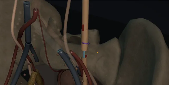
Ureteroureterostomy: how the structures relateColon reflected medially, the ureter on the psoas from kidney to bladder. Lower pole of the kidney: brought down when length is short Upper and mid ureter on the psoas. Upper and mid ureter on the psoas: runs beside it; the vein most easily torn Gonadal vein (danger). Upper and mid ureter on the psoas: ends face each other at 180 degrees Spatulated ends, the anastomosis. Spatulated ends, the anastomosis: stripped or coagulated, the join necroses Adventitia and periureteric fat (danger). Spatulated ends, the anastomosis: the external iliac vein lies beneath Iliac vessels at the brim (danger). Iliac vessels at the brim: the bladder cannot reach above the brim Bladder. Bladder: from renal pelvis through the join to here The 6 French JJ stent.KidneyBladderLower pole of the kidneyUpper and mid ureter onthe psoas!Gonadal veinSpatulated ends, theanastomosis!Adventitia andperiureteric fat!Iliac vessels at the brimBladderThe 6 French JJstentbrought down when length isshortruns beside it; thevein most easilytornends face each other at 180degreesstripped orcoagulated, thejoin necrosesthe external iliac vein liesbeneaththe bladder cannot reach abovethe brimfrom renal pelvisthrough the join tohereTargetDangerLandmarkContext
Colon reflected medially, the ureter on the psoas from kidney to bladder. Schematic, not to scale; relationships only.
  • With the colon reflected medially the ureter lies on the psoas, the gonadal vein beside it and the iliac vessels crossing below, and the strictured segment sits above the pelvic brim where the bladder cannot reach.
  • The two ends are held on stay sutures with their fat still on them, the spatulations facing each other at 180 degrees, and the stent runs from the renal pelvis above through the join to the bladder below.
  • Omentum or fat closes over the anastomosis and the drain lies beside it.
  • ›The upper and mid ureter on the psoas - medial to the gonadal vein above the brim, crossing the iliac vessels below; found by its peristalsis, picked up with a loop.
  • ›The adventitia and periureteric fat - the longitudinal blood supply, from the medial side above the brim; mobilised with the ureter, never stripped from it.
  • ›The gonadal vein - runs beside the ureter above the brim; the landmark to find it and the vessel most easily torn.
  • ›The lower pole of the kidney - mobilised and brought down when length is short, in preference to stripping the ureter.
  • ›The spatulated ends - about a centimetre each, on opposite sides, so the join is an ellipse and not a ring.
  • ›The stent - 6 French JJ through the anastomosis, one end in the pelvis and one in the bladder, out at four to six weeks.
  • ›The bowel and its mesentery - the colon reflected medially to reach the ureter; kept off the anastomosis by fat, peritoneum or omentum.
Click me next Section 3 of 8 Steps The operation in order, with the landmark that guards each move.

Operative steps

  1. Who: the short stricture in the mid or upper ureter A defect of up to two or three centimetres in the upper or mid ureter, above the iliac vessels, where the bladder cannot be brought up to reach: a stricture after ureteroscopy or an impacted stone, a segment injured and stented at another operation that has narrowed at six weeks, a segment excised for a small low-grade tumour in a patient who must keep the kidney, a retrocaval ureter, or the crossing ureter of a duplex system. The kidney has function worth keeping, above about 15 to 20 per cent on the renogram. A defect longer than a few centimetres, a distal stricture, or an irradiated field is a different operation: reimplantation with a hitch or flap, a transureteroureterostomy, an ileal ureter or an autotransplant.
  2. Imaging the length The operation is planned on a length, not a description. A retrograde pyelogram from below and, where there is a nephrostomy, an antegrade study from above, taken together on the same screen, show the upper and lower limits of the stricture; a CT urogram shows the kidney, the course of the ureter and what sits beside it. A MAG3 renogram gives the split function that decides whether the kidney is worth the operation. Any stent is removed a week or two before the studies so that the true calibre is seen, and the studies are done as close to the operation as possible. Two centimetres on the film becomes three on the table once the diseased ends are cut back.
  3. Approach Open through a flank or a Gibson incision for the upper and mid ureter, extraperitoneal, or through a midline when the abdomen is already open; laparoscopic or robotic transperitoneal in the elective case, with the colon reflected medially and the ureter found on the psoas below the lower pole of the kidney, which is now the usual route for a planned stricture. The patient is positioned so that the kidney can be mobilised downward if length is short. A ureteric catheter or stent placed at the start, if it passes, makes the ureter easier to find and the stricture easier to feel. The gonadal vein is the landmark that runs beside the ureter above the brim and is protected or divided early.
  4. Finding and mobilising the ureter The ureter is identified by its peristalsis when it is touched and picked up with a vessel loop, never with forceps on its wall. It is freed above and below the stricture only as far as is needed for the ends to meet without pull, and it is freed with its adventitia and a cuff of periureteric fat intact: the blood supply runs longitudinally in the adventitia, from the medial side above the brim, and a ureter that has been skeletonised looks fine at the end of the operation and necroses at ten days. Diathermy is kept off the wall. Where more length is needed the kidney is mobilised and brought down, or the bladder brought up, rather than the ureter stripped further.
    where it bites: the adventitia is the blood supply; a ureter picked up by its wall with forceps, skeletonised or coagulated on its surface strictures again.
  5. Excision to bleeding ends The strictured or injured segment is excised with scissors, and each end is cut back until the mucosa is pink, the wall is soft and the cut edge bleeds; a white, thickened or fibrotic end is not a partner and is trimmed again. This is where the gap grows, and the surgeon must be willing to change the operation if the healthy ends will no longer meet. The segment is sent for histology, always, because a stricture without a clear cause is a tumour until the pathologist says otherwise. A fine stay suture is placed at the end of each segment, on the adventitia not the mucosa, to hold it for the spatulation.
    where it bites: a stricture with no explanation is urothelial carcinoma until the histology says it is not; the specimen always goes to the pathologist.
  6. Spatulation Each end is spatulated for about a centimetre, on opposite sides, so that the anastomosis becomes an oblique ellipse rather than a ring: the upper end opened on its lateral wall, the lower on its medial, or the other way round, so long as the two cuts sit at 180 degrees to each other. A ring anastomosis contracts to a stricture as it heals; an elliptical one has a circumference nearly twice the calibre of the ureter and tolerates the shrinkage. The spatulation is made with fine scissors in one cut, not nibbled. The apex of each spatulation is where the first suture will go, and it must be in healthy, bleeding tissue.
  7. The anastomosis over a stent The first two sutures are placed at the apex of each spatulation into the corresponding side of the opposite end, 4-0 or 5-0 PDS, Monocryl or Vicryl, mucosa to mucosa, full thickness, knots outside. A 6 French JJ stent is passed through the anastomosis before it is closed, one end in the renal pelvis and the other in the bladder, its position checked with the image intensifier or by the curl seen in the pelvis. The anastomosis is completed with interrupted sutures or two running sutures, one for each wall, bites two millimetres from the edge, tied without tension so that the ureter lies straight and unkinked. It is watertight but not strangled: a suture line that blanches has been pulled too tight.
    where it bites: tension is not cured by tighter sutures; if the ends will not lie together without pull, mobilise the kidney or change the operation.
  8. Omentum or fat around it, and a drain The anastomosis is covered with the tissue that will feed it and keep it away from everything else: retroperitoneal fat closed over it, a tongue of omentum brought down and tacked around it with a few fine sutures, or the peritoneum closed so that the join is extraperitoneal. Where there is a bowel anastomosis or a vascular repair beside it, omentum between them is not optional. A tube or suction drain is placed close to the anastomosis but not on it and brought out through a separate stab. The wound is closed in layers. A urethral catheter stays for a day or two so that the bladder does not fill and reflux up the stent onto the fresh suture line.
  9. After: the stent, the MAG3 The drain comes out when it has been dry for a day, after the catheter is out. The stent stays for four to six weeks and comes out under local anaesthetic with a flexible cystoscope, its removal date recorded in a register. An ultrasound at three months, after the stent has been out for at least six weeks, and a MAG3 renogram show whether the anastomosis drains and what function the kidney has kept; a dilated pelvis with a normal renogram is followed, an obstructed curve is a stricture and is imaged with a retrograde. Surveillance continues with ultrasound at six and twelve months and yearly for a while, because a stricture that returns does so quietly. The histology is chased and acted on.
Click me next Section 4 of 8 Danger zones Where this operation bites, and why it bites there.
Critical relationship

Danger zones

The spatial relationships the animation is built to make automatic. Before you read each one, ask yourself: at this point in the operation, what is about to go wrong, and what is my first move? The save for each sits in the Complications tab.

  • RiskThe segmental blood supply - skeletonised, grasped or coagulated ureter necroses or strictures; the fat stays on and the loop replaces the forceps.
  • RiskThe gonadal and iliac vessels - the gonadal vein beside the ureter and the external iliac vein beneath it at the brim; both are found before the dissection, not after.
  • RiskThe contralateral ureter in a transureteroureterostomy - the healthy ureter is opened and now drains both kidneys; a leak or a stricture there is bilateral obstruction.
  • RiskBowel and the duplex pedicles - the colon and its mesentery on the ureter, and in a duplex system the shared vessels of the two ureters running together in one sheath.
Practise it Take me into the scene Opens the animation at a danger moment and asks you to predict the mechanism and your first move before it reveals.
Click me next Section 5 of 8 Complications The save for each one: the first move, the escalation trigger, the op-note line.

Complications and immediate saves

Trigger each of these live in the Animation tab, then read the save: the first move, the escalation trigger, and the line that goes in the op note.

Urine leak or urinoma

  • urine in the drain beyond the first days, or a collection on the ultrasound: tension, an ischaemic end, a stent that is not across the join, or a bladder that filled and refluxed.
The save
  1. Confirm the catheter is draining and the stent is in position on a plain film; a full bladder refluxes up the stent through the join. first move
  2. Drain fluid creatinine to confirm urine; keep the drain and the catheter.
  3. A CT urogram with delayed phase to find the leak and any collection; a large collection is drained percutaneously.
  4. Sepsis, a rising creatinine or a leak that does not settle with drainage: nephrostomy, and a return to theatre if the anastomosis has separated. escalation trigger
  5. Note: the leak, the drains and stent, the antibiotic and the date of repeat imaging. document

Anastomotic stricture

  • hydronephrosis on the three-month ultrasound or later: ischaemia, a ring anastomosis without adequate spatulation, tension, residual disease or scar.
The save
  1. Ultrasound at three months after the stent is out, and a MAG3 if the pelvis is dilated; a dilated pelvis with a normal curve is watched. first move
  2. A retrograde or antegrade study to measure the stricture and a renogram for function.
  3. A short stricture is balloon dilated or incised endoscopically and stented for six weeks, with the recurrence rate explained.
  4. A long or recurrent stricture or a kidney losing function: redo anastomosis, a Boari flap if the bladder reaches, or an ileal ureter. escalation trigger
  5. Note: the length, the split function, the treatment and the surveillance interval. document

Devascularisation and a longer defect than planned

  • the ends cut back further than expected, the ureter skeletonised in a difficult dissection, or the segment that looked healthy on the film is not: the gap will not close.
The save
  1. Stop before tying under tension; measure the gap and mobilise the kidney downward or the bladder upward first. first move
  2. A dusky end is trimmed again until it bleeds; a join to a dusky end is a leak in a week.
  3. Mid-ureteric gap beyond reach: a Boari flap with a psoas hitch reaches most of the mid ureter.
  4. A gap beyond a flap: transureteroureterostomy, ileal ureter, autotransplant or, in a poor kidney, nephrectomy, with senior help and the plan changed rather than forced. escalation trigger
  5. Op note: the length excised, the length of healthy ureter, what was mobilised and why the operation changed. document

Bleeding from the gonadal or iliac vessels

  • the gonadal vein torn during mobilisation, or the external iliac vein beneath the ureter at the brim opened in a scarred field.
The save
  1. Pressure with a swab, suction and light, and a call for help before any clamp or clip goes on blind. first move
  2. The gonadal vein is ligated or clipped above and below the tear; it is expendable.
  3. An iliac vein tear is repaired with fine Prolene under proximal and distal control with a vascular surgeon. escalation trigger
  4. Check the ureter after the bleeding is controlled; the anastomosis is not made in a pool of blood.
  5. Op note: the vessel, the repair, the blood loss and who helped. document

Stent problems and infection

  • stent symptoms, a migrated stent, encrustation, a urinary infection above a fresh join, and the stent that is forgotten because no one wrote the date down.
The save
  1. A plain film on the first day to confirm the stent position, and the removal date written in the stent register before the patient leaves. first move
  2. Stent symptoms are treated with an alpha blocker and an anticholinergic; a migrated stent is repositioned or replaced under screening.
  3. Fever with a stent in: culture, antibiotics and an ultrasound; an obstructed infected system is drained.
  4. Sepsis above the anastomosis that does not settle: nephrostomy, and the stent changed rather than left. escalation trigger
  5. Note: the stent size, the date in, the date due out, and the culture result. document
Click me next Section 6 of 8 Troubleshooting When the case turns: the problem, then the next move.

Intra-operative troubleshooting

When the case turns: the problem, then the next move.

  • ActionThe ureter cannot be found - find it at the brim where it crosses the iliac vessels, or on the psoas below the lower pole, and follow it; a ureteric catheter placed at the start makes it palpable.
  • ActionThe gap is longer than the film suggested - expected; cut back to bleeding ends anyway, then mobilise the kidney downward or the bladder upward before deciding on a flap or a substitution.
  • ActionThe end is dusky after mobilisation - trim it until it bleeds; if the whole segment is dusky the ureter has been stripped and the repair is not made today.
  • ActionThe stent will not pass to the bladder - a guidewire first, the stent over it, screening for the curl; if the distal ureter is blocked it needs its own study before the join is made.
  • ActionThe anastomosis leaks on inspection - one interrupted suture at the point, not a purse-string; if the tissue tears the end is ischaemic and is cut back again.
  • ActionHydronephrosis on the first ultrasound with the stent out - MAG3 before anything; oedema settles by three months, an obstructed curve is a stricture and gets a retrograde.
Click me next Section 7 of 8 FRCS viva Say it out loud, then mark yourself against the model answer.

FRCS viva questions

The examiner stems this scene prepares you for. Try to answer out loud first, then reveal, and learn the discriminator, the detail that turns a pass into a strong pass.

QWhen is a ureteroureterostomy the right operation for a ureteric defect, and when is it not?reveal answer
  • I would consider ureteroureterostomy for a short defect, up to about two or three centimetres, in the upper or mid ureter above the iliac vessels, where the bladder cannot be brought up to reach, in a kidney with function worth preserving, above about 15 to 20 per cent on the renogram.
  • The typical causes are a stricture after ureteroscopy or an impacted stone, an iatrogenic injury recognised at the time or stented and found strictured later, a segment excised for a small low-grade urothelial tumour in a patient who must keep the kidney, a retrocaval ureter and the duplex system.
  • It is not the right operation for a distal defect, where reimplantation with a psoas hitch or Boari flap is more reliable than a low anastomosis to a stump of doubtful blood supply, nor for a defect longer than a few centimetres after the ends have been cut back to healthy tissue, nor in a heavily irradiated field.
  • For those I would consider transureteroureterostomy, an ileal ureter, autotransplantation or nephrectomy.
  • I would measure the defect on combined antegrade and retrograde studies close to the operation, confirm function with a MAG3, and consent for leak, stricture, a change of operation and a stent.
Strong pass

Say both

  • The length threshold with the reason it grows on the table
  • The distal ureter excluded in favour of reimplantation
Examiner note
The length threshold with the reason it grows on the table, and the distal ureter excluded in favour of reimplantation.
QDescribe the technique of ureteroureterostomy.reveal answer
  • Through a flank or Gibson incision, or laparoscopically with the colon reflected, I would find the ureter on the psoas by its peristalsis and pick it up with a vessel loop, never with forceps on the wall.
  • I would mobilise it above and below the stricture only as far as needed for the ends to meet without tension, keeping the adventitia and periureteric fat intact because the blood supply runs longitudinally in the adventitia, and keeping diathermy off the wall.
  • I would excise the diseased segment and trim each end back until it bleeds, sending the segment for histology.
  • I would spatulate each end for about a centimetre on opposite sides so that the anastomosis is an oblique ellipse rather than a ring, place the first sutures at the apices, and pass a 6 French JJ stent across the join with its position confirmed.
  • I would complete the anastomosis with interrupted or two running 4-0 or 5-0 absorbable sutures, mucosa to mucosa, full thickness, watertight but not tight.
  • I would cover it with retroperitoneal fat, peritoneum or omentum, place a drain beside it, and leave a urethral catheter for a day or two so that the bladder does not fill and reflux up the stent.
  • The stent comes out at four to six weeks.
Strong pass

The loop not the forceps, the fat left on, the histology sent, and the spatulation on opposite sides with the ring-versus-ellipse reason.

QWhy do ureteric anastomoses fail, and how do you avoid it?reveal answer
  • They fail for three reasons, and each is a technical decision.
  • Ischaemia: the ureter has a longitudinal adventitial blood supply from the medial side above the brim and the lateral side below, so a ureter that is skeletonised, grasped with forceps, coagulated on its surface or joined at a dusky end necroses and leaks at about ten days or strictures at three months;
  • I avoid it by mobilising with the fat on, using a loop, keeping the diathermy off the wall and trimming until the end bleeds.
  • Tension: a join under pull ischaemes its own edges and tears;
  • I avoid it by mobilising only what is needed, bringing the kidney down or the bladder up, and by changing the operation, to a flap or a substitution, rather than tying tighter.
  • Technique: a circular anastomosis contracts as it heals, so I spatulate for a centimetre on opposite sides to make an ellipse with nearly twice the circumference, use fine absorbable suture mucosa to mucosa without strangling the edge, stent it, drain it, and cover it with fat or omentum so that a small leak is absorbed rather than becoming a fistula or a collection.
  • Residual disease is the fourth cause, which is why the specimen goes to histology and the ends are healthy.
Strong pass

Say both

  • The blood supply described by side and level
  • The ring-to-ellipse arithmetic given as the reason for spatulation
Examiner note
The blood supply described by side and level, and the ring-to-ellipse arithmetic given as the reason for spatulation.
QHow do you follow up a ureteroureterostomy, and what would make you intervene?reveal answer
  • I would keep the drain until it has been dry for a day after the urethral catheter is removed, and the stent for four to six weeks, with its removal date entered in the stent register before discharge.
  • I would confirm the stent position on a plain film and treat stent symptoms with an alpha blocker and an anticholinergic.
  • At three months, with the stent out for at least six weeks, I would arrange an ultrasound and a MAG3 renogram: a dilated pelvis with a normal drainage curve is oedema or a capacious system and is watched;
  • an obstructed curve or a fall in split function is a stricture, and I would define it with a retrograde or antegrade study.
  • I would continue ultrasound at six and twelve months and then yearly for a period, because recurrent strictures are silent.
  • I would intervene for a leak that persists with drainage in place, for sepsis above the join, for a symptomatic or migrated stent, and for any obstruction: a short recurrent stricture is dilated or incised endoscopically and stented, with the recurrence rate explained, and a long or recurrent one, or a kidney losing function, is revised with a redo anastomosis, a Boari flap or an ileal ureter.
  • I would chase the histology and act on it.
Strong pass

Say both

  • The renogram interpreted as curve and split function rather than as dilatation alone
  • The stent register as part of the operation
Examiner note
The renogram interpreted as curve and split function rather than as dilatation alone, and the stent register as part of the operation.
Click me next Section 8 of 8 Evidence The guidance each of these answers is built on.

Evidence and guidance

Two minutes

Close the lap: recall it before you leave.

Say it aloud, then reveal. Reading is not remembering.

  1. 01 The relationship Say the critical spatial relationship out loud, then reveal. reveal
    Memory frame

    With the colon reflected medially the ureter lies on the psoas, the gonadal vein beside it and the iliac vessels crossing below, and the strictured segment sits above the pelvic brim where the bladder cannot reach.

    Why?
    The two ends are held on stay sutures with their fat still on them, the spatulations facing each other at 180 degrees, and the stent runs from the renal pelvis above through the join to the bladder below. Omentum or fat closes over the anastomosis and the drain lies beside it.
  2. 02 The first move Urine leak or urinoma. Your first move? reveal
    First move

    Confirm the catheter is draining and the stent is in position on a plain film; a full bladder refluxes up the stent through the join.

  3. 03 The escalation trigger Urine leak or urinoma. What makes you call for help? reveal
    Escalate when

    Sepsis, a rising creatinine or a leak that does not settle with drainage: nephrostomy, and a return to theatre if the anastomosis has separated.

  4. 04 The discriminator What turns a pass into a strong pass? reveal
    Strong pass

    Say both

    • The length threshold with the reason it grows on the table
    • The distal ureter excluded in favour of reimplantation
    Examiner note
    The length threshold with the reason it grows on the table, and the distal ureter excluded in favour of reimplantation.
  5. 05 The pitfall Where does this operation bite first? reveal
    Pitfall

    The segmental blood supply

    Why?
    skeletonised, grasped or coagulated ureter necroses or strictures; the fat stays on and the loop replaces the forceps.

Revision practice with self-marked answers, not an assessment of clinical competence. Kept in this browser only; it does not sync between devices.