
Revision sheet
Perineal urethrostomy: inverted-U flap
8 steps, each one the authored title and caption from the interactive animation, with the frame captured from that step. Read it before the case, or print it and work through the steps in order.
Teaching only, not for diagnosis or treatment decisions. The text on this sheet is the animation's own step copy, reproduced verbatim.
UrOops3D · UroRef · © Nity G
- 01
Mark the flap
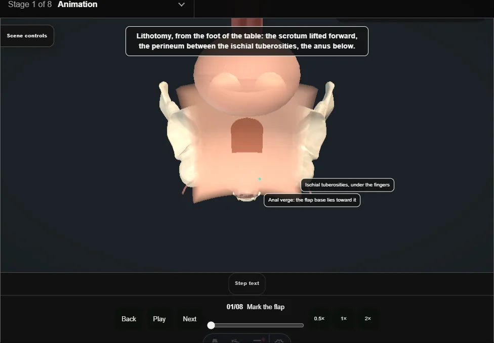
- Exaggerated lithotomy, the scrotum lifted forward.
- Two fingers on the ischial tuberosities; the marker draws an inverted U between them, apex toward the scrotum and its wide base toward the anus.
- The base is the blood supply.
- 02
Raise the flap
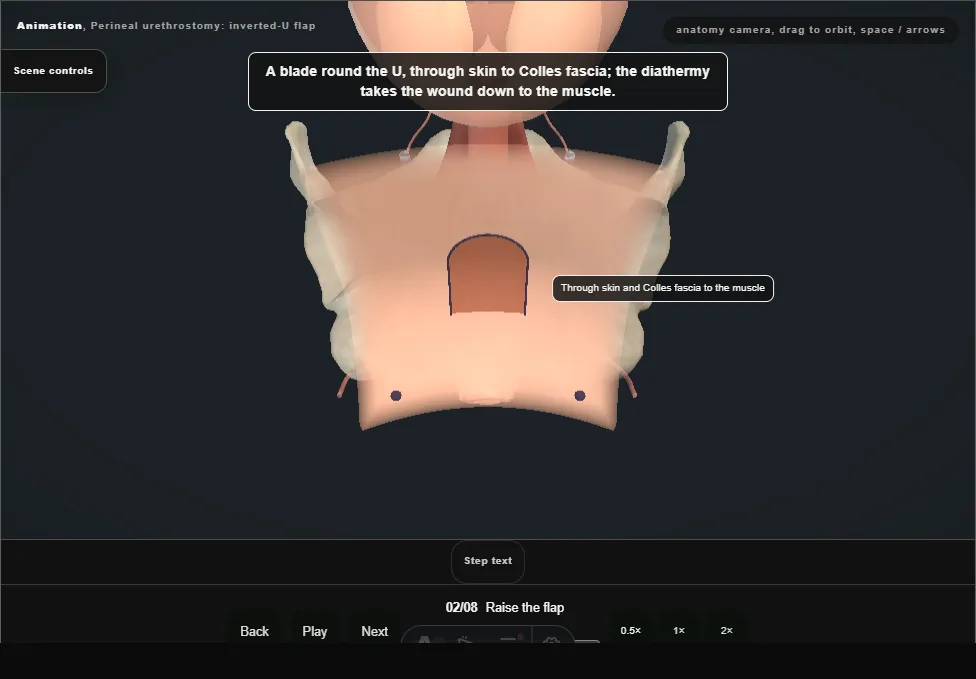
- A blade round the U through skin to Colles fascia, the diathermy down to bulbospongiosus.
- The flap lifts full thickness on its hinge, the fat kept on its underside; a thin tip is the part that dies.
- 03
Expose the bulb
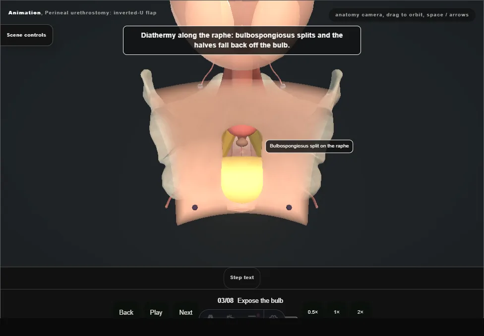
- Diathermy along the raphe: bulbospongiosus splits and the halves are dissected off the bulb without breaching the tunica.
- A self-retaining ring on the wound edges.
- The bare bulb lies across the field.
- 04
Ventral urethrotomy
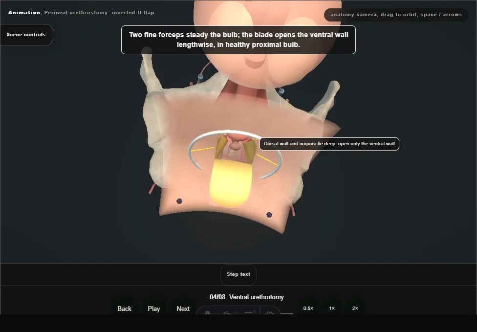
- Two fine forceps steady the bulb and the blade opens the ventral wall lengthwise in healthy proximal bulb, between them.
- Stay sutures lift both edges.
- Three layers in the cut face: mucosa within, spongy tissue, tunica without.
- The dorsal wall and the corpora lie deep and are not entered.
- 05
Calibrate and inspect
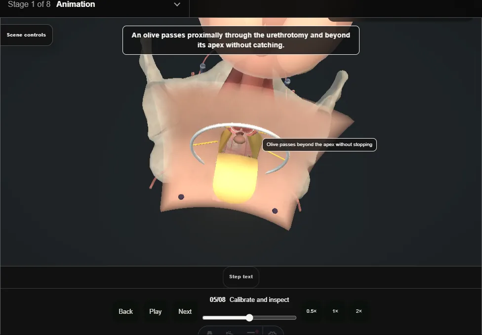
- An olive passes proximally through the urethrotomy and beyond its apex without catching; a flexible scope follows to the bladder to confirm a healthy proximal urethra, a normal bladder neck and bladder.
- A missed proximal narrowing fails early.
- 06
Apex inset
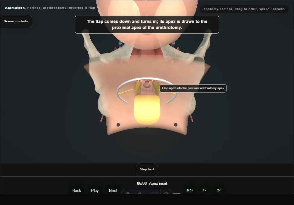
- The flap comes down and turns in.
- One stitch through the flap apex, then the proximal urethral apex mucosa and tunica; the knot draws the tip into the apex without tension.
- The flap tip in the proximal apex breaks the circular scar that would otherwise ring the outlet.
- 07
Mucocutaneous stitches
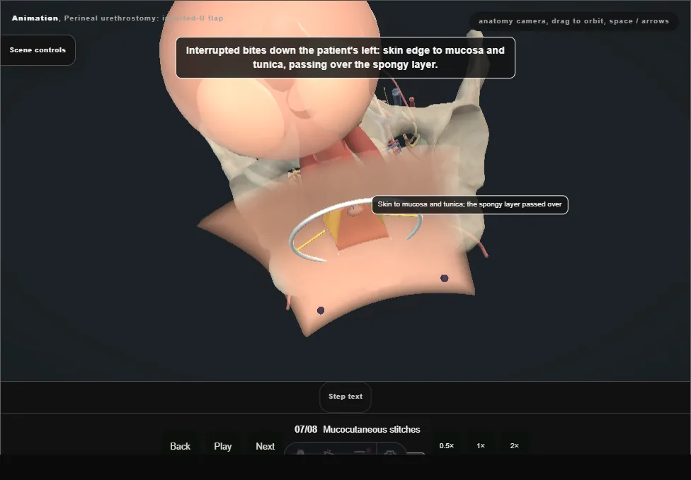
- Interrupted bites down each side: skin edge to mucosa and tunica, passing over the spongy layer, so no spongy tissue is in a bite to bleed or scar.
- The lateral edges of the flap are closed to the skin in two layers.
- Mucosa everted to skin all round.
- 08
Patent outlet
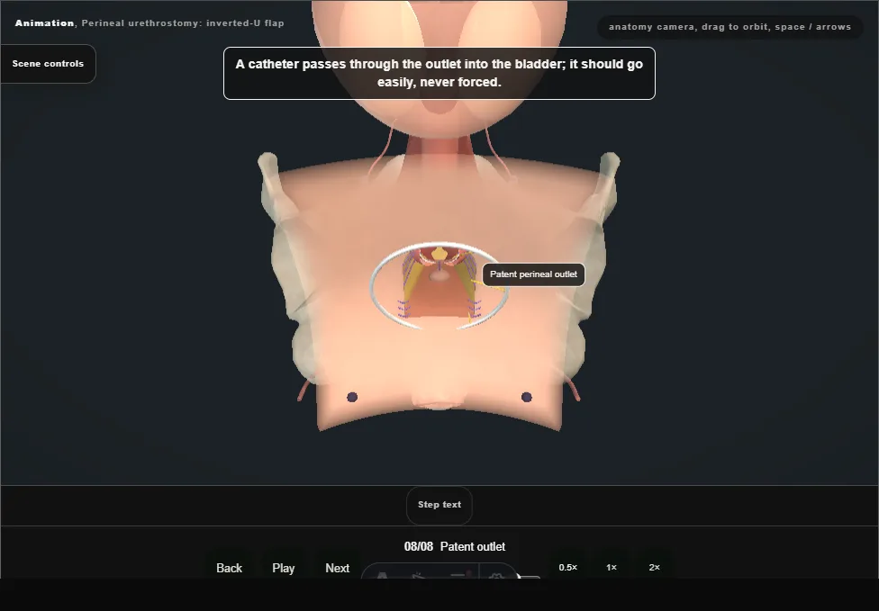
- A catheter passes through the outlet into the bladder; it should go easily and is never forced.
- Haemostasis checked and the flap confirmed viable before the dressing.
- Head-on, the outlet is an open ellipse ringed by everted mucosa.
Derived from PerinealUrethrostomyScene.tsx at build time: every step title and caption above is the authored
ProcedureDef text, reproduced verbatim, and this page shares its readable-points renderer with the animation
itself. 8 of 8 steps have a captured frame.
Text fingerprint 697cb1b7cd162748. A caption edit without a re-capture fails the production
check, so the sheet and the animation cannot drift apart silently.