
Revision sheet
Radical cystectomy + PLND (extirpative phase)
7 steps, each one the authored title and caption from the interactive animation, with the frame captured from that step. Read it before the case, or print it and work through the steps in order.
Teaching only, not for diagnosis or treatment decisions. The text on this sheet is the animation's own step copy, reproduced verbatim.
UrOops3D · UroRef · © Nity G
- 01
Read the pelvis
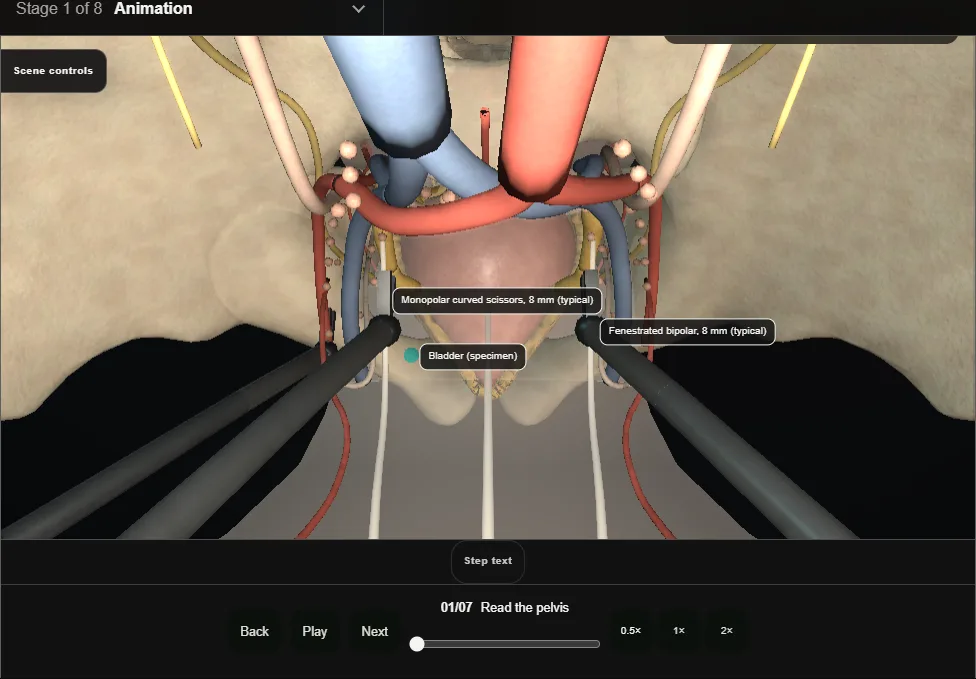
- Console view through the camera port, male pelvis; the arms are 8 mm instruments, the assistant works through a 12 mm port (typical sizes).
- Peritoneum over the dome, the medial umbilical ligaments converging on the urachus, both ureters crossing the common iliac bifurcation under the peritoneum of the sidewall.
- Rectum behind, dorsal venous plexus below.
- 02
Ureteric dissection + division
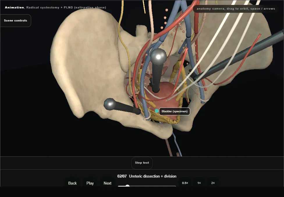
- The peritoneum is opened over the iliac vessels and each ureter is followed down, under the vas, to within 2-3 cm of the bladder.
- Clips proximal and distal, divided between them: the distal stump travels with the specimen, the proximal end waits for the diversion.
- 03
Posterior plane
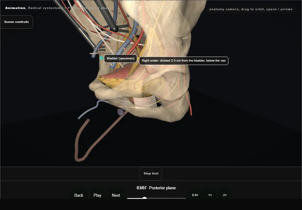
- The rectovesical pouch is incised and the plane is dropped behind the vasa and seminal vesicles onto Denonvilliers' fascia, then swept to the apex with the rectum in view.
- Avascular when the layer is right; fat or blood means the wrong layer.
- 04
Vesical pedicles
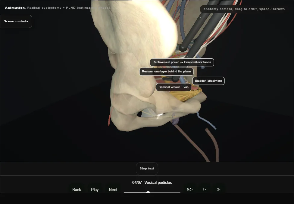
- From the divided ureter down toward the apex: the superior vesical pedicle (umbilical artery origin) first, then the inferior vesical pedicle to the base and vesicle.
- Sealed or clipped on the bladder side of the anterior division; the obturator nerve lies in the same lateral gutter.
- 05
Apex + dorsal venous plexus
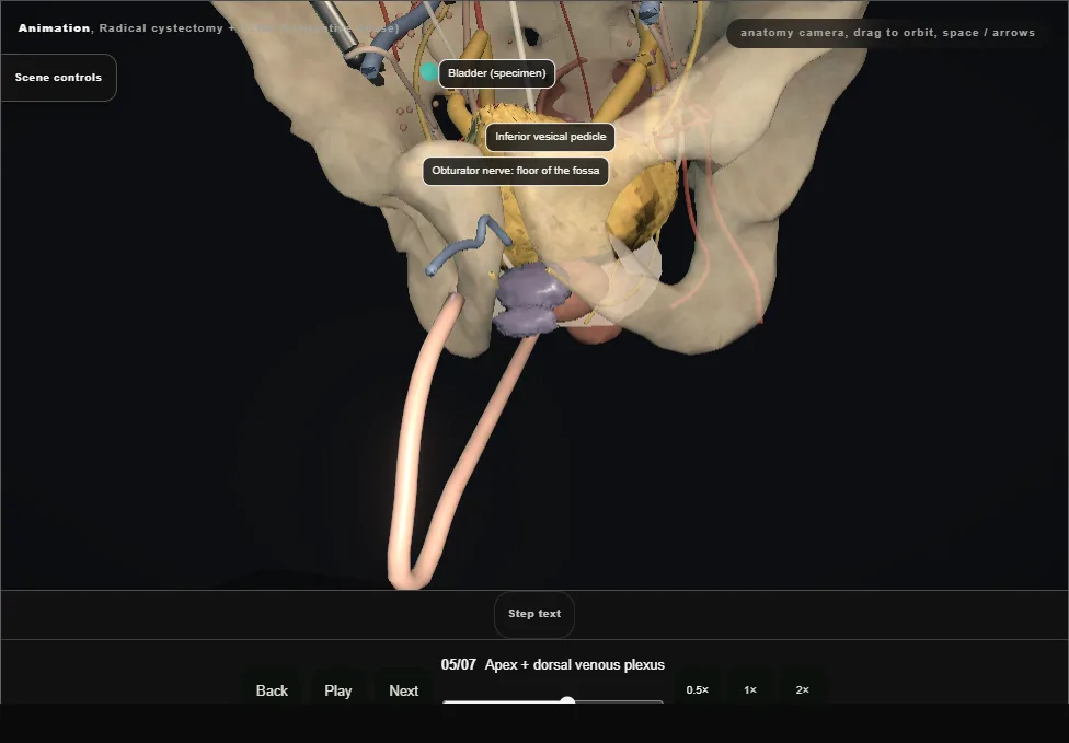
- The bladder is dropped first: the urachus and medial umbilical ligaments taken down into the space of Retzius.
- Then the endopelvic fascia incised beside the prostate and the dorsal venous plexus secured with a figure-of-eight before the urethra is divided at the apex.
- The last attachment comes away without a venous tear.
- 06
Specimen out
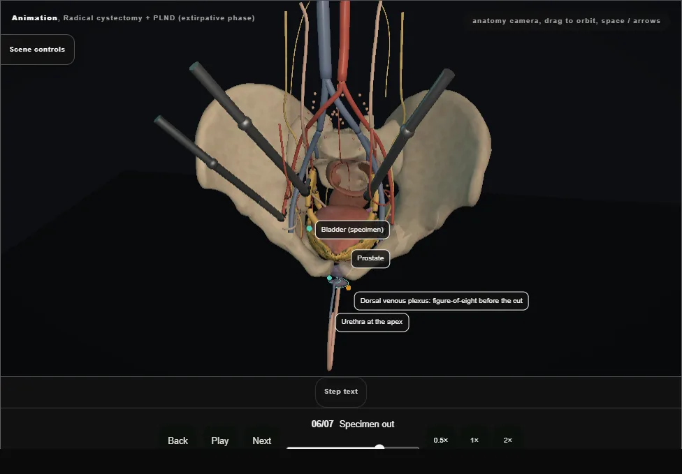
- Bladder, prostate, seminal vesicles, distal ureters and the perivesical fat leave as one block.
- What remains is the template: skeletonised iliac vessels, the obturator nerve on the floor of the fossa and the node packets still in place.
- 07
Pelvic lymphadenectomy
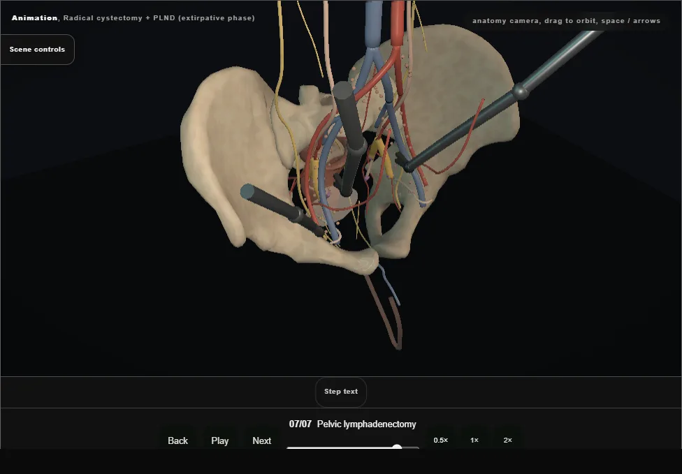
- Standard template: external iliac packet from the circumflex iliac vein (node of Cloquet) to the bifurcation, then the obturator fossa between the external iliac vein and the obturator nerve, then the internal iliac nodes.
- Extended templates continue to the aortic bifurcation.
- Each packet is lifted off the vessels and handed out through the assistant port.
- The diversion follows in its own modules: ileal conduit, neobladder or continent diversion.
Derived from cystectomy/RadicalCystectomy3dScene.tsx at build time: every step title and caption above is the authored
ProcedureDef text, reproduced verbatim, and this page shares its readable-points renderer with the animation
itself. 7 of 7 steps have a captured frame.
Text fingerprint 701ddfd59ab88261. A caption edit without a re-capture fails the production
check, so the sheet and the animation cannot drift apart silently.