
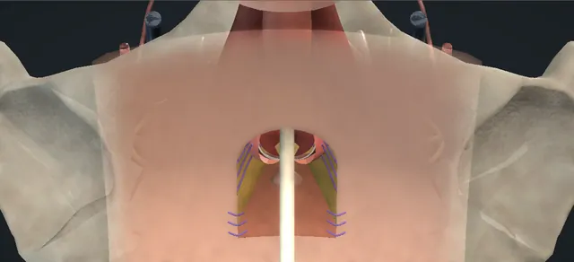
UrOops3D, danger-zone atlas

Schematic model.

Operative animation, Perineal urethrostomy

Perineal urethrostomy: the inverted-U flap outlet

The definitive perineal outlet for the man whose anterior urethra cannot be reconstructed, or who does not want it reconstructed.

A full-thickness inverted-U flap is raised between the ischial tuberosities, the ventral wall is opened in healthy proximal bulb, the flap apex is set into the proximal apex, and mucosa is sutured to skin with bites that leave the spongy tissue alone.

The lesson is the blood supply of the flap, where the urethra is opened, what the needle picks up, and why the flap tip goes where it goes.

Advanced tier FRCS reconstruction, urethral stricture, andrology Interactive 3D

Operative visualisation

Schematic scene preview: Perineal urethrostomy: inverted-U flap
Preparing the 3D anatomy…

UrOops3D · UroRef · © Nity G

How to use this module. Play through the eight steps, or use Back and Next. Watch the flap lift on its hinge, the bulb bared, the three layers of the cut face, the flap tip drawn into the apex, and the bites that pass over the spongy layer. The triggers show a narrowed outlet, an oozing cut face and a dusky flap tip.

Click me next Section 2 of 8 Anatomy The structures this operation works between, and the one it must not cross.

The structures in the scene

Perineal urethrostomy: how the structures relateThe perineum from the foot of the table, scrotum lifted, down onto the bulb. Inverted-U flap, base toward the anus: drawn between them Ischial tuberosities either side. Inverted-U flap, base toward the anus: lies under the flap; parted to the bulb Bulbospongiosus, split on the raphe. Inverted-U flap, base toward the anus: the apex goes into its proximal end Ventral bulbar wall: the urethrotomy. Inverted-U flap, base toward the anus: bites take the outer and inner, not the middle Cut face: mucosa, spongy tissue, tunica (danger). Inverted-U flap, base toward the anus: deep to the opening; never entered Dorsal wall and corpora, deep (danger). Inverted-U flap, base toward the anus: calibrated and seen before any stitch Proximal urethra and bladder neck.Inverted-U flap, basetoward the anusIschial tuberositieseither sideBulbospongiosus,split on the rapheVentral bulbar wall:the urethrotomy!Cut face: mucosa,spongy tissue,tunica!Dorsal wall andcorpora, deepProximal urethra andbladder neckdrawn between themlies under theflap; parted to thebulbthe apex goes intoits proximal endbites take theouter and inner,not the middledeep to theopening; neverenteredcalibrated and seenbefore any stitchTargetDangerLandmarkContext
The perineum from the foot of the table, scrotum lifted, down onto the bulb. Schematic, not to scale; relationships only.
  • Seen from the foot of the table with the scrotum lifted, the bulbar urethra runs under the perineal skin between the tuberosities, the anus below.
  • An inverted U drawn between the tuberosities with its base toward the anus is a flap that keeps its blood supply.
  • Bulbospongiosus split on the raphe bares the bulb; the ventral wall is opened lengthwise in healthy proximal urethra, the dorsal wall and corpora left deep.
  • The flap turns in so its tip sits in the proximal apex of the opening, and the mucosa is sutured to skin round the rest: an outlet whose floor is skin and whose roof is urethra.
  • ›Ischial tuberosities - the bony landmarks the fingers find; the inverted U is drawn between them.
  • ›Perineal skin flap - full thickness, fat on its underside, a wide base toward the anus; its apex goes into the proximal urethrotomy apex.
  • ›Colles fascia and perineal fat - the layer between skin and bulbospongiosus; the diathermy goes through it to the muscle.
  • ›Bulbospongiosus - split along its raphe and dissected off the bulb; the halves fall laterally.
  • ›Corpus spongiosum and the bulb - the tunica is thin on the bulb; the ventral wall is what is opened, in healthy proximal urethra.
  • ›The cut face: mucosa, spongy tissue, tunica - three layers; the stitches take the outer and the inner and pass over the middle.
  • ›Dorsal urethral wall and corpora cavernosa - deep to the opened urethra; not entered.
  • ›Proximal urethra and bladder neck - calibrated with the olive and seen with the scope before the stitches; must be healthy.
  • ›Anal verge - below the field; the flap base lies toward it and the wound stays clear of it.
Click me next Section 3 of 8 Steps The operation in order, with the landmark that guards each move.

Operative steps

  1. Mark the flap Exaggerated lithotomy on padded stirrups, a bolster under the buttocks, the scrotum lifted forward. The man has complex anterior stricture disease after failed urethroplasty, or is not fit or not willing to have another reconstruction, and cystoscopy and imaging have shown a healthy proximal bulbar urethra. Two fingers find the ischial tuberosities. The marker draws an inverted U between them: the apex toward the scrotum, the limbs running back, the wide base toward the anus. The base is the flap's blood supply.
    where it bites: a narrow base or a long thin tip is a flap that dies; the width of the base is decided before the knife.
  2. Raise the flap A blade follows the U through skin and Colles fascia; the diathermy takes the wound down to bulbospongiosus. The flap is lifted full thickness on its hinge with the fat kept on its underside, and reflected out of the field. The fat is not trimmed off the flap to make it lie flatter.
    where it bites: a flap thinned to make it supple is a flap with no circulation at its tip.
  3. Expose the bulb Diathermy along the raphe divides bulbospongiosus in the midline; the halves are dissected off the corpus spongiosum sharply, taking care not to perforate the tunica albuginea. A self-retaining ring retractor is seated on the wound edges. The bare bulb now lies across the field with the muscle halves lateral.
    where it bites: the tunica of the spongiosum is thin on the bulb; a breach here bleeds into the field before the urethra is even opened.
  4. Ventral urethrotomy Two fine forceps hold the bulb either side of the midline and the blade opens the ventral wall lengthwise between them, in healthy proximal bulbar urethra. Stay sutures on the lateral edges lift them. The cut face shows three layers: the mucosa within, the spongy tissue, and the tunica without. The dorsal wall and the corpora lie deep and are not entered; the distal urethra is left where it is.
    where it bites: open healthy urethra only; a urethrotomy carried into scarred bulb makes a scarred outlet.
  5. Calibrate and inspect A bougie with an olive tip is passed proximally through the urethrotomy and beyond its apex; it should pass without catching. A flexible cystoscope follows through the urethrotomy to the bladder to confirm a healthy proximal urethra, a normal bladder neck and no bladder pathology. This is done before any stitch is placed.
    where it bites: a proximal narrowing that is not found now is an outlet that fails early.
  6. Apex inset The flap is brought down and turned in. One stitch passes through the apex of the flap, then through the mucosa and tunica at the proximal apex of the urethrotomy, and the knot draws the tip into the apex without tension. The flap now forms the posterior floor of the new outlet, and its tip in the proximal apex breaks the ring of scar that would otherwise form round the opening.
    where it bites: tension at the apex narrows the outlet; the flap must reach comfortably, which is decided at the marking.
  7. Mucocutaneous stitches Interrupted absorbable sutures approximate the perineal skin to the incised ventral urethral edges on both sides. Each bite takes skin, then the urethral mucosa and the tunica albuginea of the corpus spongiosum, and passes over the spongy tissue so that no spongy tissue is in any bite. The lateral edges of the flap are then closed to the skin in two layers. Haemostasis with the diathermy; the flap is checked and seen to be viable.
    where it bites: spongy tissue in a bite bleeds now and scars later; the needle picks up mucosa and tunica, nothing between.
  8. Patent outlet A urethral catheter is advanced through the urethrostomy into the bladder; it goes easily and is never forced. A dressing, gauze fluffs and a supporter to limit swelling. Head-on, the finished outlet is an open ellipse ringed by everted mucosa with the flap below it. The operation note records the flap, the urethrotomy, the calibration and cystoscopy findings, the outlet and the catheter.
    where it bites: an outlet that will not take the catheter easily at the end is an outlet that is already too small.
Click me next Section 4 of 8 Danger zones Where this operation bites, and why it bites there.
Critical relationship

Danger zones

The spatial relationships the animation is built to make automatic. Before you read each one, ask yourself: at this point in the operation, what is about to go wrong, and what is my first move? The save for each sits in the Complications tab.

  • RiskThe flap base - the blood supply; a narrow base or a thinned tip is necrosis in the first days.
  • RiskThe ventral wall, not the dorsal - the urethrotomy stays ventral in healthy proximal bulb; the dorsal wall and the corpora lie deep and are not entered.
  • RiskThe spongy layer in a bite - bleeding now, scar later; the bite takes mucosa and tunica and passes over it.
  • RiskThe proximal urethra - calibrated and seen before the stitches; a missed proximal narrowing fails early.
  • RiskTension at the apex - the flap tip must reach the proximal apex comfortably; tension narrows the outlet.
Practise it Take me into the scene Opens the animation at a danger moment and asks you to predict the mechanism and your first move before it reveals.
Click me next Section 5 of 8 Complications The save for each one: the first move, the escalation trigger, the op-note line.

Complications and immediate saves

Trigger each of these live in the Animation tab, then read the save: the first move, the escalation trigger, and the line that goes in the op note.

Outlet stenosis

  • the urethrostomy narrows over months: an ischaemic flap tip, tension at the apex, spongy tissue in the bites, or the disease recurring in the proximal urethra.
The save
  1. Calibrate gently to confirm the narrowing and to see where it is: the skin ring, or the urethra above it. first move
  2. A flexible scope through the outlet separates a stenosed skin ring from recurrent urethral disease; the two are revised differently.
  3. Revision of a failed perineal urethrostomy: V-Y plasty of the ring, augmentation, or a redo, in a reconstructive unit. escalation trigger
  4. The patient is told what has narrowed and why; a self-dilatation holding pattern is a bridge, not the answer.
  5. Op note and clinic letter: the outlet calibre, where the narrowing lies, what the scope showed, and the revision plan. document

Spongiosal bleeding

  • ooze from the cut spongy layer at the urethrotomy edges, between or beneath the stitches.
The save
  1. Pressure on the edge for a timed interval before anything else. first move
  2. Bites that take the tunica albuginea with the mucosa and close the spongy layer under them; a bite into the spongy tissue itself bleeds more.
  3. Bleeding that continues after the stitch line is complete, or a field that is being lost, needs a senior reconstructive surgeon in the room now. escalation trigger
  4. A catheter across the outlet and a pressure dressing once the line is dry; no blind diathermy into the spongy tissue.
  5. Op note: where it bled, what stopped it, the estimated loss and the dressing plan. document

Flap tip necrosis

  • the tip of the flap darkens in the first days: a base too narrow, a flap thinned of its fat, or a tip set in under tension.
The save
  1. Inspect the flap and dress it; a dusky tip is watched daily, not cut back at once. first move
  2. Keep the catheter in so the outlet is splinted while the tip declares itself.
  3. A dead tip is debrided when it has demarcated and the outlet is revised; this is a reconstructive decision, not a ward one. escalation trigger
  4. The lesson for the next flap: base width and the fat on the flap keep it alive, not the tightness of the sutures.
  5. Op note at the time: flap viability confirmed at the end of the operation, the base width and thickness kept; then each review. document
Click me next Section 6 of 8 Troubleshooting When the case turns: the problem, then the next move.

Intra-operative troubleshooting

When the case turns: the problem, then the next move.

  • ActionThe flap will not reach the proximal apex without tension - shorten the urethrotomy at its proximal end rather than pull the flap; a flap under tension at its tip narrows the outlet and dies. If the plan needs a more proximal apex, this is the augmented technique with an oral graft, not a stretched flap.
  • ActionThe olive catches at the proximal apex - the urethrotomy is not in healthy urethra or the disease extends proximally; extend the urethrotomy proximally into healthy urethra, or convert to the augmented Gil-Vernet-Blandy with a graft onto the dorsal wall. Do not place stitches round a narrowing.
  • ActionThe bulb is deep and the field is poor in an obese man - extend the inverted U cephalad in the midline for exposure; the 7-flap is the technique described for obesity.
  • ActionThe ventral wall is scarred where the urethrotomy was planned - move proximally into healthy bulb; if the dorsal wall is extensively scarred, it is replaced with a buccal graft as an onlay.
  • ActionThe man is not sure he wants to give up the distal urethra - a staged urethroplasty keeps the decision open; the guideline names it for the man who is unsure. Loop urethrostomy is the temporary alternative.
Click me next Section 7 of 8 FRCS viva Say it out loud, then mark yourself against the model answer.

FRCS viva questions

The examiner stems this scene prepares you for. Try to answer out loud first, then reveal, and learn the discriminator, the detail that turns a pass into a strong pass.

QDescribe the inverted-U perineal urethrostomy and where it sits in the management of anterior urethral stricture.reveal answer
  • A perineal urethrostomy is a definitive outlet in the perineum, the distal urethra left in place but bypassed.
  • The inverted-U technique raises a full-thickness flap of perineal skin between the ischial tuberosities, based toward the anus so its blood supply is wide, exposes the bulb through a midline split of bulbospongiosus, opens the ventral wall of the healthy proximal bulbar urethra lengthwise, sets the apex of the flap into the proximal apex of that urethrotomy and sutures the urethral mucosa to the perineal skin on both sides, taking mucosa and tunica and passing over the spongy tissue.
  • The European guideline offers it for complex anterior stricture disease, strongly, and to men who are unfit for or do not want reconstruction, weakly;
  • the choice of technique rests on the surgeon's experience and the patient.
  • The alternatives I would name are the Johanson, the augmented Gil-Vernet-Blandy with an oral graft when the disease extends proximally, the 7-flap in obesity, a loop urethrostomy as a temporary outlet and a staged urethroplasty when the man is undecided.
Strong pass

Say both

  • Naming the flap base as the blood supply and the apex inset into the proximal apex
  • Placing the operation against the guideline's indications rather than as a salvage afterthought
Examiner note
Naming the flap base as the blood supply and the apex inset into the proximal apex, and placing the operation against the guideline's indications rather than as a salvage afterthought.
QTalk me through the operation step by step.reveal answer
  • Exaggerated lithotomy, padded, a bolster under the buttocks, antibiotics, the scrotum lifted.
  • I palpate both ischial tuberosities and mark an inverted U between them, apex toward the scrotum, wide base toward the anus.
  • I incise the U through skin and Colles fascia and take the wound down to bulbospongiosus with the diathermy, raising the flap full thickness with its fat and reflecting it.
  • I divide bulbospongiosus in the midline and dissect the halves off the spongiosum without breaching the tunica, then seat a self-retaining retractor.
  • With two fine forceps on the bulb I open the ventral wall lengthwise in healthy proximal urethra and place stay sutures on the edges.
  • I calibrate proximally with a bougie and pass a flexible cystoscope through the urethrotomy to confirm a healthy proximal urethra and a normal bladder.
  • Then I bring the flap in and suture its apex to the proximal urethral apex, mucosa and tunica, and run interrupted absorbable sutures from skin to the urethral edges on each side, each bite taking mucosa and tunica and passing over the spongy tissue.
  • The lateral edges of the flap are closed in two layers, haemostasis is secured, the flap is confirmed viable, and a catheter goes through the urethrostomy into the bladder easily.
  • Dressing, fluffs and a supporter.
Strong pass

Say both

  • Calibration and cystoscopy before any stitch
  • The bite described as mucosa plus tunica with the spongy tissue passed over
Examiner note
Calibration and cystoscopy before any stitch, and the bite described as mucosa plus tunica with the spongy tissue passed over.
QWhy does the flap apex go into the proximal end of the urethrotomy?reveal answer
  • Because a circular suture line between skin and urethra contracts into a ring.
  • Setting a tongue of skin into the proximal apex of the opening breaks that circle: the posterior wall of the outlet is skin flap rather than a seam, so the scar cannot close as a ring round the opening.
  • It also moves the deepest part of the outlet, where the lumen begins, onto well-vascularised full-thickness skin, which is why the flap must be based widely and kept thick.
  • The same principle is behind the V-Y plasty used to revise a stenosed urethrostomy: interrupt the ring with a flap of skin.
  • If the flap cannot reach the proximal apex without tension, the answer is a shorter urethrotomy or the augmented technique with a graft, not a stretched flap.
Strong pass

Say both

  • Explaining the apex inset as breaking the circular scar
  • Linking it to the V-Y revision
Examiner note
Explaining the apex inset as breaking the circular scar, and linking it to the V-Y revision.
QWhat are the complications and how would you manage a urethrostomy that has narrowed?reveal answer
  • The operative text lists bleeding, infection, stomal stenosis and obstructive uropathy;
  • the ones that come back to the operation itself are flap tip necrosis and outlet stenosis, and the causes overlap: a narrow or thinned flap, tension at the apex, spongy tissue in the bites, and recurrent disease in the proximal urethra.
  • A narrowed urethrostomy I would calibrate gently and look at with a flexible scope to decide whether the skin ring or the urethra above it has narrowed, because they are revised differently.
  • The guideline describes revision by V-Y plasty, augmentation or a redo, and that is a reconstructive unit's decision.
  • Obstructive uropathy is the reason a man with a tight outlet has his upper tracts checked and is not simply told to dilate himself.
  • Prognostic work on the Blandy technique points to age, body mass and a suprapubic catheter as predictors of failure, which belong in the consent conversation.
Strong pass

Say both

  • Separating ring stenosis from recurrent urethral disease before choosing the revision
  • Naming obstructive uropathy as the reason a tight outlet is not left alone
Examiner note
Separating ring stenosis from recurrent urethral disease before choosing the revision, and naming obstructive uropathy as the reason a tight outlet is not left alone.
Click me next Section 8 of 8 Evidence The guidance each of these answers is built on.

Evidence and guidance

Operative Dictations in Urologic Surgery
Armenakas NA, Fracchia JA, Golan R. Perineal urethrostomy. Chapter 63, pp 233-234. Wiley, 2019
Indications, the five essential steps (full-thickness infrascrotal inverted-U flap with a viable blood supply; ventral urethrotomy in the proximal bulb; skin to mucosa and tunica with spongiosal preservation; lateral flap edges closed in two layers; catheter through the urethrostomy), the variations (7-flap, inverted Y, cephalad extension in obesity, buccal onlay for a scarred dorsal wall) and the complication list. Book numbers kept off screen: a 4 cm ventral urethrotomy, calibration to 30 Fr, 4-0 chromic stays, 2-0 polyglactin sutures, a 20 Fr catheter.
Open guideline
EAU Guidelines
Urethral Strictures: Disease Management in Males, section 6.3.4 Perineal urethrostomy (indications, types, outcomes and recommendations) and 6.3.2.f staged repair, read 8 October 2026
Offer perineal urethrostomy in complex anterior stricture disease (strong) and to men unfit or unwilling to undergo reconstruction (weak); choose the type by experience and patient (weak); the Gil-Vernet-Blandy described as a posteriorly based flap through an inverted U or lambda incision; augmented Gil-Vernet-Blandy or the 7-flap for proximal bulbar extension and the 7-flap in obesity (weak); revision of a failed urethrostomy by V-Y plasty, augmentation or redo; staged repair when the man is unsure (weak). Tissue-transfer section 9.2 cautions against hair-bearing perineal skin in urethral reconstruction; whether that applies to a urethrostomy outlet is not stated.
Open guideline
World Journal of Urology
Ponce de Leon J et al. Analysis of prognostic factors of failure in perineal urethrostomy. World J Urol 2023;41:1109-15
Blandy technique with an inverted-U perineal flap; age, body mass index and a suprapubic catheter predicted failure (abstract read 8 October 2026).
Open guideline
Japanese Journal of Urology
Takekawa K et al. Perineal urethrostomy (Blandy) after penectomy. Jpn J Urol 2020;111:164-8
The flap apex sutured to the proximal urethrotomy margin; supports the shared outlet with the total penectomy module (English abstract only).
Open guideline

Curriculum. Supports learning relevant to the ISCP Urology Curriculum syllabus topic Urethral stricture disease. Not a workplace-based assessment and not evidence of competence.

Two minutes

Close the lap: recall it before you leave.

Say it aloud, then reveal. Reading is not remembering.

  1. 01 The relationship Say the critical spatial relationship out loud, then reveal. reveal
    Memory frame

    Seen from the foot of the table with the scrotum lifted, the bulbar urethra runs under the perineal skin between the tuberosities, the anus below.

    Why?
    An inverted U drawn between the tuberosities with its base toward the anus is a flap that keeps its blood supply. Bulbospongiosus split on the raphe bares the bulb; the ventral wall is opened lengthwise in healthy proximal urethra, the dorsal wall and corpora left deep. The flap turns in so its tip sits in the proximal apex of the opening, and the mucosa is sutured to skin round the rest: an outlet whose floor is skin and whose roof is urethra.
  2. 02 The first move Outlet stenosis. Your first move? reveal
    First move

    Calibrate gently to confirm the narrowing and to see where it is: the skin ring, or the urethra above it.

  3. 03 The escalation trigger Outlet stenosis. What makes you call for help? reveal
    Escalate when

    Revision of a failed perineal urethrostomy: V-Y plasty of the ring, augmentation, or a redo, in a reconstructive unit.

  4. 04 The discriminator What turns a pass into a strong pass? reveal
    Strong pass

    Say both

    • Naming the flap base as the blood supply and the apex inset into the proximal apex
    • Placing the operation against the guideline's indications rather than as a salvage afterthought
    Examiner note
    Naming the flap base as the blood supply and the apex inset into the proximal apex, and placing the operation against the guideline's indications rather than as a salvage afterthought.
  5. 05 The pitfall Where does this operation bite first? reveal
    Pitfall

    The flap base

    Why?
    the blood supply; a narrow base or a thinned tip is necrosis in the first days.

Revision practice with self-marked answers, not an assessment of clinical competence. Kept in this browser only; it does not sync between devices.