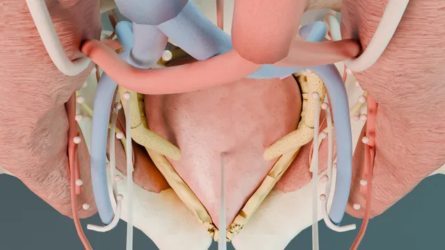
Radical cystectomy and pelvic lymphadenectomy: the pelvic atlas
A male pelvis built from reference anatomy, seen from the console. Both ureters cross the common iliac bifurcation and are divided distal to the vas. The posterior plane drops behind the vesicles onto Denonvilliers' fascia with the rectum one layer deeper.
The vesical pedicles come off the anterior division of the internal iliac with the obturator nerve in the same gutter. The dorsal venous plexus is secured before the urethra is cut, and the node template is cleared from the external iliac vein to the obturator nerve.
Advanced tierFRCS oncology, pelvic surgery, pelvic anatomyInteractive 3D
From the console, the pelvis as a bowl around the bladder. Schematic, not to scale; relationships only.
From the console the pelvis is a bowl.
Both ureters run down its rim over the common iliac bifurcation, under the vas, to the base of the bladder.
Behind the bladder the peritoneal pouch, the seminal vesicles and Denonvilliers' fascia are stacked in front of the rectum.
On each sidewall the anterior division of the internal iliac sends the superior and inferior vesical pedicles medially to the bladder, with the obturator nerve running lateral to them along the floor of the obturator fossa and the external iliac vein above.
Under the pubis the dorsal venous plexus covers the apex and the urethra.
›Ureter at the bifurcation - crosses anterior to the common iliac bifurcation, then runs under the vas to the base; divided distal to the vas with clips either side.
›Rectovesical pouch and Denonvilliers' fascia - the peritoneal reflection reaches the top of the vesicles; the fascia lies behind them with the rectal wall one layer deeper.
›Superior and inferior vesical pedicles - branches of the anterior division of the internal iliac; the umbilical artery marks the superior pedicle, the inferior runs to the base and vesicle.
›Obturator nerve - leaves the pelvis through the obturator canal along the floor of the fossa; the packet lies on it, the external iliac vein above it.
›Dorsal venous plexus - behind the pubis, anterior to the apex and urethra; secured with a figure-of-eight before the urethra is cut.
›External iliac and obturator node packets - the standard template from the node of Cloquet to the bifurcation plus the obturator fossa and the internal iliac group.
Read the pelvisFrom the camera port the peritoneum lies over the dome with the urachus and both medial umbilical ligaments converging on the umbilicus. Each ureter is a peritoneal ridge on the sidewall where it crosses the common iliac bifurcation, and the rectosigmoid sits in the sacral hollow behind the bladder. The bony pelvis, obturator internus and levator are the walls of the field; everything the operation touches lies inside that bowl.
Ureteric dissection + divisionThe peritoneum is incised over the iliac vessels and the ureter is followed down with its strip of adventitia, passing under the vas, to within two to three centimetres of the bladder. It is clipped proximally and distally and divided between the clips. The distal stump stays with the specimen; the proximal end is left long and untouched for the diversion. where it bites: the ureter lies directly on the iliac vessels at the brim: traction avulses it and a blind clip bites the vein.
Posterior planeThe peritoneum of the rectovesical pouch is incised and the plane is developed behind the vasa and seminal vesicles onto Denonvilliers' fascia, then dropped to the apex with the rectum in view. When the layer is right the plane is bloodless; fat or bleeding means it has been lost and the dissection returns to a clean area. where it bites: the rectal wall is one layer behind the fascia; a posterior tumour or previous radiotherapy thins that margin to nothing.
Vesical pediclesThe lateral pedicles are taken in order from the divided ureter toward the apex: the superior vesical pedicle at the patent umbilical artery first, then the inferior vesical pedicle running to the base and the vesicle. Each is skeletonised, clipped or sealed on the bladder side of the anterior division, and divided. where it bites: the obturator nerve shares the lateral gutter with the pedicle; a deep bite or a broad seal takes the nerve.
Apex + dorsal venous plexusThe urachus is divided high, the bladder is dropped and the endopelvic fascia is incised beside the prostate. The dorsal venous plexus is secured with a figure-of-eight before the urethra is divided at the apex, so the last attachment comes away without a venous tear. The catheter is withdrawn before the cut, and the specimen side of the urethra is clipped or tied as it is divided so nothing spills from the bladder into the pelvis. The urethral margin goes for frozen section where a neobladder is planned. where it bites: the plexus retracts behind the pubis when it tears; the save is a deep suture, never blind diathermy.
Specimen outBladder, prostate, seminal vesicles, distal ureters and the perivesical fat leave as one block. What remains is the template: the skeletonised iliac vessels, the obturator nerve on the floor of the fossa and the node packets still in place against the sidewall.
Pelvic lymphadenectomyThe standard template clears the external iliac packet from the circumflex iliac vein down at the node of Cloquet up to the bifurcation, the obturator fossa between the external iliac vein and the obturator nerve, and the internal iliac nodes. The genitofemoral nerve on psoas is the lateral limit. An extended template continues over the common iliacs to the aortic bifurcation. where it bites: an obturator vein or a tributary of the external iliac vein torn under traction bleeds into the deepest corner of the field.
The spatial relationships the animation is built to make automatic. Before you read each one, ask yourself: at this point in the operation, what is about to go wrong, and what is my first move? The save for each sits in the Complications tab.
RiskRectal wall behind Denonvilliers' fascia - one layer deep from the plane; the injury that turns a cystectomy into a colorectal reconstruction.
RiskObturator nerve in the lateral gutter - shares the gutter with the vesical pedicles and forms the floor of the node packet; a deep bite or traction injures it.
RiskExternal iliac vein at the obturator fossa - the roof of the fossa; a torn tributary bleeds dark blood into the deepest corner while the packet is lifted.
RiskDorsal venous plexus under the pubis - torn before it is secured, it retracts and bleeds from a space no clamp reaches.
Trigger each of these live in the Animation tab, then read the save: the first move, the escalation trigger, and the line that goes in the op note.
Rectal injury
the posterior plane entered one layer too deep, or a posterior tumour or previous radiotherapy leaving no plane; recognised by direct vision or the appearance of gas or stool.
The save
Recognise it the moment the plane stops being avascular: call it, improve the light and exposure, and control contamination.first move
Assess site, size and context: prepared or unprepared bowel, irradiated pelvis, degree of contamination.
A small clean injury is repaired at once in two layers with an omental or tissue interposition between the repair and the pelvis.
Larger, irradiated, contaminated or fragile-bowel injuries are repaired or resected and defunctioned with a loop stoma; colorectal input early for a low injury.
Copious irrigation, a pelvic drain and antibiotics; watch for pelvic sepsis and drain any collection radiologically.escalation trigger
Op note: site and size, layers of repair, interposition, diversion or not, and the review plan.document
Obturator nerve injury
the nerve on the floor of the fossa clipped, sealed or divided while the pedicle or the packet is taken; adductor weakness and medial thigh numbness afterwards.
The save
Recognise it: a visible clip or cut on the nerve, or the leg adducting under energy.first move
Remove the clip at once; a clean sharp division is repaired end to end with fine epineural sutures, without tension.
A thermal or crush injury is left, documented and referred for early physiotherapy; most partial injuries recover.
A complete division that cannot be repaired primarily needs a nerve surgeon's opinion for grafting.escalation trigger
Op note: mechanism, level, repair or not, and the rehabilitation plan communicated to the patient.document
External iliac vein injury
a tear of the vein or an obturator tributary while the packet is lifted out of the fossa, bleeding dark blood into the deepest corner of the template.
The save
Pressure with a swab or the assistant's instrument; do not chase it with clips in a pool of blood.first move
Tell the anaesthetist, increase the pneumoperitoneum if laparoscopic, and improve suction and exposure.
Gain proximal and distal control of the vein before the second look, then lateral venorrhaphy with a fine vascular suture.
A tributary avulsed flush with the vein is sutured, not clipped, because the clip enlarges the hole.
Failure to control, haemodynamic instability or a laceration beyond a lateral repair: convert and call vascular help.escalation trigger
Op note: vessel, method of control, estimated loss, products given, and the thromboprophylaxis plan.document
Dorsal venous plexus bleed
the plexus tears at the apex before it is secured, retracts behind the pubis and bleeds briskly exactly when the specimen is nearly free.
The save
Direct pressure with a pack and do not keep lifting it to look; tell the anaesthetist and call for help early.first move
Improve the exposure before the second look: light, retraction, suction, a clear field.
A deep figure-of-eight taken behind the retracted plexus; adjuncts supplement a stitch, they do not replace it.
If the field cannot be reached, pack the pelvis, resuscitate and plan a staged return.
Ongoing loss, coagulopathy or instability: definitive control in theatre with senior help, not repeated attempts.escalation trigger
Op note: plexus, method of control, estimated loss, blood products given.document
When the case turns: the problem, then the next move.
ActionThe ureter is hard to find at the brim - find it where it crosses the common iliac bifurcation under the peritoneum; the vas crosses over it lower down and marks the way to the base.
ActionBleeding in the posterior plane - the layer has been lost; stop, return to a clean area behind the vesicles and re-enter on Denonvilliers' fascia under direct vision.
ActionThe obturator nerve is not seen before the packet is lifted - find it at the obturator canal below the packet and follow it back; never lift a packet whose floor is unseen.
ActionThe pedicle bleeds after the seal - a second seal or clip on the sidewall side, taken only on skeletonised tissue with the nerve in view.
The examiner stems this scene prepares you for. Try to answer out loud first, then reveal, and learn the discriminator, the detail that turns a pass into a strong pass.
QDescribe the standard and extended pelvic lymphadenectomy templates for bladder cancer.reveal answer
The standard template clears the external iliac, obturator and internal iliac nodes bilaterally.
Its boundaries are the genitofemoral nerve laterally on psoas, the bladder and the umbilical artery medially, the circumflex iliac vein and the node of Cloquet distally, the common iliac bifurcation proximally, and the obturator nerve as the floor.
The obturator fossa packet lies between the external iliac vein and the obturator nerve and is the group most often positive.
The extended template continues up the common iliac vessels to the aortic bifurcation and includes the presacral nodes; a super-extended dissection carries on to the inferior mesenteric artery.
The evidence for the extended template is still argued: the German LEA trial did not show a survival benefit for extended over standard dissection, and the SWOG S1011 trial reported no improvement in disease-free survival with more morbidity, so the standard template is the minimum and the extended template is a case-by-case decision.
What is not argued is that a thorough bilateral dissection improves staging, that node yield reflects the surgeon and the pathologist as much as the patient, and that the template is cleared with the same discipline as the cystectomy itself.
Strong pass
Say both
Naming the obturator nerve as the floor and the genitofemoral nerve as the lateral limit
Stating the trial evidence on extended templates honestly rather than asserting a benefit
Examiner note
Naming the obturator nerve as the floor and the genitofemoral nerve as the lateral limit, and stating the trial evidence on extended templates honestly rather than asserting a benefit.
QWhere are the vesical pedicles and how do you control them?reveal answer
The bladder is supplied from the anterior division of the internal iliac artery.
The superior vesical artery arises from the patent proximal part of the umbilical artery and reaches the superolateral bladder;
the obliterated distal part continues as the medial umbilical ligament, which is why the ligament marks the pedicle from the console.
The inferior vesical artery, sometimes shared with the middle rectal or the internal pudendal, runs to the bladder base, the seminal vesicle and the prostate.
Their veins drain to the vesical plexus and the internal iliac vein.
I control the pedicles in order after the ureter is divided, working from the divided ureter toward the apex so that each pedicle is skeletonised before it is sealed or clipped on the bladder side, leaving the anterior division itself intact.
The obturator nerve runs in the same lateral gutter, so bites are shallow and applied only to tissue that has been seen through.
Bleeding after a seal is managed on the sidewall side with a second clip on skeletonised tissue, never by a broad blind grasp.
Strong pass
Say both
Linking the medial umbilical ligament to the superior vesical pedicle
Controlling the pedicles on the bladder side of the anterior division with the nerve in view
Examiner note
Linking the medial umbilical ligament to the superior vesical pedicle and controlling the pedicles on the bladder side of the anterior division with the nerve in view.
QTake me through the posterior dissection in a male cystectomy.reveal answer
With both ureters divided, the peritoneum of the rectovesical pouch is incised and the vasa are followed to the seminal vesicles.
The plane is developed behind the vesicles onto Denonvilliers' fascia and swept toward the apex with the rectum kept in view throughout.
Done in the right layer this is an avascular plane, and the moment fat or bleeding appears the layer has been lost, so I return to a clean area and re-enter under direct vision.
The fascia can be taken with the specimen for a posterior tumour, which puts the dissection directly on the rectal wall and is the situation in which previous radiotherapy or a bulky posterior tumour leaves no plane at all.
The lateral extent of the posterior dissection defines the pedicles, which are then taken from the ureter downward.
The rectal injury that this plane risks is recognised by direct vision or by gas and stool;
a small clean injury on prepared bowel is repaired in two layers with interposition, a large, irradiated or contaminated injury is repaired and defunctioned.
Strong pass
Say both
Treating loss of the avascular plane as the warning sign
Describing the decision to take Denonvilliers' fascia with the specimen as a deliberate oncological choice with its own rectal risk
Examiner note
Treating loss of the avascular plane as the warning sign and describing the decision to take Denonvilliers' fascia with the specimen as a deliberate oncological choice with its own rectal risk.
QHow do you avoid and manage injury to the obturator nerve during cystectomy?reveal answer
The nerve leaves the lumbar plexus, runs along the pelvic sidewall medial to psoas and leaves the pelvis through the obturator canal, forming the floor of the obturator fossa.
It is at risk at two moments: when the lateral pedicle is taken, because it shares the gutter, and when the obturator packet is lifted, because the nerve lies directly beneath it.
Prevention is exposure: I identify the nerve at the canal before any packet is lifted and keep it in view along the floor, I skeletonise before I clip or seal, and I keep energy away from it.
If a clip has been placed on the nerve it is removed immediately.
A clean sharp transection is repaired end to end with fine epineural sutures without tension;
a thermal or crush injury is left, documented and referred for early physiotherapy, and most partial injuries recover with time.
The functional consequence is adductor weakness with a leg that swings out when walking and numbness of the medial thigh, which the patient is told about honestly.
The op note records the mechanism, the level and the repair.
Strong pass
Say both
Identifying the nerve at the canal before lifting the packet
Separating a repairable sharp division from a thermal injury that is managed expectantly
Examiner note
Identifying the nerve at the canal before lifting the packet, and separating a repairable sharp division from a thermal injury that is managed expectantly.
Say it aloud, then reveal. Reading is not remembering.
01The relationshipSay the critical spatial relationship out loud, then reveal.reveal
Memory frame
From the console the pelvis is a bowl.
Why?
Both ureters run down its rim over the common iliac bifurcation, under the vas, to the base of the bladder. Behind the bladder the peritoneal pouch, the seminal vesicles and Denonvilliers' fascia are stacked in front of the rectum. On each sidewall the anterior division of the internal iliac sends the superior and inferior vesical pedicles medially to the bladder, with the obturator nerve running lateral to them along the floor of the obturator fossa and the external iliac vein above. Under the pubis the dorsal venous plexus covers the apex and the urethra.
02The first moveRectal injury. Your first move?reveal
First move
Recognise it the moment the plane stops being avascular: call it, improve the light and exposure, and control contamination.
03The escalation triggerRectal injury. What makes you call for help?reveal
Escalate when
Copious irrigation, a pelvic drain and antibiotics; watch for pelvic sepsis and drain any collection radiologically.
04The discriminatorWhat turns a pass into a strong pass?reveal
Strong pass
Say both
Naming the obturator nerve as the floor and the genitofemoral nerve as the lateral limit
Stating the trial evidence on extended templates honestly rather than asserting a benefit
Examiner note
Naming the obturator nerve as the floor and the genitofemoral nerve as the lateral limit, and stating the trial evidence on extended templates honestly rather than asserting a benefit.
05The pitfallWhere does this operation bite first?reveal
Pitfall
Rectal wall behind Denonvilliers' fascia
Why?
one layer deep from the plane; the injury that turns a cystectomy into a colorectal reconstruction.
Revision practice with self-marked answers, not an assessment of clinical competence. Kept in
this browser only; it does not sync between devices.